[Preprint]. 2024 Dec 11:2024.05.03.592468.
doi: 10.1101/2024.05.03.592468.

Transient interdomain interactions modulate the monomeric structural ensemble and self-assembly of Huntingtin Exon 1

Affiliations

Transient interdomain interactions modulate the monomeric structural ensemble and self-assembly of Huntingtin Exon 1

Priyesh Mohanty et al. bioRxiv. .

Update in

Abstract

Polyglutamine expansion (≥ 36 residues) within the N-terminal exon-1 of Huntingtin (Httex1) leads to Huntington's disease, a neurodegenerative condition marked by the presence of intranuclear Htt inclusions. Notably, the polyglutamine tract in Httex1 is flanked by an N-terminal coiled-coil domain - N17 (17 amino acids), which undergoes self-association to promote the formation of soluble Httex1 oligomers and brings the aggregation-prone polyQ tracts in close spatial proximity. However, the mechanisms underlying the subsequent conversion of soluble oligomers into insoluble β-rich aggregates with increasing polyQ length, remain unclear. Current knowledge suggests that expansion of the polyQ tract increases its helicity, and this favors its oligomerization and aggregation. In addition, studies utilizing photocrosslinking, conformation-specific antibodies and a stable coiled-coil heterotetrametric system fused to polyQ indicate that domain "cross-talk" (i.e., interdomain interactions) may play a role in the emergence of toxic conformations and the conversion of Httex1 oligomers into fibrillar aggregates. Here, we performed extensive atomistic molecular dynamics (MD) simulations (aggregate time ~ 0.7 ms) to uncover the interplay between structural transformation and domain "cross-talk" on the conformational ensemble and oligomerization landscape of Httex1. Notably, our MD-derived ensembles of N17-polyQ monomers validated against 13C NMR chemical shifts indicated that in addition to elevated α-helicity, polyQ expansion also favors transient, interdomain (N17-polyQ) interactions which result in the emergence of β-sheet conformations. Further, interdomain interactions competed with increased polyQ tract α-helicity to modulate the stability of N17-mediated dimers and thereby promoted a heterogenous dimerization landscape. Finally, we observed that the C-terminal proline-rich domain (PRD) promoted condensation of Httex1 through self-interactions involving its P10/P11 tracts while also interacting with N17 to suppress its α-helicity. In summary, our study demonstrates a significant role for domain "cross-talk" in modulating the monomeric structural ensemble and self-assembly of Httex1.


Figures
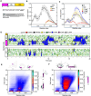
Figure 1.
Figure 1.. Analysis of N17-Q16 conformational ensemble illustrates the structural cooperativity between N17 and polyQ regions.
A. Schematic showing the organization of Httex1 into three regions - N17, polyQ and PRD (top), and the corresponding sequences of N17/PRD domains (bottom). The length of polyQ region in the wild-type Huntingtin protein is ~23 residues. B. Comparison of per-residue α-helix fractions for N17-Q16 wild-type and its deletion constructs with respect to NMR. Helical fractions were computed using the DSSP algorithm and compared to SSP scores for the helical propensity (grey) computed from experimental 13C chemical shifts. Error bars denote std. error of mean calculated over three independent trajectories. C. Same as in panel B for N17-Q16 wild-type and 14LKSF17 mutants. D. Secondary structure variation as a function of time in a representative N17-Q16 (top) and Q16 (bottom) trajectory. E. SS-map of N17-Q16 wild-type computed from an aggregate trajectory (~63 μs) indicating the probability of various helical length across N17 and polyQ regions. Representative α-helical conformations corresponding to numbered regions of the SS-map are shown in cartoon representation. F. Same as in E for 14LLLF17 mutant.
Figure 2.
Figure 2.. Conformational ensemble of N17-Q46.
A. Comparison of per-residue α-helix fractions for N17-polyQn constructs with increasing polyQ length (Q16–46) against NMR-derived helical propensities (SSP scores). Error bars denote std. error of mean calculated over three independent trajectories for N17-Q16–32 constructs and over four intervals (150 ns each) of the PT-WTE replica trajectory (293 K) for N17-Q46. B. SS-map of N17-Q46 wild-type (left) from the PT-WTE aggregate trajectory (600 ns) indicating the probability of α-helices formed of varying lengths across N17 and polyQ regions. Representative helical conformations corresponding to numbered regions of the SS-map are shown in cartoon representation. C. Secondary structure variation as a function of time for two representative N17-Q46 trajectories showing the formation of two-stranded β-sheet structures (black boxes). D. Two-dimensional intramolecular contact maps calculated over six N17-Q46 independent trajectories indicating the low population of β-conformations (marked as *) relative to α-helices. E. Representative β-sheet structures (5 in total) from the trajectory periods highlighted in C (black boxes) involving either direct (i) and indirect (ii) contacts between the N17 and Q46 domains. The coloring scheme for the structures is as follows: N17 - purple, Q46 - white and 5P - Gold, β-strand - red. F. Contact maps calculated for the two β-sheet structural ensembles shown in E.
Figure 3.
Figure 3.. Competition between structural transformation and interdomain interactions shape the dimerization landscape of N17-polyQ.
A. NMR structure of Httex1-Q7 (H7) tetramer showing interactions (sidechains shown as sticks) in the hydrophobic core. The anti-parallel dimer was extracted from the structure for MD simulations. B. Comparison of per-residue α-helix fractions for H7/16 chains from dimer simulations against monomer simulations and NMR. Error bars denote std. error of mean calculated over six independent trajectories. The comparison indicates significantly elevated helicity of N17 region in H7/16 dimers compared to their respective monomers. C. Two-dimensional PMF (potential of mean force) plots as a function of two order parameters - (i) total number of intermolecular N17 contacts and (ii) the inter-helical angle (°) between N17 domains, which characterize the association free energy landscape for four N17 dimer variants. The plots shows the combined data derived from 6 independent dimer trajectories (~2.3 μs) for each variants. The position of NMR - N17 dimer (initial structure) is marked on the PMF plots as (*). Representative dimer MD structures from the basins marked (1, 2) in PMF plots are shown on the left with interfacial hydrophobic residues shown as grey sticks.
Figure 4.
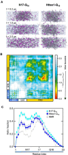
Figure 4.. PRD-mediated interactions stabilize the Httex1-Q16 condensate and suppress N17 ɑ-helicity.
A. All-atom MD simulation snapshots of N17-Q16 (left) and Httex1-Q16 (right) condensate systems over 2 μs of their respective trajectories. Protein chains are show as ribbons and ions (Na+/Cl-) are shown as spheres. The snapshots indicate destabilization of the N17-Q16 dense phase (0.2 – 1.0 μs) and its complete dissolution (2.0 μs), resulting in a homogeneous system. B. Pairwise 2D-intermolecular contact map averaged over all chain pairs reveals significant contributions of the PRD towards stabilization of the Httex1-Q16. C. Mean fractional ɑ-helicity per-residue (computed over all dense phase chains) shows reduced ɑ-helical formation within N17 for the Httex1-Q16 condensate compared to a homogenous N17-Q16 condensate system (i.e., lacking protein-solvent interface). Error bars denote std. error of mean over all chains.
Figure 5.
Figure 5.. polyQ expansion promotes “domain crosstalk” which modulates the structural ensemble and oligomerization landscape of Httex1 to favor fibrillar aggregation.
Schematic illustrating the effect of polyQ length expansion on the conformation and oligomerization of Httex1. polyQ expansion promotes inter-domain N17-polyQ interactions. These interactions can (i) induce the emergence of transient β-sheet conformers in monomers which may fold to form the critical nucleus and initiate protofibril formation (oligomer-independent pathway), and (ii) counteract stabilization of N17-mediated oligomers to favor polyQ-polyQ interactions which lead to nucleation and fibrillation. The nucleus size is shown as a monomer (n=1) and is representative of Httex1-Q>23.

Similar articles

References

    1. Ross C. A. Polyglutamine Pathogenesis. Neuron 2002, 35 (5), 819–822. 10.1016/S0896-6273(02)00872-3. - DOI - PubMed
    1. Shao J.; Diamond M. I. Polyglutamine Diseases: Emerging Concepts in Pathogenesis and Therapy. Hum Mol Genet 2007, 16 (R2), R115–R123. 10.1093/hmg/ddm213. - DOI - PubMed
    1. Adegbuyiro A.; Sedighi F.; Pilkington A. W.; Groover S.; Legleiter J. Proteins Containing Expanded Polyglutamine Tracts and Neurodegenerative Disease. Biochemistry 2017, 56 (9), 1199–1217. 10.1021/acs.biochem.6b00936. - DOI - PMC - PubMed
    1. DiFiglia M.; Sapp E.; Chase K. O.; Davies S. W.; Bates G. P.; Vonsattel J. P.; Aronin N. Aggregation of Huntingtin in Neuronal Intranuclear Inclusions and Dystrophic Neurites in Brain. Science (1979) 1997, 277 (5334), 1990–1993. 10.1126/science.277.5334.1990. - DOI - PubMed
    1. Sieradzan K. A.; Mechan A. O.; Jones L.; Wanker E. E.; Nukina N.; Mann D. M. A. Huntington’s Disease Intranuclear Inclusions Contain Truncated, Ubiquitinated Huntingtin Protein. Exp Neurol 1999, 156 (1), 92–99. 10.1006/exnr.1998.7005. - DOI - PubMed

Publication types

LinkOut - more resources