[Preprint]. 2024 May 6:2024.05.06.592747.
doi: 10.1101/2024.05.06.592747.

Structural basis for Rab6 activation by the Ric1-Rgp1 complex

Affiliations

Structural basis for Rab6 activation by the Ric1-Rgp1 complex

J Ryan Feathers et al. bioRxiv. .

Update in

Abstract

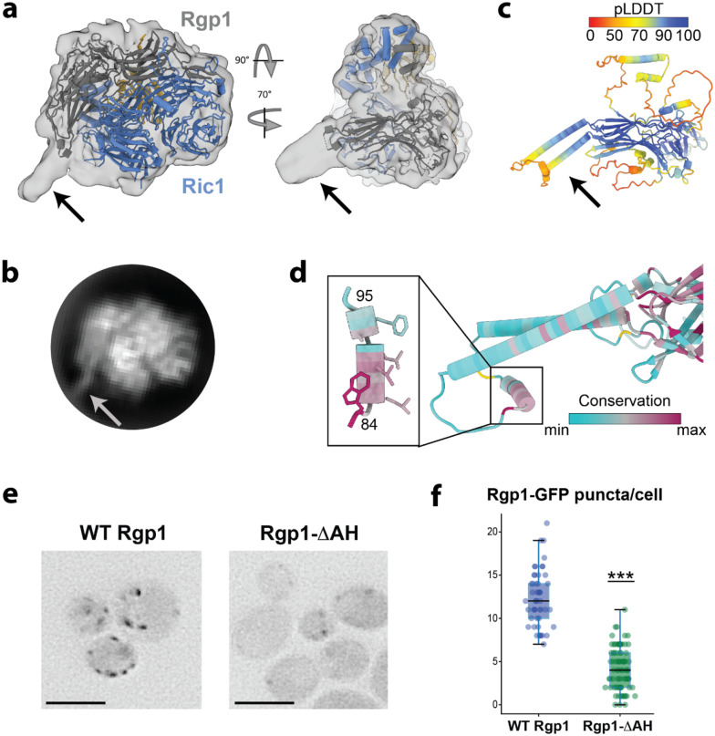
Rab GTPases act as molecular switches to regulate organelle homeostasis and membrane trafficking. Rab6 plays a central role in regulating cargo flux through the Golgi and is activated via nucleotide exchange by the Ric1-Rgp1 protein complex. Ric1-Rgp1 is conserved throughout eukaryotes but the structural and mechanistic basis for its function has not been established. Here we report the cryoEM structure of a Ric1-Rgp1-Rab6 complex representing a key intermediate of the nucleotide exchange reaction. This structure reveals the overall architecture of the complex and enabled us to identify interactions critical for proper recognition and activation of Rab6 on the Golgi membrane surface. Ric1-Rgp1 interacts with the nucleotide-binding domain of Rab6 using an uncharacterized helical domain, which we establish as a novel RabGEF domain by identifying residues required for Rab6 nucleotide exchange. Unexpectedly, the complex uses an arrestin fold to interact with the Rab6 hypervariable domain, indicating that interactions with the unstructured C-terminal regions of Rab GTPases may be a common specificity mechanism used by their activators. Collectively, our findings provide a detailed mechanistic understanding of regulated Rab6 activation at the Golgi.


Conflict of interest statement

COMPETING INTERESTS The authors declare no competing interests.

Figures

Figure 1:
Figure 1:. Architecture of the Ric1-Rgp1-Rab6 activation intermediate complex
a) CryoEM reconstruction of the Ric1-Rgp1 complex ‘caught in the act’ of performing nucleotide exchange on Rab6. b) Atomic model of the complex built using the cryoEM density, shown in cartoon format, and highlighting several structural elements including some newly defined in this work (see panel e). c) Same as in (b), from a different perspective. d) Same as in (b) and (c), from a different perspective. e) Structural elements of the three proteins in the complex. Newly established elements are the GEF domain, the GBE (GEF binding element); and an amphipathic helix (AH).
Figure 2:
Figure 2:. Structural basis for Rab6 activation by nucleotide exchange
a) View of the interface between the Ric1 GEF domain (blue) and Rab6 (gold), with the Rab6 ‘switch I’ element (residues 32–44) colored red. The GBE of Rgp1 is colored gray. b) Overlay of the crystal structure of Rab6 in its GDP-bound state (colored purple, PDB: 1D5C), with the nucleotide-free state from the Ric1-Rgp1-bound cryoEM structure (colored gold). Equivalent positions in primary sequence are marked with asterisks. c) Close-up view of the Rab6-GEF domain interface, highlighting the Ric1 residues subjected to mutational analysis. d) Results from an in vitro GEF assay using purified Ric1-Rgp1, prenylated-Rab6-GDI complex, and liposomes. The change in fluorescence is due to exchange of the Rab6-bound mant-GDP for GTP, representing activation of Rab6. n = 3 traces are shown for each condition (WT or F860A/R912A Ric1-Rgp1 complex). e) Complementation test (cells lacking Ric1-Rgp1 function are temperature sensitive). f). Comparison of the localization patterns of WT and F860A/R912A Ric1-mNeonGreen.
Figure 3:
Figure 3:. Comparison of Ric1-Rgp1 to other RabGEF domains
a) Comparison of the Ric1 GEF-Rab6 structure to structures of other known RabGEF domain-Rab structures. Note for many of these structures, while the structure of the GEF domain is known, the structure of the intact GEF has not been determined. Each Rab is colored gray and positioned in the same orientation. Ric1-Rgp1 (this work, PDB 9AYR), Sec2/Rabin8, (PDB 2OCY); DrrA/SidM, (PDB 3JZA); Mon1-Ccz1, (PDB 5LDD); TRAPP complexes, (PDB 7U05); DENND1B-S, (PDB 3TW8); RABEX-5,, (PDB 4Q9U); ARA7/Vps9a, (PDB 4G01); SH3BP5, (PDB 6DJL). b) Comparison of the Rab6-bound Ric1 GEF domain to the Arf1-bound Sec7 GEF domain (PDB: 1RE0). c) Overlay of the nine GEF-bound Rab structures shown in panel (a), with switch I regions colored red and switch II regions colored blue.
Figure 4:
Figure 4:. The Ric1-Rgp1 complex binds to the HVD of Rab6
a) Surface view of the Ric1-Rgp1-Rab6 atomic model. b) Close-up view of the portion of the Rab6 HVD bound to the arrestin-C subdomain of Rgp1. The HVD adopts a β-strand conformation to integrate into the β-sheet on the surface of the arrestin-C subdomain β-sandwich fold. c) Same view as in panel (b), with the surface of Rgp1 colored by sequence conservation. d) Live-cell imaging to measure the colocalization of WT and chimeric GFP-tagged Rab6 constructs with endogenous RFP-Rab6. Scale bar, 5 microns. e) Quantitation of the data in (c), with all individual data points overlaid on box-and-whiskers plots. ***, p=2.6×10⁻⁹. WT: n=13 images; mutant: n=16 images.
Figure 5:
Figure 5:. A predicted amphipathic helix in Rgp1 is important for Golgi membrane association
a) CryoEM density map shown at low threshold, together with the atomic model of the Ric1-Rgp1-Rab6 complex. The arrow denotes unmodeled density projecting from the Rgp1 subunit. b) CryoEM 2D class average representing the 2D projection of the complex from the same approximate orientation as the perspective depicted in panel (a). The arrow corresponds to the unmodeled density in the 3D reconstruction. c) AlphaFold predicted structure of the Rgp1 subunit, colored by confidence score. The arrow corresponds to the region of the prediction that is unmodeled in the cryoEM structure due to the low-resolution of that portion of the cryoEM density. d) A conserved amphipathic helix lies at the distal tip of this structural element. e) Live-cell imaging of WT and ΔAH Rgp1-GFP constructs. Scale bar, 5 microns. f) Quantitation of the experiment shown in panel (e) with all individual data points overlaid with box-and-whiskers. ***, p=2.2×10⁻¹⁶. WT: n=50 cells; mutant: n=83 cells. See Methods for descriptions of statistics.
Figure 6:
Figure 6:. Structural model for Rab6 activation on the Golgi membrane surface
The expected orientation of the Ric1-Rgp1 complex relative to the Golgi membrane surface is shown. GDI must transiently release inactive, GDP-bound Rab6 before it can be activated by the Ric1-Rgp1 complex. The GDI-Rab-GDP complex is depicted using the crystal structure of a GDI-Rab1/Ypt1 complex (PDB: 2BCG).

Similar articles

References

    1. Pfeffer S. R. Rab GTPases: master regulators that establish the secretory and endocytic pathways. Mol. Biol. Cell 28, 712–715 (2017). - PMC - PubMed
    1. Hutagalung A. H. & Novick P. J. Role of Rab GTPases in membrane traffic and cell physiology. Physiol. Rev. 91, 119–149 (2011). - PMC - PubMed
    1. Barr F. A. Review series: Rab GTPases and membrane identity: causal or inconsequential? J. Cell Biol. 202, 191–199 (2013). - PMC - PubMed
    1. Goody R. S., Rak A. & Alexandrov K. The structural and mechanistic basis for recycling of Rab proteins between membrane compartments. Cell. Mol. Life Sci. 62, 1657–1670 (2005). - PMC - PubMed
    1. Lee P. L., Ohlson M. B. & Pfeffer S. R. Rab6 regulation of the kinesin family KIF1C motor domain contributes to Golgi tethering. Elife 4, (2015). - PMC - PubMed

Publication types