[Preprint]. 2024 May 11:2024.05.10.593592.
doi: 10.1101/2024.05.10.593592.

Asesino: a nucleus-forming phage that lacks PhuZ

Affiliations

Asesino: a nucleus-forming phage that lacks PhuZ

Amy Prichard et al. bioRxiv. .

Update in

Abstract

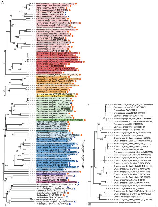
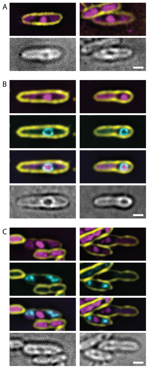
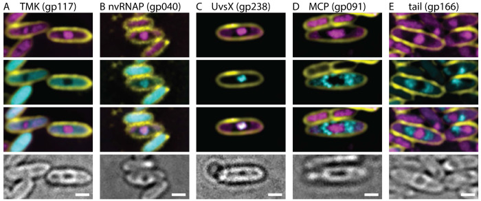
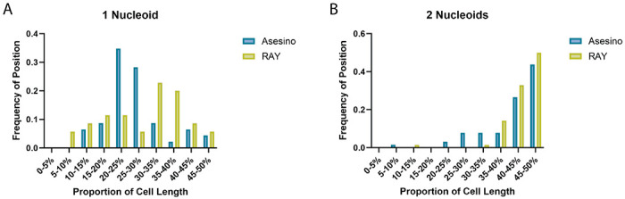
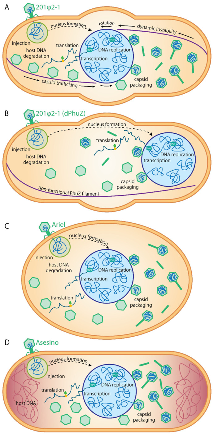
As nucleus-forming phages become better characterized, understanding their unifying similarities and unique differences will help us understand how they occupy varied niches and infect diverse hosts. All identified nucleus-forming phages fall within the proposed Chimalliviridae family and share a core genome of 68 unique genes including chimallin, the major nuclear shell protein. A well-studied but non-essential protein encoded by many nucleus-forming phages is PhuZ, a tubulin homolog which aids in capsid migration, nucleus rotation, and nucleus positioning. One clade that represents 24% of all currently known chimalliviruses lacks a PhuZ homolog. Here we show that Erwinia phage Asesino, one member of this PhuZ-less clade, shares a common overall replication mechanism with other characterized nucleus-forming phages despite lacking PhuZ. We show that Asesino replicates via a phage nucleus that encloses phage DNA and partitions proteins in the nuclear compartment and cytoplasm in a manner similar to previously characterized nucleus-forming phages. Consistent with a lack of PhuZ, however, we did not observe active positioning or rotation of the phage nucleus within infected cells. These data show that some nucleus-forming phages have evolved to replicate efficiently without PhuZ, providing an example of a unique variation in the nucleus-based replication pathway.


Conflict of interest statement

COMPETING INTERESTS STATEMENT J.P. has an equity interest in Linnaeus Bioscience Incorporated and receives income. The terms of this arrangement have been reviewed and approved by the University of California, San Diego, in accordance with its conflict-of-interest policies.

Figures

Figure 1.
Figure 1.. Phylogenetic trees of chimalliviruses.
(A) A whole-genome phylogenetic tree made using VICTOR of the chimalliviruses, highlighted in rainbow colors according to predicted genus assignment (Figure S1), plus 10 outgroup representatives. Phages encoding a PhuZ homolog are marked with orange circles, and phages without PhuZ homologs are marked with blue X marks. The only clade where every member lacks PhuZ (besides singleton Vibrio phage Yong XC31) is highlighted in blue and labeled “Phuzzywuzzyviruses.” (B) A whole genome phylogenetic tree made using VICTOR of all currently known members of the phuzzywuzzyvirus clade. For both trees, bootstrap values are displayed on the branches.
Figure 2.
Figure 2.. Asesino makes a nucleus during its infection cycle.
(A) DAPI staining shows a bright circular region consistent with a phage nucleus, as well as extranuclear DNA consistent with undegraded bacterial DNA. (B) Asesino’s chimallin homolog (gp039) tagged with GFP encloses the bright DAPI signal. (C) E. amylovora H-NS tagged with GFP localizes with the extranuclear DNA during infection, supporting the idea that this is undegraded host DNA. For all panels, yellow is FM4-64 (membrane stain), magenta is DAPI (DNA stain), cyan is GFP, and grayscale is brightfield. Scale bars represent 1μm.
Figure 3.
Figure 3.. Protein localization in Asesino is consistent with other nucleus-forming phages.
(A) Thymidylate kinase is localized in the cytoplasm. (B) A nonvirion RNA polymerase subunit and (C) RecA homolog UvsX are localized in the nucleus. (D) The major capsid protein and (E) a tail protein localize around the phage nucleus. For all panels, yellow is FM4-64 (membrane stain), magenta is DAPI (DNA stain), cyan is GFP, and grayscale is brightfield. Scale bars represent 1μm.
Figure 4.
Figure 4.. Phage nucleus positioning in Asesino compared to RAY.
(A) When only one bacterial nucleoid is present, the Asesino phage nucleus has a bias away from midcell positioning, with most phage nuclei being found between the pole and midcell (20-30% of the cell length, blue), but the RAY phage nucleus has a slight bias toward midcell positioning (light green). (B) When two bacterial nucleoids are present, both the Asesino (blue) and RAY (light green) phage nuclei are positioned at or near midcell, with Asesino having a slightly wider distribution.
Figure 5.
Figure 5.. Infection Models.
Diagrams comparing (A) a PhiKZ-like Pseudomonas phage 201phi2-1 infected cell; (B) a 201phi2-1 infected cell expressing a catalytically dead, dominant negative PhuZ mutant; (C) a Vibrio phage Ariel infected cell; and (D) an Erwinia phage Asesino infected cell.

References

    1. Knipe D. M., Prichard A., Sharma S. & Pogliano J. Replication Compartments of Eukaryotic and Bacterial DNA Viruses: Common Themes Between Different Domains of Host Cells. Annu Rev Virol 9, 307–327 (2022). - PMC - PubMed
    1. Chaikeeratisak V. et al. Assembly of a nucleus-like structure during viral replication in bacteria. Science 355, 194–197 (2017). - PMC - PubMed
    1. Chaikeeratisak V. et al. The Phage Nucleus and Tubulin Spindle Are Conserved among Large Pseudomonas Phages. Cell Rep. 20, 1563–1571 (2017). - PMC - PubMed
    1. Malone L. M. et al. A jumbo phage that forms a nucleus-like structure evades CRISPR-Cas DNA targeting but is vulnerable to type III RNA-based immunity. Nat Microbiol 5, 48–55 (2020). - PubMed
    1. Mendoza S. D. et al. A bacteriophage nucleus-like compartment shields DNA from CRISPR nucleases. Nature 577, 244–248 (2020). - PMC - PubMed

Publication types