[Preprint]. 2024 May 12:2024.05.11.593568.
doi: 10.1101/2024.05.11.593568.

Functional interrogation of cellular Lp(a) uptake by genome-scale CRISPR screening

Affiliations

Functional interrogation of cellular Lp(a) uptake by genome-scale CRISPR screening

Taslima G Khan et al. bioRxiv. .

Update in

Abstract

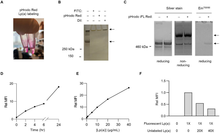
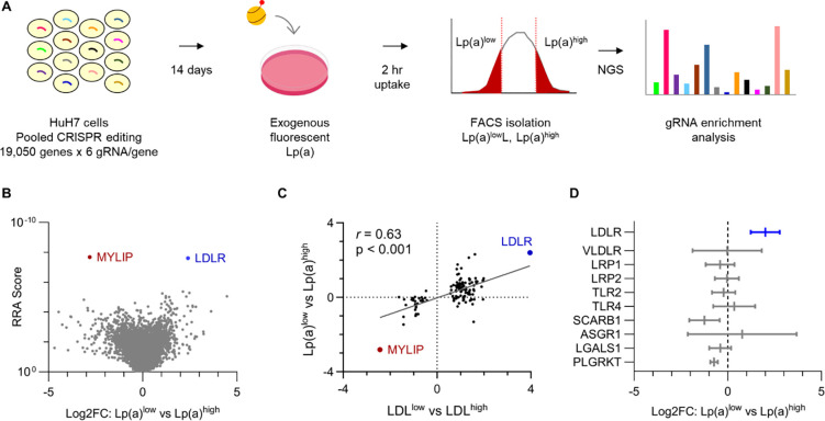
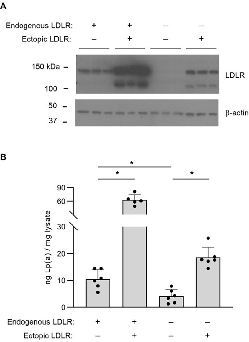
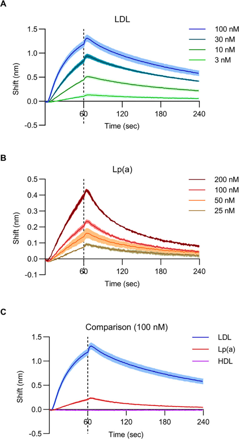
An elevated level of lipoprotein(a), or Lp(a), in the bloodstream has been causally linked to the development of atherosclerotic cardiovascular disease and calcific aortic valve stenosis. Steady state levels of circulating lipoproteins are modulated by their rate of clearance, but the identity of the Lp(a) uptake receptor(s) has been controversial. In this study, we performed a genome-scale CRISPR screen to functionally interrogate all potential Lp(a) uptake regulators in HuH7 cells. Strikingly, the top positive and negative regulators of Lp(a) uptake in our screen were LDLR and MYLIP, encoding the LDL receptor and its ubiquitin ligase IDOL, respectively. We also found a significant correlation for other genes with established roles in LDLR regulation. No other gene products, including those previously proposed as Lp(a) receptors, exhibited a significant effect on Lp(a) uptake in our screen. We validated the functional influence of LDLR expression on HuH7 Lp(a) uptake, confirmed in vitro binding between the LDLR extracellular domain and purified Lp(a), and detected an association between loss-of-function LDLR variants and increased circulating Lp(a) levels in the UK Biobank cohort. Together, our findings support a central role for the LDL receptor in mediating Lp(a) uptake by hepatocytes.


Conflict of interest statement

Declaration of interests The authors have no relevant competing financial interests to declare.

Figures

Figure 1.
Figure 1.. Development of a method for fluorescent labeling of purified Lp(a) and quantification of its cellular uptake with flow cytometry.
(A) Overt sample precipitation during attempted fluorescent labeling of purified Lp(a) preparations with pHrodo Red. (B) SDS-PAGE and silver straining of purified Lp(a) preparations before and after attempted fluorescent labeling with FITC, pHrodo Red, and DiI. Upper arrow corresponds to precipitated protein remaining in loading well. Lower arrow corresponds to expected size (~500 kDa) of co-migrating apo(a) and apolipoprotein B proteins. (C) SDS-PAGE under reducing and nonreducing conditions of a purified Lp(a) preparation before and after fluorescent labeling with pHrodo iFL Red followed by silver staining or in-gel fluorescence scanning. Upper arrow corresponds to expected migration of disulfide-linked apo(a)-apolipoprotein B complexes under nonreducing conditions; lower arrow corresponds to expected comigration of noncomplexed apo(a) and apolipoprotein B under reducing conditions. (D) Time course analysis of HuH7 cellular uptake of 10 μg/mL pHrodo iFL Red-labeled Lp(a). (E) Concentration-dependence of HuH7 cellular uptake of pHrodo iFL Red-labeled Lp(a) over 2 hrs. (F) Dose-dependent competitive inhibition of HuH7 cellular fluorescent Lp(a) uptake by co-incubation with a molar excess of unlabeled Lp(a). Representative flow cytometry plots are provided in Supplemental Figure 1.
Figure 2.
Figure 2.. A genome-scale CRISPR screen for modifiers of HuH7 cellular Lp(a) uptake.
(A) Schematic overview of screening strategy, with pools of HuH7 cells transduced with the GeCKOv2 genome-wide CRISPR knockout library at low MOI, passaged for 14 days, incubated with fluorescent Lp(a) for 2 hrs, and sorted into Lp(a)low and Lp(a)high subpopulations for extraction of genomic DNA and quantification of individual gRNA abundance by next-generation sequencing (NGS). (B) Volcano plot of aggregate gene-level Robust Rank Aggregation (RRA) scores (y-axis) relative to log2 fold change of normalized gRNA counts in Lp(a)low relative to Lp(a)high subpopulations. Genes whose disruption was associated with a significant (FDR<5%) decrease or increase in Lp(a) uptake are labeled and highlighted in blue and red, respectively. (C) Analysis of genes previously identified in our analogous prior screen of cellular LDL uptake, with the aggregate log2 fold change for gRNAs targeting each gene in LDLlow relative to LDLhigh cells (x-axis) in that screen plotted relative to gRNA log2 fold change in Lp(a)low relative to Lp(a)high cells in this study. (D) Mean and 95% confidence intervals of aggregate gRNA log2 fold enrichment in Lp(a)low relative to Lp(a)high cells across 3 independent biologic replicates for genes encoding receptors previously proposed as mediators of cellular Lp(a) uptake. RRA scores, aggregate log2 fold change, and FDR values were calculated by MAGeCK; 95% confidence intervals of independent replicates were calculated using GraphPad Prism. Source data are provided in Supplemental Tables 1 and 2.
Figure 3.
Figure 3.. Analysis of Lp(a) uptake in LDLR-disrupted and LDLR-overexpressing HuH7 cells.
(A) Immunoblot analysis of LDLR protein abundance in wild-type HuH7 cells and a clonal HuH7 cell line harboring a homozygous frameshift-causing insertion in exon 6 of LDLR, each with and without transduction of a lentiviral expression construct of a LDLR cDNA. (B) Quantification of cellular apolipoprotein(a) internalization for the cell lines indicated in (A) after incubation with 50 μg/mL Lp(a) for 3 hrs. Individual data points represent the mean of technical duplicates for each independent biologic replicate. Asterisks indicates p<0.05 by Student’s t-test and error bars depict standard deviation.
Figure 4.
Figure 4.. In vitro binding between Lp(a) and the LDLR extracellular domain.
(A) Biolayer interferometry measurements for LDLR binding with LDL at the indicated concentrations. (B) Biolayer interferometry measurements for LDLR binding with Lp(a) at the indicated concentrations. (C) Comparison of biolayer interferometry measurements for LDLR binding to LDL, Lp(a), and HDL each at 100 nM. For all panels, solid lines indicate mean values and shaded regions indicate 1 standard deviation above and below the mean. Sensors were incubated with lipoprotein suspensions at t = 0 seconds and transferred to buffer only at t = 60 seconds, indicated by the dashed line.
Figure 5.
Figure 5.. Analysis of Lp(a) levels for carriers of LDLR pathogenic alleles in the UK Biobank cohort.
(A-B) Adjusted LDL and Lp(a) levels aggregated for 225 carriers and 358,405 noncarriers of 25 expert-curated pathogenic LDLR alleles in UK Biobank. Statistical analysis for (A) and (B) was performed with the burden testing framework in Regenie. (C) Correlation between LDL and Lp(a) beta coefficients for all single LDLR variants with the indicated annotations and a minor allele count of at least 25 in UK Biobank. Individual data points represent single variants and are colored by functional annotation. Regression analysis was performed with GraphPad Prism. (D) Lp(a) beta coefficients for single variants represented by at least 25 individuals in UK Biobank, grouped by ClinVar annotation with statistical analysis between groups performed by Student’s t-test assuming equal variance. For all panels, horizontal lines represent mean values and error bars indicate standard error of the mean.

References

    1. Berg K. A New Serum Type System in Man--the Lp System. Acta Pathol Microbiol Scand 59, 369–382, doi:10.1111/j.1699-0463.1963.tb01808.x (1963). - DOI - PubMed
    1. Berg K., Dahlen G. & Frick M. H. Lp(a) lipoprotein and pre-beta1-lipoprotein in patients with coronary heart disease. Clin Genet 6, 230–235, doi:10.1111/j.1399-0004.1974.tb00657.x (1974). - DOI - PubMed
    1. Djurovic S. & Berg K. Epidemiology of Lp(a) lipoprotein: its role in atherosclerotic/thrombotic disease. Clin Genet 52, 281–292, doi:10.1111/j.1399-0004.1997.tb04345.x (1997). - DOI - PubMed
    1. Seman L. J. et al. Lipoprotein(a)-cholesterol and coronary heart disease in the Framingham Heart Study. Clin Chem 45, 1039–1046 (1999). - PubMed
    1. Bostom A. G. et al. Elevated plasma lipoprotein(a) and coronary heart disease in men aged 55 years and younger. A prospective study. JAMA 276, 544–548, doi:10.1001/jama.1996.03540070040028 (1996). - DOI - PubMed

Publication types