[Preprint]. 2024 May 10:rs.3.rs-4350156.
doi: 10.21203/rs.3.rs-4350156/v1.

Radiomics-Based Predictive Nomogram for Assessing the Risk of Intracranial Aneurysms

Affiliations

Radiomics-Based Predictive Nomogram for Assessing the Risk of Intracranial Aneurysms

Sricharan S Veeturi et al. Res Sq. .

Update in

Abstract

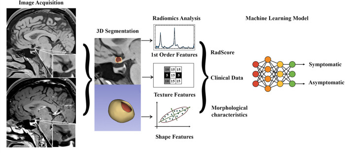
Background: Aneurysm wall enhancement (AWE) has the potential to be used as an imaging biomarker for the risk stratification of intracranial aneurysms (IAs). Radiomics provides a refined approach to quantify and further characterize AWE's textural features. This study examines the performance of AWE quantification combined with clinical information in detecting symptomatic IAs.

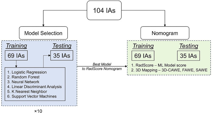
Methods: Ninety patients harboring 104 IAs (29 symptomatic and 75 asymptomatic) underwent high-resolution magnetic resonance imaging (HR-MRI). The assessment of AWE was performed using two different methods: 3D-AWE mapping and composite radiomics-based score (RadScore). The dataset was split into training and testing subsets. The testing set was used to build two different nomograms using each modality of AWE assessment combined with patients' demographic information and aneurysm morphological data. Finally, each nomogram was evaluated on an independent testing set.

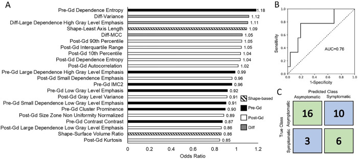
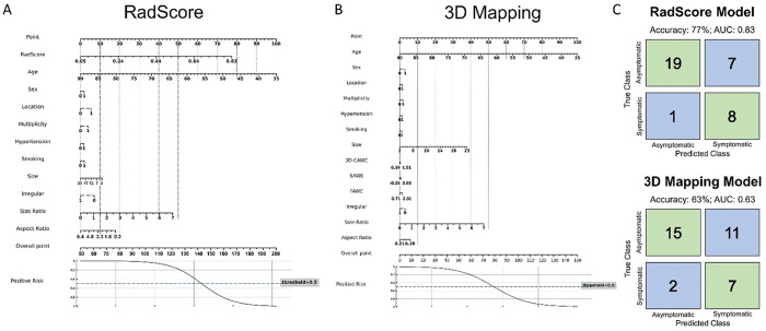
Results: A total of 22 radiomic features were significantly different between symptomatic and asymptomatic IAs. The 3D-AWE Mapping nomogram achieved an area under the curve (AUC) of 0.77 (63% accuracy, 78% sensitivity and 58% specificity). The RadScore nomogram exhibited a better performance, achieving an AUC of 0.83 (77% accuracy, 89% sensitivity and 73% specificity).

Conclusions: Combining AWE quantification through radiomic analysis with patient demographic data in a clinical nomogram achieved high accuracy in detecting symptomatic IAs.

Keywords: Aneurysms; aneurysm wall enhancement; radiomics.


Figures

Figure 1
Figure 1. Analysis Pipeline.
Images were acquired with a 3T high resolution magnetic resonance imaging with and without contrast gadolinium. Segmentation of the aneurysm wall (shell) and aneurysm sac were performed in 3D Slicer. Radiomic analysis included extraction of enhancement (1st order features), texture and shape features of the aneurysm wall. Transform-based RFs were not included in the figure, as these were not used for analysis. Significant RFs, clinical data and morphological aneurysm characteristics were used to build a machine learning model that accurately identified symptomatic status.
Figure 2
Figure 2. Overall workflow.
We split the dataset into training and testing datasets. The training dataset was used to evaluate six different machine learning models. We then evaluate the best machine learning model that can identify symptomatic IAs. We then use the probability of this model as an input to build two nomograms: RadScore-based nomogram and 3D Mapping-based nomogram.
Figure 3
Figure 3. Final trained RadScore model.
The bar plot shows odds ratios for all the radiomics features used in the RadScore model in descending order of importance. The overall AUC was 0.76 and the accuracy was 63% (67% sensitivity and 62% specificity).
Figure 4
Figure 4. Nomograms for RadScore (Composite Radiomics-based score) and 3D mapping models.
Two nomograms were constructed using RadScore, AWE metrics from 3D mapping pipeline and patient demographics (gender = female 1 and male 0; location = 0 for low-risk location MCA or ICA, and = 1 for high-risk location ACom or posterior circulation; multiplicity = 0 for a single aneurysm in the patient and 1 for more than one aneurysm; hypertension = 0 for no hypertension and 1 for hypertension; and smoking = 0 for never smoked and 1 for past or current smoker) and IA characteristics (size, SR, AR, irregularity = 0 for smooth regular shape and 1for multi-lobulated irregular shape or has blebs). We observed that the RadScore-based nomogram had a higher accuracy, sensitivity and specificity compared to the 3D mapping pipeline-based nomogram.

References

    1. Wiebers DO, Whisnant JP, Huston J 3rd, Meissner I, Brown RD Jr., Piepgras DG et al. Unruptured intracranial aneurysms: natural history, clinical outcome, and risks of surgical and endovascular treatment. Lancet. 2003;362(9378):103–10. doi:10.1016/s0140-6736(03)13860-3. - DOI - PubMed
    1. Sanchez S, Hickerson M, Patel RR, Ghazaleh D, Tarchand R, Paranjape GS et al. Morphological Characteristics of Ruptured Brain Aneurysms: A Systematic Literature Review and Meta-Analysis. Stroke: Vascular and Interventional Neurology. 2023;3(2):e000707. doi:doi:10.1161/SVIN.122.000707. - DOI
    1. Waqas M, Chin F, Rajabzadeh-Oghaz H, Gong AD, Rai HH, Mokin M et al. Size of ruptured intracranial aneurysms: a systematic review and meta-analysis. Acta Neurochir (Wien). 2020;162(6):1353–62. doi:10.1007/s00701-020-04291-z. - DOI - PubMed
    1. Bijlenga P, Gondar R, Schilling S, Morel S, Hirsch S, Cuony J et al. PHASES Score for the Management of Intracranial Aneurysm. Stroke. 2017;48(8):2105–12. doi:doi:10.1161/STROKEAHA.117.017391. - DOI - PubMed
    1. Backes D, Rinkel GJE, Greving JP, Velthuis BK, Murayama Y, Takao H et al. ELAPSS score for prediction of risk of growth of unruptured intracranial aneurysms. Neurology. 2017;88(17):1600–6. doi:10.1212/wnl.0000000000003865. - DOI - PubMed

Publication types

LinkOut - more resources