Review
. 2024 May 15;68(2):3954.
doi: 10.4081/ejh.2024.3954.

A candidate projective neuron type of the cerebellar cortex: the synarmotic neuron

Affiliations
Review

A candidate projective neuron type of the cerebellar cortex: the synarmotic neuron

Paolo Flace et al. Eur J Histochem. .

Abstract

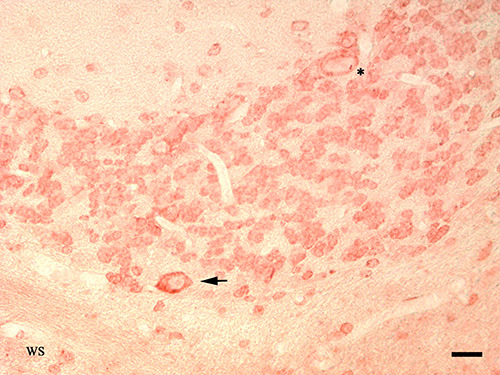
Previous studies on the granular layer of the cerebellar cortex have revealed a wide distribution of different subpopulations of less-known large neuron types, called "non-traditional large neurons", which are distributed in three different zones of the granular layer. These neuron types are mainly involved in the formation of intrinsiccircuits inside the cerebellar cortex. A subpopulation of these neuron types is represented by the synarmotic neuron, which could play a projective role within the cerebellar circuitry. The synarmotic neuron cell body map within the internal zone of the granular layer or in the subjacent white substance. Furthermore, the axon crosses the granular layer and runs in the subcortical white substance, to reenter in an adjacent granular layer, associating two cortico-cerebellar regions of the same folium or of different folia, or could project to the intrinsic cerebellar nuclei. Therefore, along with the Purkinje neuron, the traditional projective neuron type of the cerebellar cortex, the synarmotic neuron is candidate to represent the second projective neuron type of the cerebellar cortex. Studies of chemical neuroanatomy evidenced a predominant inhibitory GABAergic nature of the synarmotic neuron, suggesting that it may mediate an inhibitory GABAergic output of cerebellar cortex within cortico-cortical interconnections or in projections towards intrinsic cerebellar nuclei. On this basis, the present minireview mainly focuses on the morphofunctional and neurochemical data of the synarmotic neuron, and explores its potential involvement in some forms of cerebellar ataxias.


Figures

Figure 1.
Figure 1.
The Anatomic Table by Eber Landau on the synarmotic neuron in the human cerebellar cortex (modified by Paolo Flace, 2021). Table of transverse section of human cerebellar cortex according to the information provided by the Golgi and by Ramon y Cajal silver staining. Morphological structure of the cerebellar cortex: ML, molecular layer; PL, Purkinje neuron layer; GL, granular layer; WS, white substance. Cortico-cerebellar neurons present in the modified anatomic table: P, Purkinje neuron; G, Golgi neuron; S, synarmotic neuron. The cell bodies of different synarmotic neurons are distributed in the internal zone of the granular layer and in the subcortical white substance.
Figure 2.
Figure 2.
The Anatomic Table by Eber Landau on the synarmotic neuron in the human cerebellar cortex (modified by Paolo Flace, 2021). Table of transverse section of human cerebellar cortex according to the information provided by the Golgi silver staining. Morphological structure of the cerebellar cortex: ML, molecular layer; PL, Purkinje neuron layer; GL, granular layer; WS, white substance. Cortico-cerebellar neurons present in the modified anatomic table: P, Purkinje neuron; G, Golgi neuron; S, synarmotic neuron. A synarmotic neuron cell body localized in the subcortical white substance interposed between two adjacent granular layers, its processes are distributed in both the granular layers.
Figure 3.
Figure 3.
The Anatomic Table by Eber Landau on the synarmotic neuron in the dog cerebellar cortex (modified by Paolo Flace, 2021). Table of transverse section of dog cerebellar cortex according to the information provided by the Golgi and by Ramon y Cajal silver staining. Morphological structure of the cerebellar cortex: ML, molecular layer; PL, Purkinje neuron layer; GL, granular layer; WS, white substance. Cortico-cerebellar neurons present in the modified anatomic table: S, synarmotic neuron. A synarmotic neuron cell body localized in the internal zone of the granular layer, its processes are distributed in two adjacent granular layers.
Figure 4.
Figure 4.
GAD65/67 immunoreactive synarmotic neurons in the granular layer of the human cerebellar cortex. GAD65/67 immunoreactive synarmotic neuron cell body (arrow) localized in the granular layer in close proximity to the subcortical white substance (WS), from the cell body originates a thin axon-like process. GAD65/67 immunoreactive Purkinje neuron cell body and primary dendritic trunk (asterisk); magnification 40x; scale bar: 15 μm. Method: streptavidin-biotin-peroxidase immunoenzymatic technique; chromogen: 3-amino-9-ethyl-carbazole; type of antibody: GAD65/67 rabbit polyclonal antibody; Chemicon International, Temecula, CA, USA. Negative control: the sections are covered with normal rabbit serum or with anti-GAD antiserum absorbed with an excess of synthetic GAD (Chemicon International) in place of the primary antibody (original unpublished micrographs; for protocol details, see).
Figure 5.
Figure 5.
5-HT immunoreactive synarmotic neuron in the granular layer of the human cerebellar cortex. 5-HT immunoreactive synarmotic neuron cell body localized in the internal zone of the granular layer (arrow) among immunonegative granules, in proximity to the subcortical white substance (WS); magnification 40x; scale bar: 20 μm. Method: streptavidin-biotin-peroxidase immunoenzymatic technique; chromogen: 3,3′-Diaminobenzidine; type of antibody: 5-HT rabbit polyclonal antobody, Covance Lab., Inc., Princeton, NJ, USA. Positive control: fragments of rat duodenum and brainstem are subjected to the same experimental procedures (original unpublished micrographs; for protocol details, see).
Figure 6.
Figure 6.
Contactin-1 immunoreactive synarmotic neuron in the mouse cerebellar cortex. Contactin-1 immunoreactive synarmotic neuron cell body (arrow) localized in the internal zone of the granular layer, in close proximity to the subcortical white substance (WS). Contactin-1 immunoreactive Purkinje neuron cell body (asterisk); magnification 40x; scale bar: 20 μm. Method: streptavidin-biotin-peroxidase immunoenzymatic technique; chromogen: 3-amino-9-ethyl-carbazole; type of antibody: Contactin-1 rabbit polyclonal antibody (original unpublished micrographs; for protocol details, see).
Figure 7.
Figure 7.
GAD65-auto-antibodies in the cerebellum. Scheme on the distribution of the autoantibodies against the GABA biosynthetic enzymatic isoform, glutamic acid decarboxylase isoform 65 (GAD65-Auto-Abs) related to the GABAergic synaptic sites of the synarmotic neuron in the cerebellum. BN, basket neuron; PN, Purkinje neuron, SN, stellate neuron; GN, Golgi neuron; GrN, granule; Syn, synarmotic neuron; CNu, cerebellar nuclei; CB, climbing fibers; MF, mossy fibers.

References

    1. Jansen J, Brodal A. [Das Kleinhirn] In: Bargman W, editor. [Handbuch der Mikroskopiscen Anatomie des Menschen].[Book in German]. Berlin, Springer; 1958. pp 91-149.
    1. Eccles JC, Ito M, Szentàgothai J. The cerebellum as a new neuronal machine. Springer, Berlin; 1967.
    1. Fox CA, Snider RS. The cerebellum. Amsterdam, Elsevier; 1967.
    1. Mugnaini E. The histology and cytology of the cerebellar cortex. In: Larsell O, Jansen J. editors. The comparative anatomy and histology of the cerebellum: the human cerebellum, cerebellar connections and cerebellar cortex. Minneapolis, Minnesota Press; 1972. pp 201-64.
    1. Ito M. The cerebellum and neural control. New York, Raven Press; 1984.