. 2024 Sep 2;223(9):e202402006.
doi: 10.1083/jcb.202402006. Epub 2024 May 20.

NEKL-4 regulates microtubule stability and mitochondrial health in ciliated neurons

Affiliations

NEKL-4 regulates microtubule stability and mitochondrial health in ciliated neurons

Kaiden M Power et al. J Cell Biol. .

Abstract

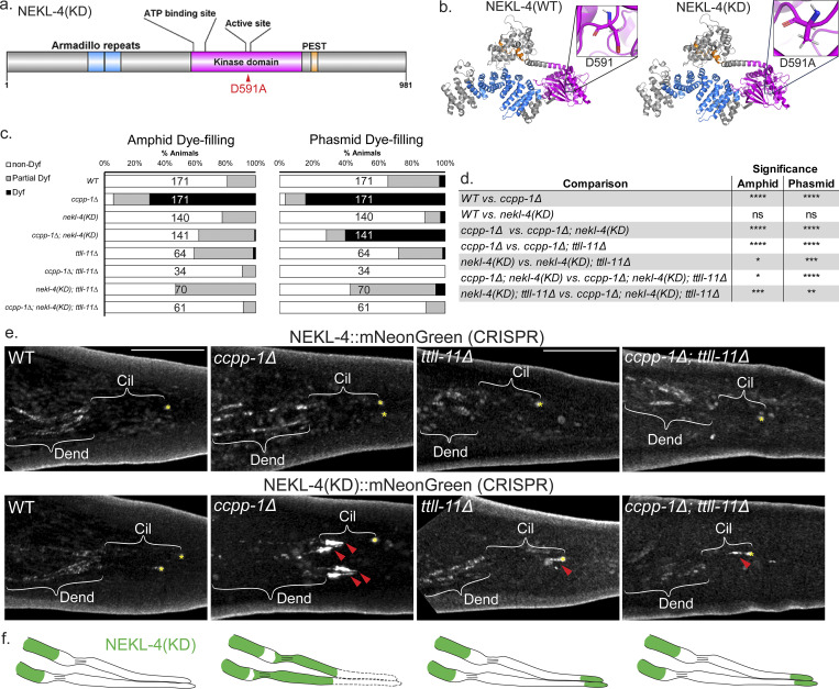
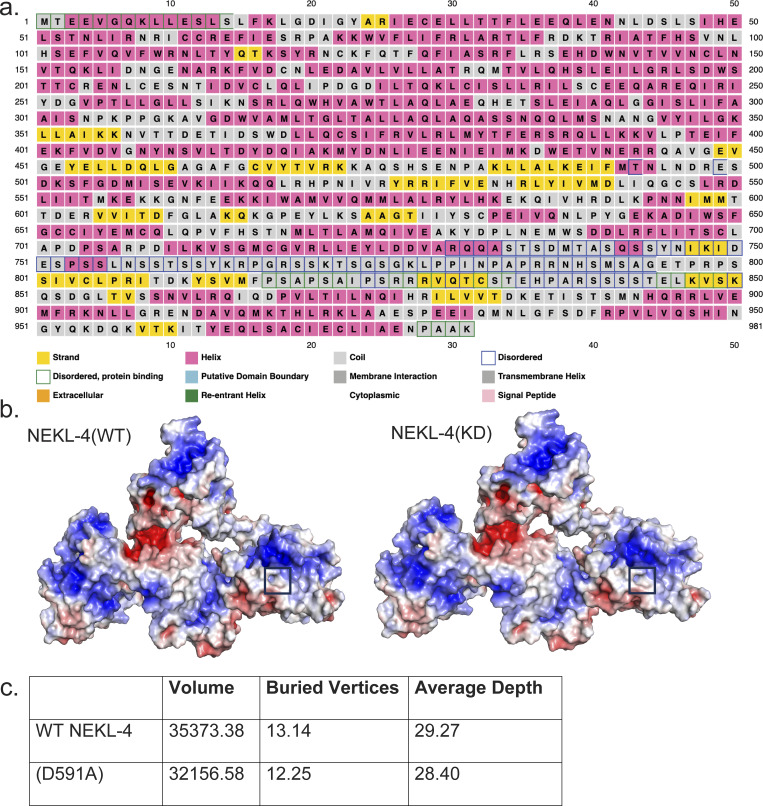
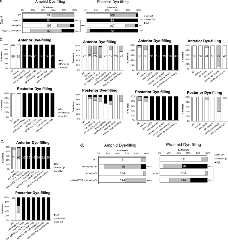
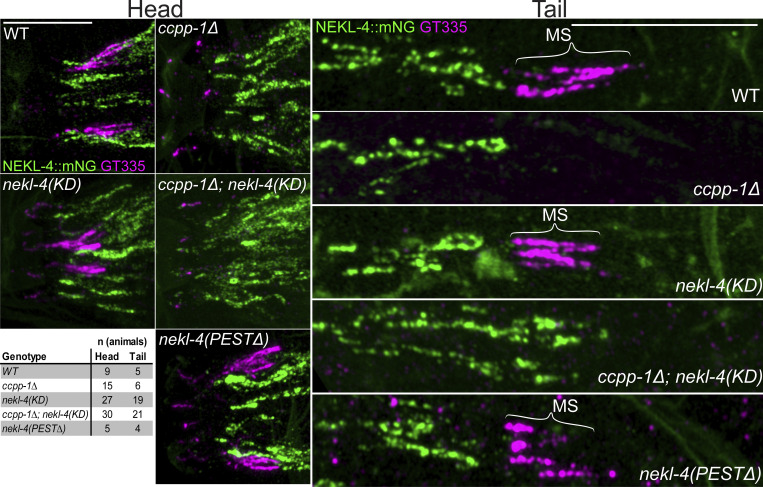
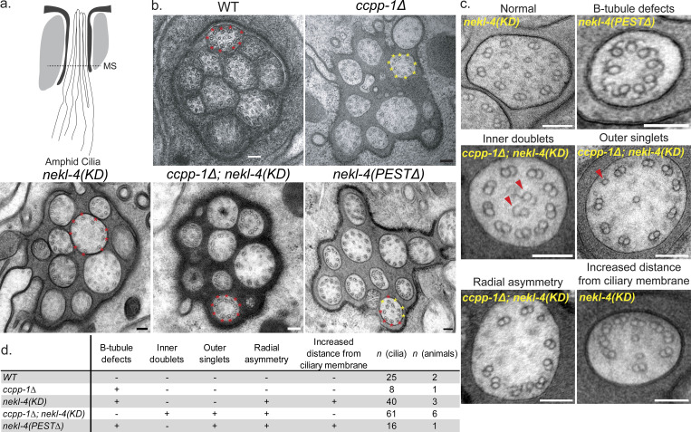
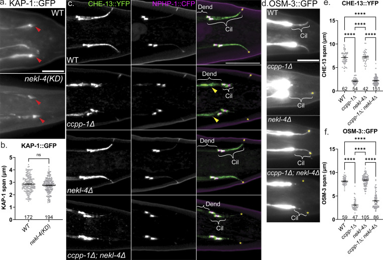
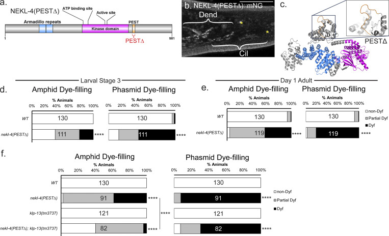
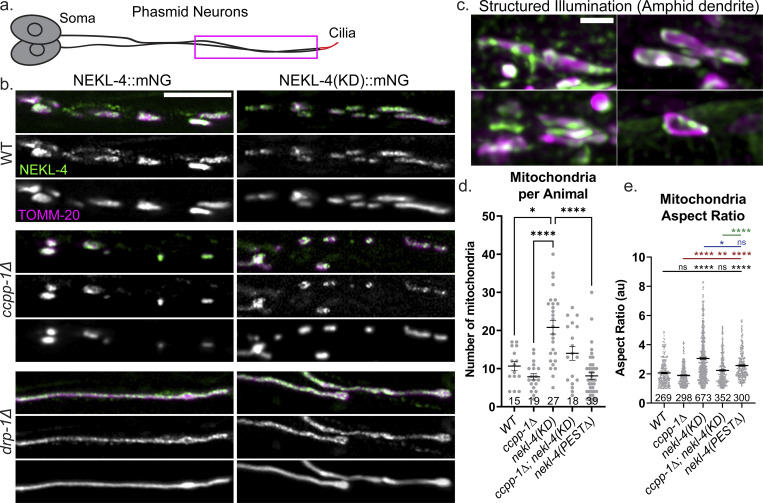
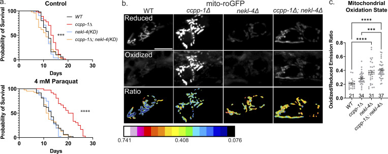
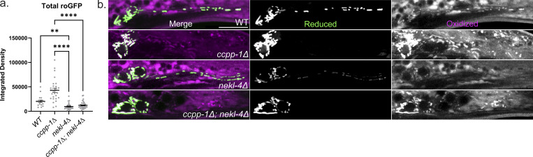
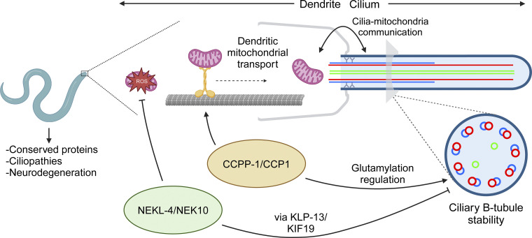
Ciliopathies are often caused by defects in the ciliary microtubule core. Glutamylation is abundant in cilia, and its dysregulation may contribute to ciliopathies and neurodegeneration. Mutation of the deglutamylase CCP1 causes infantile-onset neurodegeneration. In C. elegans, ccpp-1 loss causes age-related ciliary degradation that is suppressed by a mutation in the conserved NEK10 homolog nekl-4. NEKL-4 is absent from cilia, yet it negatively regulates ciliary stability via an unknown, glutamylation-independent mechanism. We show that NEKL-4 was mitochondria-associated. Additionally, nekl-4 mutants had longer mitochondria, a higher baseline mitochondrial oxidation state, and suppressed ccpp-1∆ mutant lifespan extension in response to oxidative stress. A kinase-dead nekl-4(KD) mutant ectopically localized to ccpp-1∆ cilia and rescued degenerating microtubule doublet B-tubules. A nondegradable nekl-4(PEST∆) mutant resembled the ccpp-1∆ mutant with dye-filling defects and B-tubule breaks. The nekl-4(PEST∆) Dyf phenotype was suppressed by mutation in the depolymerizing kinesin-8 KLP-13/KIF19A. We conclude that NEKL-4 influences ciliary stability by activating ciliary kinesins and promoting mitochondrial homeostasis.


Conflict of interest statement

Disclosures: All authors have completed and submitted the ICMJE Form for Disclosure of Potential Conflicts of Interest. C. Rongo reported grants from FiteBac Pharma outside the submitted work. No other disclosures were reported.

Figures

Figure 1.
Figure 1.
Loss of NEKL-4 kinase activity suppresses ccpp-1∆ dye-filling defects (Dyf), and NEKL-4(KD) localizes to cilia in ccpp-1∆, ttll-11∆, and ccpp-1∆; ttll-11∆ mutant animals. (a) Schematic diagram of NEKL-4 domains showing the location of the D591A active site mutation. (b) Full-length NEKL-4 and NEKL-4(KD) predicted protein structures based on the amino-acid sequence and homolog structure information. Magenta = kinase domain, blue = armadillo repeat domain, orange = PEST sequence. The positions of the active site residue D591 and the mutant D591A are highlighted. (c) Dye-filling assays of Day 2 adult animals. (d) Significance of relevant comparisons in c. * indicates P ≤ 0.05, ** indicates P ≤ 0.01, *** indicates P ≤ 0.001, **** indicates P ≤ 0.0001 by Kruskal–Wallis one-way ANOVA with post hoc Dunn’s correction for multiple comparisons. (e) Confocal images of endogenously-tagged NEKL-4::mNeonGreen in the phasmid neurons. Scale bar = 10 µm. Cil = cilia, Dend = dendrites, red arrowhead = NEKL-4 ciliary localization, * = phasmid pore. (f) Schematics of NEKL-4(KD) localization in WT, ccpp-1∆, ttll-11∆, and ccpp-1∆; ttll-11∆ mutants. Dashed lines in the ccpp-1∆ panel represent ciliary degeneration.
Figure S1.
Figure S1.
In silico modeling of NEKL-4 and NEKL-4(KD). (a) Secondary structure prediction and prediction of intrinsically disordered regions of NEKL-4. (b) Surface Electrostatic Potential of NEKL-4 and NEKL-4(KD). Blue = Positive, Red = Negative (−5 to +5 kT/e). Boxed area is residue D591 or D591A. (c) Cleft analysis of NEKL-4 and NEKL-4(KD).
Figure S2.
Figure S2.
Additional dye-filling data for nekl-4(KD), nekl-4∆, and nekl-4(PEST∆) mutants. (a) nekl-4(KD) mutation continues to suppress the ccpp-1∆ dye-filling defect into later adulthood (Day 4). (b) Negative dye-filling interactions for nekl-4∆ and nekl-4(KD). (c) Negative dye-filling interactions for nekl-4(PEST∆). (d) Mutation of kinesin-3 KLP-4/KIF13 suppresses the nekl-4(PEST∆) Dyf phenotype. For all panels, * indicates P ≤ 0.05, ** indicates P ≤ 0.01, **** indicates P ≤ 0.0001 by Kruskal–Wallis one-way ANOVA with post hoc Dunn’s correction for multiple comparisons.
Figure S3.
Figure S3.
Loss of NEKL-4 kinase activity does not suppress the loss of GT335 staining in ccpp-1∆ mutant amphid and phasmid cilia. Images of endogenously-tagged NEKL-4::mNeonGreen and GT335, a monoclonal antibody that detects branch point glutamylation, in the amphid, labial, and cephalic cilia. In most images, labial and cephalic cilia are stained but not visible since the images are adjusted to best show the amphids, which stain much brighter. Scale bars = 10 µm. MS = middle segment.
Figure 2.
Figure 2.
nekl-4(KD) mutation suppresses B-tubule instability in ccpp-1∆ amphid cilia. (a) Diagram of the amphid cilia indicating the position of the cross-sections examined. MS = middle segment. (b) Examples of microtubule doublet structure in WT, ccpp-1∆, nekl-4(KD), ccpp-1∆; nekl-4(KD), and nekl-4(PEST∆) mutant amphid channels. The WT and ccpp-1∆ panels are reproduced from O’Hagan et al. (2011) and show a male amphid. All other genotypes show a hermaphrodite amphid. Red * = normal microtubule doublet, yellow * = microtubule doublet with broken or missing B-tubule in a representative cilium. Scale bars = 100 nm. (c) Examples of phenotypes seen in nekl-4(KD), ccpp-1∆; nekl-4(KD), and nekl-4(PEST∆) mutant cilia. Scale bars = 100 nm. (d) Table of phenotypes observed in each genotype and numbers of cilia and animals analyzed. The WT and ccpp-1∆ data are reproduced from O’Hagan et al. (2011).
Figure S4.
Figure S4.
KAP-1, CHE-13, and OSM-3 spans are not altered in nekl-4 mutants. (a) Examples of KAP-1::GFP localization. Red arrowhead = phasmid cilia. Scale bar = 10 µm. (b) Quantification of KAP-1 span in the phasmid cilia. Not significant by Mann–Whitney test. (c) Confocal images of CHE-13 and NPHP-1, a transition zone protein. (d) Widefield images of OSM-3 in the phasmid cilia. (e) Quantification of the span of CHE-13 in the phasmid cilia. (f) Quantification of the span of OSM-3 in the phasmid cilia. For c and d, Cil = cilia, Dend = dendrites, yellow arrowhead = dendritic CHE-13 accumulation, * = phasmid pore. Scale bar = 10 µm. For e and f, mean ± SEM; **** indicates P ≤ 0.0001 by Kruskal–Wallis one-way ANOVA with post hoc Dunn’s correction for multiple comparisons. Note that in c, the dendritic accumulation of CHE-13 in ccpp-∆ mutants is suppressed by nekl-4∆.
Figure 3.
Figure 3.
NEKL-4(PEST∆) causes a progressive Dyf phenotype that is suppressed by mutation of kinesin-8 KLP-13/KIF19. (a) Schematic diagram of NEKL-4 showing the location of the PESTΔ mutation. (b) Fluorescence microscope image showing expression of NEKL-4(PEST∆)::mNG. Scale = 10 µm. Cil = cilia, Dend = dendrites, * = phasmid pore. (c) NEKL-4(PEST∆) modeled and predicted protein structure. Magenta = kinase domain, blue = armadillo repeat domain, orange = residues surrounding the removed PEST sequence. The removal of the residues within the PEST domain is highlighted. (d) Dye-filling assays of nekl-4(PEST∆) L3 larvae. (e) Dye-filling assays of nekl-4(PEST∆) Day 1 adults. For d and e, **** indicates P ≤ 0.0001 by Mann–Whitney test. (f) Dye-filling assays of nekl-4(PEST∆); klp-13 Day 1 adults. **** indicates P ≤ 0.0001 by Kruskal–Wallis one-way ANOVA with post hoc Dunn’s correction for multiple comparisons.
Figure 4.
Figure 4.
NEKL-4 is mitochondria-associated and influences mitochondrial morphology. (a) Cartoon of one set of C. elegans phasmid neurons. Imaged area is boxed in magenta. (b) Confocal images of mutants showing NEKL-4::mNG and TOMM-20::tagRFP labeling the outer mitochondrial membrane. Scale bar = 5 µm. (c) Representative SIM images of NEKL-4::mNG and TOMM-20 colocalization on individual mitochondria in the amphid dendrites. Scale bar = 1 µm. (d and e) Quantification of the number of mitochondria in the phasmid dendrites per animal and the aspect ratio of each mitochondrion (length/width). Mean ± SEM; * indicates P ≤ 0.05, ** indicates P ≤ 0.01, **** indicates P ≤ 0.0001 by Kruskal–Wallis one-way ANOVA with post hoc Dunn’s correction for multiple comparisons.
Figure 5.
Figure 5.
ccpp-1∆ and nekl-4 mutations affect both sensitivity to oxidative stress and baseline oxidative stress in C. elegans ciliated sensory neurons. (a) Survival curves of control (top) and paraquat-exposed (bottom) worms, where Day 0 represents when Day 1 adult hermaphrodites were placed on plates. Control n = 50, 54, 52, 47; PQ n = 47, 36, 47, 40; *** indicates P ≤ 0.001, **** indicates P ≤ 0.0001 by Log-rank Mantel-Cox test. (b) Uniformly adjusted images of phasmid soma in the “reduced” channel (top) and “oxidized” channel (middle), as well as the ratio of oxidized/reduced (bottom). Scale bar = 5 µm. (c) Quantification of oxidized/reduced emission ratios, where one point represents one set of phasmid soma. Mean ± SEM; *** indicates P ≤ 0.001, **** indicates P ≤ 0.0001 by Kruskal–Wallis one-way ANOVA with post hoc Dunn’s correction for multiple comparisons.
Figure S5.
Figure S5.
Differences in roGFP levels and dendritic localization in ccpp-1∆ and nekl-4∆ mutants. (a) Total roGFP signal measured in phasmid soma, calculated by adding the integrated densities of the 488 and 405 nm channels. Mean ± SEM; ** indicates P ≤ 0.01, **** indicates P ≤ 0.0001 by Kruskal–Wallis one-way ANOVA with post hoc Dunn’s correction for multiple comparisons. (b) Dendritic mitochondrial localization is defective in ccpp-1∆ and ccpp-1∆; nekl-4∆ mutants. Uncropped images of roGFP in the phasmid neurons. Scale bar = 10 µm.
Figure 6.
Figure 6.
Model for NEKL-4 function in relation to cilia, mitochondria, and CCPP-1. NEKL-4 functions in the ciliated neurons to reduce mitochondrial oxidative stress and destabilize ciliary B-tubules through the action of ciliary kinesins such as KLP-13. This ciliary function opposes the B-tubule stabilizing action of CCPP-1 through a pathway distinct from glutamylation regulation. CCPP-1 also promotes dendritic transport in the ciliated neurons, specifically the transport of mitochondria. Created with https://BioRender.com (license BE26DR2HV3).

Update of

References

    1. Al Mutairi, F., Alkhalaf R., Alkhorayyef A., Alroqi F., Yusra A., Umair M., Nouf F., Khan A., Meshael A., Hamad A., et al. . 2020. Homozygous truncating NEK10 mutation, associated with primary ciliary dyskinesia: A case report. BMC Pulm. Med. 20:141. 10.1186/s12890-020-1175-1 - DOI - PMC - PubMed
    1. Alvarez Viar, G., Klena N., Martino F., Nievergelt A., and Pigino G.. 2023. The tubulin nano-code: A protofilament-specific pattern of tubulin post-translational modifications regulates ciliary beating mechanics. bioRxiv. 10.1101/2023.06.28.546853 (Preprint posted June 28, 2023). - DOI - PMC - PubMed
    1. Apfeld, J., and Kenyon C.. 1999. Regulation of lifespan by sensory perception in Caenorhabditis elegans. Nature. 402:804–809. 10.1038/45544 - DOI - PubMed
    1. Bae, J.-E., Jang S., Kim J.B., Hyung H., Park N.Y., Kim Y.H., Kim S.H., Kim S.H., Ha J.M., Oh G.S., et al. . 2023. Enhanced primary ciliogenesis via mitochondrial oxidative stress activates AKT to prevent neurotoxicity in HSPA9/mortalin-depleted SH-SY5Y cells. Mol. Brain. 16:41. 10.1186/s13041-023-01029-7 - DOI - PMC - PubMed
    1. Bae, J.-E., Kang G.M., Min S.H., Jo D.S., Jung Y.-K., Kim K., Kim M.-S., and Cho D.-H.. 2019. Primary cilia mediate mitochondrial stress responses to promote dopamine neuron survival in a Parkinson’s disease model. Cell Death Dis. 10:952. 10.1038/s41419-019-2184-y - DOI - PMC - PubMed

MeSH terms

LinkOut - more resources