Review
. 2024 Apr;116(2):78-92.
doi: 10.32074/1591-951X-993.

Cardiovascular pathology in vasculitis

Affiliations
Review

Cardiovascular pathology in vasculitis

Monica De Gaspari et al. Pathologica. 2024 Apr.

Abstract

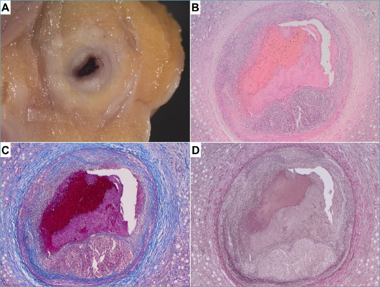
Vasculitides are diseases that can affect any vessel. When cardiac or aortic involvement is present, the prognosis can worsen significantly. Pathological assessment often plays a key role in reaching a definite diagnosis of cardiac or aortic vasculitis, particularly when the clinical evidence of a systemic inflammatory disease is missing. The following review will focus on the main histopathological findings of cardiac and aortic vasculitides.

Keywords: aorta; autopsy; cardiovascular pathology; coronary arteries; vasculitis.


Conflict of interest statement

The authors declare no competing interests.

Figures

Figure 1.
Figure 1.
Aortitis in giant cell arteritis. A 73-year-old male with aneurysmatic dilation of the ascending aorta. (A) The panoramic view of the aorta reveals a massive granulomatous inflammation located in the outer third of the media, coupled with mild adventitial inflammation and thickening. (B) At higher magnification, numerous giant cells are admixed with activated macrophages and lymphocytes. (C) Elastic stain demonstrates the complete disruption of the tunica media. (A, B) Hematoxylin-eosin stain, (C) Weigert van Gieson stain.
Figure 2.
Figure 2.
Aortitis in Ig4-related disease. A 56-year-old woman underwent surgery for an abdominal aortic aneurysm. (A) Marked adventitial thickening consistent in lymphoplasmacytic inflammation coupled with fibrosis in a storiform pattern is demonstrated at histology. (B) Abundant plasma cells are evident at higher magnification. C) Masson trichrome stain highlights storiform fibrosis that completely replaced tunica media. (A, B) Hematoxylin-eosin stain, (C) Masson trichrome stain.
Figure 3.
Figure 3.
Coronary artery vasculitis in Takayasu’s disease. A 14-year-old girl was rescued after a sudden cardiac arrest. Aortography showed mild aortic dilation with aortic valve insufficiency and sub-occlusion of coronary ostia. The girl died and the autopsy revealed massive myocardial infarction with thickening of the ascending aorta, aortic arch, pulmonary arteries, left external carotid artery, and coronary ostia (modified from ). (A) The left anterior descending coronary artery shows severe luminal narrowing because of concentric intimal hyperplasia. The tunica media appears destroyed and the adventitia is fibrotic. (B) The involved segments reveal necrotizing granulomatous inflammation with giant cells, consistent with Takayasu’s arteritis. (A) Masson trichrome stain; (B) Hematoxylin-eosin stain.
Figure 4.
Figure 4.
Coronary artery vasculitis in Kawasaki disease. A 6-year-old child recently diagnosed with Hodgkin lymphoma died after some episodes of chest pain. The autopsy revealed aneurysmal dilatation of both the left and right coronary arteries, with suspected luminal thrombosis. (A) A tortuous and dilated left descending coronary artery is indicated by arrows. (B-C) The lumen of coronary artery is occluded by a thrombus. (D-E) Histological analysis confirms the presence of aneurysm with thinning of the vessel wall, polymorphic inflammation and elastic disruption (D). (C, D) Hematoxylin-eosin stain; (E) Weigert van Gieson stain.
Figure 5.
Figure 5.
Coronary artery vasculitis in panarteritis nodosa. A 49-year-old man presenting with chest pain due to a suspected myocardial infarction died after a short hospitalization. Panarteritis nodosa was diagnosed only at autopsy. (A) Gross analysis of coronary arteries revealed diffuse concentric lesions. (B-D) At histology, transmural necrotizing polymorphic inflammation was detected with thrombotic occlusion of the right coronary artery. (B) Hematoxylin-eosin stain, (C) Heidenhain’s trichrome stain, (D) Weigert van Gieson stain.
Figure 6.
Figure 6.
Coronary artery dissection and eosinophilic coronary periarteritis. A 35-year-old woman was admitted to the emergency room because of acute thoracic pain. She died shortly afterward. (A, B) At autopsy, coronary artery dissection of the left anterior descending and diagonal branches was revealed with serial sections of the coronary tree. (C) Histological analysis demonstrated a marked eosinophilic infiltrate at the level of the adventitia in the culprit vessel (C, Hematoxylin-eosin stain). A diagnosis of eosinophilic coronary periarteritis in the setting of coronary artery dissection was made.

Similar articles

Cited by

References

    1. Watts RA, Robson J. Introduction, epidemiology and classification of vasculitis. Best Pract Res Clin Rheumatol. 2018;32(1):3-20. https://doi.org/10.1016/j.berh.2018.10.003 10.1016/j.berh.2018.10.003 - DOI - PubMed
    1. Ponte C, Águeda AF, Luqmani RA. Clinical features and structured clinical evaluation of vasculitis. Best Pract Res Clin Rheumatol. 2018;32(1):31-51. https://doi.org/10.1016/j.berh.2018.10.001 10.1016/j.berh.2018.10.001 - DOI - PubMed
    1. Silveira LH. Cardiovascular Manifestations of Systemic Vasculitides. Curr Rheumatol Rep. 2020;22(10):72. https://doi.org/10.1007/s11926-020-00952-1 10.1007/s11926-020-00952-1 - DOI - PubMed
    1. Jennette JC, Falk RJ, Bacon PA, et al. . 2012 revised International Chapel Hill Consensus Conference Nomenclature of Vasculitides. Arthritis Rheum. 2013;65(1):1-11. https://doi.org/10.1002/art.37715 10.1002/art.37715 - DOI - PubMed
    1. Domínguez-Quintana M, Alba MA, Hinojosa-Azaola A. Classification of ANCA-associated vasculitis: differences based on ANCA specificity and clinicopathologic phenotype. Rheumatol Int. 2021;41(10):1717-1728. https://doi.org/10.1007/s00296-021-04966-5 10.1007/s00296-021-04966-5 - DOI - PubMed