Review
. 2024 Apr;116(2):104-118.
doi: 10.32074/1591-951X-990.

Systemic vasculitis involving the kidney: the nephropathologist's point of view

Affiliations
Review

Systemic vasculitis involving the kidney: the nephropathologist's point of view

Vincenzo L'Imperio et al. Pathologica. 2024 Apr.

Abstract

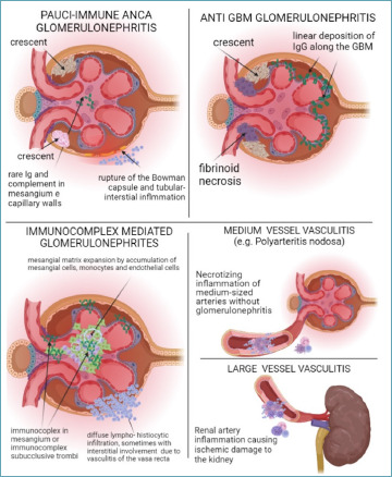
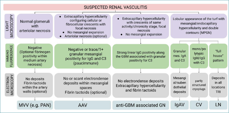
Kidneys are often targets of systemic vasculitis (SVs), being affected in many different forms and representing a possible sentinel of an underlying multi-organ condition. Renal biopsy still remains the gold standard for the identification, characterization and classification of these diseases, solving complex differential diagnosis thanks to the combined application of light microscopy (LM), immunofluorescence (IF) and electron microscopy (EM). Due to the progressively increasing complexity of renal vasculitis classification systems (e.g. pauci-immune vs immune complex related forms), a clinico-pathological approach is mandatory and adequate technical and interpretative expertise in nephropathology is required to ensure the best standard of care for our patients. In this complex background, the present review aims at summarising the current knowledge and challenges in the world of renal vasculitis, unveiling the potential role of the introduction of digital pathology in this setting, from the creation of hub-spoke networks to the future application of artificial intelligence (AI) tools to aid in the diagnostic and scoring/classification process.

Keywords: ANCA; IgA vasculitis; cryoglobulinemia; renal biopsy; systemic vasculitis.


Conflict of interest statement

The authors declare no conflicts of interest.

Figures

Figure 1.
Figure 1.
Differences in the pathogenesis of renal vasculitis on the different groups of disease recognized and the relative structural and immune features that characterise the pathology of these forms. GBM, glomerular basement membrane.
Figure 2.
Figure 2.
Schematic diagnostic approach to the pathology of renal vasculitis. MPGN, membranoproliferative glomerulonephritis; GBM, glomerular basement membrane; MVV, medium vessel vasculitis; PAN, polyarteritis nodosa; AAV, ANCA-associated vasculitis; IgAV, IgA vasculitis; CV, cryoglobulinemic vasculitis; LN, lupus nephritis; TRI, tubuloreticular inclusions.
Figure 3.
Figure 3.
Spectrum of age of crescents in ANCA-associated vasculitis ranging from cellular crescents (A: PAS 40X; B: Trichrome stain 40X) with large area of fibrinoid necrosis (yellow circle, C: AFOG stain 40; D: PTAH stain 40X) tofibrocellular (E: PAS 40X; F: Trichrome stain 40X) and fibrous ones (G: PAS 20X; H: Trichrome stain 20X). Special stains like PTAH and AFOG highlight the presence of fibrinoid necrosis, respectively with an orange and blue color. PAS, periodic acid of Schiff; PTAH, Phosphotungstic acid haematoxylin; AFOG, Acid Fuchsin Orange G.
Figure 4.
Figure 4.
Additional morphologic features in systemic vasculitis: PAS staining (A-B) demonstrated examples of medullary capillaritis with a moderate neutrophilic interstitial infiltrate in a case of a patient with p-ANCA positive paucimmune glomerulonephritis. Trichrome staining (C-D) highlighted RBC casts with associated acute tubular necrosis in biopsies with ANCA-associated vasculitis.
Figure 5.
Figure 5.
Male patient, 66 years old, affected by lung squamous carcinoma. One year later the patient was admitted to the emrgency room for fatigue, malaise, nausea, and vomiting. Laboratory tests showed anaemia, subnephrotic proteinuria (1400 mg/24h), microhematuria and rapidly progressive decrease of renal function (serum creatinine from 1.3 to 8 mg/dl), normal complement levels and double positivity for pANCA and anti GBM. Renal biopsy demonstrated diffuse epithelial crescents with large areas of fibrinoid necrosis with a strong linear politypic positivity for IgG on immunofluorescence related to anti-glomerular basement disease. There was also a severe plasmacellular interstitial infiltrate until 70 elements/hpf (A-B: PAS staining) with a marked increase of IgG4 positive plasma cells (C: IgG immunohistochemistry; D: IgG4 staining) with a maximum of 48 IgG4 positive plasma cells/hpf without storiform fibrosis, correlated to associated p-ANCA positivity.
Figure 6.
Figure 6.
A challenging case in differential diagnosis with ICV. A male patient in his 60s with previous lung cancer under chemotherapy treatment recently suspended for skin rash, presented with acute kidney injury (AKI, serum creatinine from 1.1-1.4 to 2.3 mg/dl), microhematuria of glomerular origin and subnephrotic proteinuria (2.8 g/24h). A renal biopsy was performed, showing on LM (A) interruption of the Bowman space due to the accumulation of cells in the extracapillary space (H&E, 40X, left), causing the formation of cellular crescents associated with GBM break and fibrinoid necrosis (asterisk, JMS, 40X, center) and focal but global endocapillary hypercellularity (PAS, 40X, right). Immunofluorescence (B) showed global and diffuse granular positivity, mainly in the mesangial space and segmental within capillary loops, to IgA (3+), C3 (2+), with immunoglobulin light chains being lambda > kappa (1/2+ vs trace) and fibrinogen (3+) within fibrinoid necrosis areas. These features, along with the recent history of skin rashes, may be indicative of a form of IgA vasculitis (e.g. Schonlein-Henoch purpura). Electron microscopy (C) was performed in this case, confirming the presence of medium-sized electron dense deposits within the mesangial space (black arrowhead, left, 3400X), but segmental subepithelial deposits with peculiar dome shape were noted (white asterisk, right, x10,500), compatible with subepithelial humps, which are more common in infection-related glomerulonephritis. Additional clinical data available after EM was performed (S. aureus isolation from the skin rash/ulcers, C3 hypocomplementemia 71 mg/dl with normal C4 29 mg/dl) allowed the final correct diagnosis of IgA-dominant infection related glomerulonephritis. ICV, immune complex vasculitis; H&E, Hematoxylin and Eosin; GBM, glomerular basement membrane; JMS, Jones methenamine silver stain; PAS, periodic acid of Schiff.
Figure 7.
Figure 7.
Male patient in his 60s presenting to the emergency room with fatigue and malaise. Laboratory tests demonstrated anemia, subnephrotic proteinuria (700 mg/24h), microhematuria and rapidly progressive decrease in renal function (serum creatinine from 0.8 to 2 mg/dl), normal complement levels and positivity to pANCA (MPO 347 IU/ml). Renal biopsy was performed, showing no significant abnormalities at the glomerular level (A, from left to right: PAS, JMS, and Masson trichrome stains, 20X), with immunofluorescence being negative for all antisera. Medium/large arteries showed transmural inflammation with massive fucsinophilic and pale/eosinophilic material at Masson and JMS stains (black arrowheads, B, left and right, 20X, respectively), compatible with fibrinoid necrosis, for which a diagnosis of necrotizing arteritis of medium/large sized vessels was made. The subsequent development of skin rash, arthralgias, muscle deafness and abdominal pain, along with the prevalent arterial involvement at renal biopsy with substantial glomerular sparing, pointed to the final clinico-pathological diagnosis of polyarteritis nodosa.

Similar articles

Cited by

References

    1. Scott DGI, Watts RA. Epidemiology and clinical features of systemic vasculitis. Clin Exp Nephrol. 2013;17(5):607-10. https://doi.org/10.1007/s10157-013-0830-8 10.1007/s10157-013-0830-8 - DOI - PubMed
    1. Savage CO, Harper L, Cockwell P, et al. .ABC of arterial and vascular disease: vasculitis. BMJ. 2000;320(7245):1325-8. https://doi.org/10.1136/bmj.320.7245.1325 10.1136/bmj.320.7245.1325 - DOI - PMC - PubMed
    1. Lie JT. Illustrated histopathologic classification criteria for selected vasculitis syndromes. American College of Rheumatology Subcommittee on Classification of Vasculitis. Arthritis Rheum. 1990;33(8):1074-87. https://doi.org/10.1002/art.1780330804 10.1002/art.1780330804 - DOI - PubMed
    1. Jennette JC, Falk RJ, Bacon PA, et al. 2012 revised International Chapel Hill Consensus Conference Nomenclature of Vasculitides. Arthritis Rheum. 2013;65(1):1-11. https://doi.org/10.1002/art.37715 10.1002/art.37715 - DOI - PubMed
    1. Sunderkötter CH, Zelger B, Chen KR, et al. .Nomenclature of Cutaneous Vasculitis: Dermatologic Addendum to the 2012 Revised International Chapel Hill Consensus Conference Nomenclature of Vasculitides. Arthritis Rheumatol. 2018;70(2):171-84. https://doi.org/10.1002/art.40375 10.1002/art.40375 - DOI - PubMed

LinkOut - more resources