

Extraskeletal Ewing Sarcoma of the Gastrointestinal and Hepatobiliary Tract: Deceptive Immunophenotype Commonly Leads to Misdiagnosis
- PMID: 38767576
- PMCID: PMC11321603
- DOI: 10.1097/PAS.0000000000002236
Extraskeletal Ewing Sarcoma of the Gastrointestinal and Hepatobiliary Tract: Deceptive Immunophenotype Commonly Leads to Misdiagnosis
Abstract
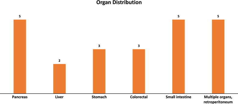
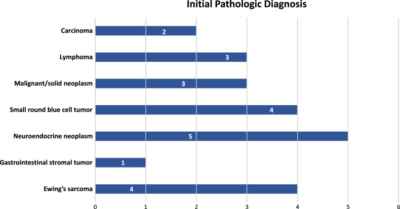
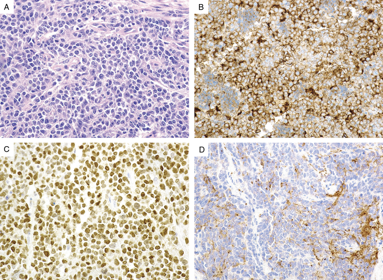
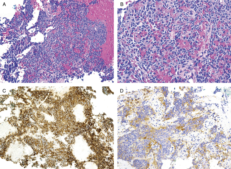
Ewing sarcoma (ES) is an uncommon mesenchymal neoplasm that typically develops as a bone mass, although up to 30% arise in extraskeletal sites. ES of the gastrointestinal (GI) and hepatobiliary tract is rare and may be misdiagnosed as other, more common neoplasms that occur in these sites. However, the correct classification of extraskeletal ES is important for timely clinical management and prognostication. We reviewed our experience of ES in the GI and hepatobiliary tract in order to further highlight the clinicopathologic features of these neoplasms and document the potential for misdiagnosis in this setting. The archives and consultation files of 6 academic institutions were retrospectively queried for cases of ES occurring in the GI and hepatobiliary tract. The histologic slides and ancillary studies were reviewed and clinical data were retrieved for each case through the electronic medical records, when available. Twenty-three patients with ES in the GI and/or hepatobiliary tract were identified from 2000 to 2022. Of these, 11 were women and 12 were men with a median age of 38 years (range, 2 to 64). Tumor locations included the pancreas (n=5), liver (n=2), stomach (n=3), colorectum (n=3), and small intestine (n=5), as well as tumors involving multiple organs, pelvis and retroperitoneum (n=5). Tumor size varied between 2 cm and 18 cm. Twenty were primary and 3 were metastases. Of the 23 cases, only 17% were initially diagnosed as ES. The most common misdiagnoses involved various forms of neuroendocrine neoplasia due to expression of synaptophysin and other neuroendocrine markers (22%). A wide variety of diagnoses including GI stromal tumor was considered due to aberrant CD117 expression (4%). The diagnosis of ES was ultimately confirmed by detection of the EWSR1 rearrangement in 22 cases. The remaining case was diagnosed using traditional immunohistochemistry. Follow-up information was available in 20 cases, with follow-up time varying between 2 and 256 months. Six patients with follow-up died of disease between 6 and 60 months following initial presentation. Our data indicate ES in the GI and hepatobiliary tract is commonly misdiagnosed leading to a delay in therapy. In light of the attendant therapeutic and prognostic implications, ES should be considered in the differential diagnosis of any GI or hepatobiliary tumor with epithelioid and/or small round cell morphology.
Copyright © 2024 The Author(s). Published by Wolters Kluwer Health, Inc.
Conflict of interest statement
Conflicts of Interest and Source of Funding: The authors have disclosed that they have no significant relationships with, or financial interest in, any commercial companies pertaining to this article.
Figures
Similar articles
-
Ewing Sarcoma of the Female Genital Tract: Clinicopathologic Analysis of 21 Cases With an Emphasis on the Differential Diagnosis of Gynecologic Round Cell, Spindle, and Epithelioid Neoplasms.Am J Surg Pathol. 2024 Aug 1;48(8):972-984. doi: 10.1097/PAS.0000000000002232. Epub 2024 May 2. Am J Surg Pathol. 2024. PMID: 38708674
-
Gastrointestinal Ewing Sarcoma: A Clinicopathological and Molecular Genetic Analysis of 25 Cases.Am J Surg Pathol. 2024 Mar 1;48(3):275-283. doi: 10.1097/PAS.0000000000002163. Epub 2023 Dec 8. Am J Surg Pathol. 2024. PMID: 38062799
-
Review with novel markers facilitates precise categorization of 41 cases of diagnostically challenging, "undifferentiated small round cell tumors". A clinicopathologic, immunophenotypic and molecular analysis.Ann Diagn Pathol. 2018 Jun;34:1-12. doi: 10.1016/j.anndiagpath.2017.11.011. Epub 2017 Nov 29. Ann Diagn Pathol. 2018. PMID: 29661713
-
Primary round cell sarcomas of the urinary bladder with EWSR1 rearrangement: a multi-institutional study of thirteen cases with a review of the literature.Hum Pathol. 2020 Oct;104:84-95. doi: 10.1016/j.humpath.2020.08.001. Epub 2020 Aug 14. Hum Pathol. 2020. PMID: 32798549 Review.
-
Clinicopathologic and Molecular Cytogenetic Analysis of 8 Cases With Uterine Cervical Ewing Sarcoma: Case Series With Literature Review.Am J Surg Pathol. 2021 Apr 1;45(4):523-530. doi: 10.1097/PAS.0000000000001674. Am J Surg Pathol. 2021. PMID: 33538423 Review.
References
-
- Esiashvili N, Goodman M, Marcus RB, Jr. Changes in incidence and survival of Ewing sarcoma patients over the past 3 decades: Surveillance Epidemiology and End Results data. J Pediatr Hematol Oncol. 2008;30:425–430. - PubMed
-
- Choi EY, Gardner JM, Lucas DR, et al. . Ewing sarcoma. Semin Diagn Pathol. 2014;31:39–47. - PubMed
-
- Hassan R, Meng LV, Ngee KT, et al. . Extraskeletal Ewing sarcoma of the duodenum presenting as duodenojejunal intussusception. Lancet. 2022;399:1265. - PubMed
