. 2024 May 20;22(5):e3002550.
doi: 10.1371/journal.pbio.3002550. eCollection 2024 May.

Alkenyl oxindole is a novel PROTAC moiety that recruits the CRL4DCAF11 E3 ubiquitin ligase complex for targeted protein degradation

Affiliations

Alkenyl oxindole is a novel PROTAC moiety that recruits the CRL4DCAF11 E3 ubiquitin ligase complex for targeted protein degradation

Ying Wang et al. PLoS Biol. .

Abstract

Alkenyl oxindoles have been characterized as autophagosome-tethering compounds (ATTECs), which can target mutant huntingtin protein (mHTT) for lysosomal degradation. In order to expand the application of alkenyl oxindoles for targeted protein degradation, we designed and synthesized a series of heterobifunctional compounds by conjugating different alkenyl oxindoles with bromodomain-containing protein 4 (BRD4) inhibitor JQ1. Through structure-activity relationship study, we successfully developed JQ1-alkenyl oxindole conjugates that potently degrade BRD4. Unexpectedly, we found that these molecules degrade BRD4 through the ubiquitin-proteasome system, rather than the autophagy-lysosomal pathway. Using pooled CRISPR interference (CRISPRi) screening, we revealed that JQ1-alkenyl oxindole conjugates recruit the E3 ubiquitin ligase complex CRL4DCAF11 for substrate degradation. Furthermore, we validated the most potent heterobifunctional molecule HL435 as a promising drug-like lead compound to exert antitumor activity both in vitro and in a mouse xenograft tumor model. Our research provides new employable proteolysis targeting chimera (PROTAC) moieties for targeted protein degradation, providing new possibilities for drug discovery.


Conflict of interest statement

The authors have declared that no competing interests exist.

Figures
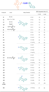
Fig 1
Fig 1. The structure and target degradation activity of JQ1-alkenyl oxindole conjugates.
aBRD4 degradation rate was relative quantification result of S1 Fig (WB). bThe Ar motif of H4 was 4-iodobenzaldehyde.
Fig 2
Fig 2. HL435 potently degrades BRD4 through the ubiquitin-proteasome system.
(A) Design and development of HL435 as a potent BRD4 degrader. (B) Representative WB results; cells were treated with gradient concentrations of HL435 for 12 hours. (C) The DC50 values of HL435 to degrade BRD4 in MDA-MB-231 and MCF-7 cells, from relative quantitative analysis. (D) Representative WB results; MDA-MB-231cells were treated with HL435 at 0.5 μM for gradient time. (E) Relative quantitative analysis for the time-dependent degradation of BRD4. (F) The relative mRNA levels of BRD4; cells were treated with HL435 at 1.0 μM for 12 hours, GAPDH used as internal reference. (G) Representative WB results; cells were cotreated with HL435 and chloroquine (CQ) or bafilomycin (Baf) for 6 hours; CQ or Baf was pretreated for 2 hours. (H) Relative quantitative analysis of BRD4 from G. (I) Representative WB results (n = 3). WT-Hela, ATG5KO-Hela, or ATG4BKO-Hela cells were treated with indicated concentration of HL435 for 6 hours. (J) WB results for BRD4 degradation; cells were pretreated with PYR-41, MG132, or PS-341 for 2 hours, followed by HL435 cotreatment for 6 hours. (K) Relative quantitative analysis of BRD4 from J. (L) WB results; HL435 and MLN4924 were cotreated at indicated concentration for 6 hours; MLN4924 was pretreated for 2 hours. (M) Representative WB results. HEK293T cells were cotransfected with Flag-BRD4 and HA-Ub plasmids for 32 hours, followed by treatment with MG132 for 8 hours and HL435 for 6 hours. Cell lysates were immunoprecipitated with anti-Flag magnetic beads and immunoblotted for ubiquitination level of BRD4. (N) Representative WB results; JQ1, HL389, or HL435 was treated for 6 hours. (O) Relative quantitative analysis BRD4 from N. The data underlying the graphs in the figure can be found in S5 Data; the raw images for WB in the figure can be found in S1 Raw Images. Data were presented as mean ± SEM. Statistical significance was determined by one-way analysis of variance (ANOVA) or Mann–Whitney test (for F). *p < 0.05, **P < 0.01, ***P < 0.001, ****P < 0.0001; ns, no statistical significance.
Fig 3
Fig 3. CRISPRi screening identified the CRL4DCAF11 complex as a potential target mediating HL435-induced proteasomal degradation of BRD4.
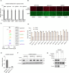
(A) Design of BRD4 BD1 dual fluorescent reporter. BRD4 BD1 domain was fused with mScarlet, followed by a P2A self-cleaving fragment and an EGFP. (B) Validation of BD1 reporter response to HL435 treatment. Representative fluorescent microscope fields for BD1 reporter levels in HEK29T cells treated with DMSO, HL435 (50 nM), or HL435 (50 nM) and MG132 (5 μM) for 24 hours. Bar = 200 μm. (C) Validation of BD1 reporter response to HL435 treatment. Relative BD1-mScarlet signal in HEK293T cells was determined by the ratio of mScarlet and EGFP as measured by flow cytometry. The cells were treated with DMSO, HL435 (50 nM), or HL435 (50 nM) and MG132 (5 μM) for 24 hours. (D) Quantification of the relative BD1-mScarlet signal in the indicated groups was shown in the bar graph (mean ± SD, n = 3 biological replicates). The data underlying this graph can be found in S5 Data. (E) CRISPRi screening strategy. CRISPRi HEK293T cells harboring BD1 reporter were transduced with an sgRNA library targeting all human E1, E2, and E3 enzymes. Cells were treated with DMSO or HL435 (50 nM, 24 hours) and 5 million cells were taken as “input.” Cells with top 25% relative BD1-mScarlet signal (BD1high) were sorted via FACS. The frequencies of BD1high and input cells expressing each sgRNA were determined by next-generation sequencing and were compared to determine sgRNAs enriched or depleted in the BD1high population. The screening was performed in duplicates. (F) Screening results analyzed by the MAGeCK-iNC pipeline were shown for HL435-treated and DMSO-treated groups. A positive phenotype indicates the corresponding sgRNA was enriched in the BD1high population and vice versa. Dots in red, blue, grey, and orange represent positive hits, negative hits, negative control, and other genes, respectively. Genes encoding components of the CRL4DCAF11 complex and the NEDD8-activating enzyme were highlighted. The data underlying the graphs can be found in S4 Data. (G) Scatter plot comparing gene scores for the screens under HL435 and DMSO treatment. Genes encoding components of the CRL4DCAF11 complex and the NEDD8-activating enzyme were highlighted. The data underlying this graph can be found in S4 Data. (H) Predicted structure of CRL4DCAF11 complex using Alphafold. Statistical significance was determined by one-way ANOVA. ****P < 0.0001.
Fig 4
Fig 4. HL435 recruits CRL4DCAF11 complex to induce proteasomal degradation of BRD4.
(A) Validation of knockdown efficiency of different hit genes in CRISPRi HEK293T reporter cells by RT-qPCR (mean ± SD, n = 3 technical replicates). (B) Representative fluorescent microscope fields for BD1 reporter levels in the reporter cells expressing sgRNAs targeting individual hit genes after DMSO or HL435 treatment (50 nM, 24 hours). Bar = 200 μm. (C) Relative BD1-mScarlet signal was quantified by flow cytometry for hit gene knockdown in BD1 reporter cells after DMSO or HL435 treatment (50 nM, 24 hours). (D) Quantification of the relative BD1 intensity in the indicated groups was shown in the bar graph (mean ± SD, n = 3 biological replicates). (E) Validation of knockdown efficiency of 2 sgRNAs targeting DCAF11 in CRISPRi HEK293T reporter cells by RT-qPCR (mean ± SD, n = 3 technical replicates). (F) WB showing endogenous protein levels of BRD4 and α-tubulin in CRISPRi HEK293T cells expressing sgRNAs targeting DCAF11 after DMSO or HL435 treatment (50 nM, 24 hours). (G) Co-immunoprecipitation analysis of HA-DCAF11 with Flag-BD1 in the absence or presence of HL435 (5 μM, 6 hours) in HEK293T cells. The data underlying the graphs in the figure can be found in S5 Data; the raw images for WB in the figure can be found in S1 Raw Images.
Fig 5
Fig 5. HL435 exhibited excellent antitumor efficacy in vitro and in vivo.
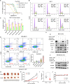
(A) IC50 values of HL435 against multiple tumor cell lines, determinated by the CCK8 assay after 48-hour treatment. (B) Representative flow cytometry analysis results of the cell cycle in MCF-7 cells, treated with indicated compounds for 24 hours before stained with PI. (C) Quantitative statistical analysis of cell cycle for B. (D) Representative flow cytometry analysis results and quantitative statistical analysis of apoptosis; MDA-MB-231 cells were treated with indicated compounds for 36 hours before stained with a 7AAD/APC Apoptosis Detection kit. (E) Representative WB results of c-Myc and cycle relevant proteins in MCF-7 cells (n = 3). (F) Representative WB results of apoptosis relevant proteins in MDA-MB-231 cells (n = 3). (G) Picture of stripped xenograft tumors at the end of experiment (day 45). NOD-SICD mice bearing the MDA-MB-231 xenograft were daily administered with vehicle (10% DMSO + 90% corn oil, IP) or HL435 (20 mg/kg, IP) 6 days per week for 27 days. (H) Growth curve of xenograft tumors after treatment. (I) Weights of stripped xenograft tumors at day 45. (J) Body weight curves during treatment. The data underlying the graphs in the figure can be found in S5 Data; the raw images for WB in the figure can be found in S1 Raw Images. Data were presented as mean ± SEM (n ≥ 3). One-way ANOVA or unpaired t test was employed to determine statistical significance. *p < 0.05, **P < 0.01, ***P < 0.001, ****P < 0.0001; ns, no statistical significance.

Similar articles

Cited by

References

    1. Deshaies RJ. Multispecific drugs herald a new era of biopharmaceutical innovation. Nature. 2020;580(7803):329–338. doi: 10.1038/s41586-020-2168-1 - DOI - PubMed
    1. Winter GE, Buckley DL, Paulk J, Roberts JM, Souza A, Dhe-Paganon S, et al.. Phthalimide conjugation as a strategy for in vivo target protein degradation. Science. 2015;348(6241):1376–1381. - PMC - PubMed
    1. Wu T, Yoon H, Xiong Y, Dixon-Clarke SE, Nowak RP, Fischer ES. Targeted protein degradation as a powerful research tool in basic biology and drug target discovery. Nat Struct Mol Biol. 2020;27(7):605–614. doi: 10.1038/s41594-020-0438-0 - DOI - PMC - PubMed
    1. Salami J, Crews CM. Waste disposal—An attractive strategy for cancer therapy. Science. 2017;355(6330):1163–1167. doi: 10.1126/science.aam7340 - DOI - PubMed
    1. Ciechanover A. Intracellular Protein Degradation: From a Vague Idea, through the Lysosome and the Ubiquitin–Proteasome System, and onto Human Diseases and Drug Targeting. Angew Chem Int Ed. 2005;44(37):5944–5967. - PubMed

MeSH terms