. 2024 May 28;121(22):e2401729121.
doi: 10.1073/pnas.2401729121. Epub 2024 May 20.

Dissecting OGT's TPR domain to identify determinants of cellular function

Affiliations

Dissecting OGT's TPR domain to identify determinants of cellular function

Sarah C Potter et al. Proc Natl Acad Sci U S A. .

Abstract

O-GlcNAc transferase (OGT) is an essential mammalian enzyme that glycosylates myriad intracellular proteins and cleaves the transcriptional coregulator Host Cell Factor 1 to regulate cell cycle processes. Via these catalytic activities as well as noncatalytic protein-protein interactions, OGT maintains cell homeostasis. OGT's tetratricopeptide repeat (TPR) domain is important in substrate recognition, but there is little information on how changing the TPR domain impacts its cellular functions. Here, we investigate how altering OGT's TPR domain impacts cell growth after the endogenous enzyme is deleted. We find that disrupting the TPR residues required for OGT dimerization leads to faster cell growth, whereas truncating the TPR domain slows cell growth. We also find that OGT requires eight of its 13 TPRs to sustain cell viability. OGT-8, like the nonviable shorter OGT variants, is mislocalized and has reduced Ser/Thr glycosylation activity; moreover, its interactions with most of wild-type OGT's binding partners are broadly attenuated. Therefore, although OGT's five N-terminal TPRs are not essential for cell viability, they are required for proper subcellular localization and for mediating many of OGT's protein-protein interactions. Because the viable OGT truncation variant we have identified preserves OGT's essential functions, it may facilitate their identification.

Keywords: O-GlcNAc transferase; OGT; TPR; cell proliferation; tetratricopeptide repeat.


Conflict of interest statement

Competing interests statement:S.J.H. has or currently serves on the SAB of Proximity Therapeutics, Psy Therapeutics, Frequency Therapeutics, Souvien Therapeutics, Sensorium Therapeutics, 4M Therapeutics, Ilios Therapeutics, Entheos Labs, and the Kissick Family Foundation FTD Grant Program, none of whom were involved in the present study. S.J.H. has also received speaking or consulting fees from Amgen, AstraZeneca, Biogen, Merck, Regenacy Pharmaceuticals, Syros Pharmaceuticals, Juvenescence Life, Neumora Therapeutics, and Biohaven Pharmaceuticals, as well as sponsored research or gift funding from AstraZeneca, JW Pharmaceuticals, Lexicon Pharmaceuticals, Vesigen Therapeutics, Compass Pathways, Atai Life Sciences, and Stealth Biotherapeutics. The funders had no role in the design or content of this article, or the decision to submit this review for publication. S.C.P., B.E.G., F.A.H., C.M.J., J.A.P., J.J, Z.G.L., G.Q.F., and S.W. declare no competing interests.

Figures

Fig. 1.
Fig. 1.
OGT performs three functions. (A) OGT uses UDP-GlcNAc as a donor to modify Ser/Thr residues of many nuclear and cytoplasmic proteins. OGA removes O-GlcNAc. (B) OGT cleaves HCF-1 via addition of O-GlcNAc on key glutamate residues. (C) OGT forms multiple protein complexes. (D) A composite crystal structure illustrates OGT’s catalytic domain (gray) and TPR domain (pink) bound to UDP (blue) and an HCF-1-derived peptide (yellow); PDB: 1W3B adapted from ref. and 4N3B adapted from ref. .
Fig. 2.
Fig. 2.
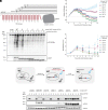
OGT-8 rescues viability in Ogt knockout iCre MEFs. (A) The TPR domain of OGT was truncated from the N terminus resulting in OGT variants with 8, 7, 6, 5, or 4 complete TPRs compared to 13 TPRs in OGT-WT. (B) TPR truncation variants show decreased global glycosylation in vitro in HeLa cell extracts. * denotes low level of endogenous OGT in extracts. (C) Ectopic expression of OGT complements the loss of the endogenous Ogt gene; replacement with OGT variants can test which OGT features are required for cell survival. (D) Ectopic OGT variants are expressed at different levels in iCre MEFs. Except for OGT-4 and OGT-5, which are expressed at levels comparable to endogenous OGT, all variants are expressed below the level of endogenous OGT. OGT-4 and OGT-5 appear at the same molecular weight as endogenous OGT so the α-FLAG blot was also included. (E) Live-cell imaging demonstrates rescue of growth by OGT truncation containing 8 complete TPRs. (F) CellTiter-Glo assay supports live-cell imaging results confirming the ability of OGT-8 to sustain cellular viability.
Fig. 3.
Fig. 3.
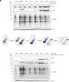
TPR truncation attenuates O-GlcNAcylation in cells. (A) iCre MEFs expressing the panel of OGT-TPR truncations were harvested at day 0, 2, or 4 after endogenous OGT knockout. A pan-O-GlcNAc antibody reveals broadly diminished O-GlcNAc signal for each of the TPR truncation variants compared to ectopic OGT-WT. (B) Schematic showing the generation of iCre-dTAG MEFs and subsequent introduction of an ectopic OGT variant of interest. (C) iCre-dTAG MEFs expressing the panel of OGT-TPR truncations were harvested at day 0, 1, or 2 after initiating FKBP12F36V-OGT-WT degradation. A pan-O-GlcNAc antibody reveals broadly diminished O-GlcNAc signal for each of the TPR truncation variants compared to ectopic OGT-WT.
Fig. 4.
Fig. 4.
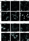
OGT’s N-terminal TPRs are necessary for nuclear localization. Live-cell confocal fluorescence microscopy was used to determine the subcellular localization of an ectopic mKate2-2×FLAG-tagged TPR-only variant (A) or OGT-WT (B) in iCre-dTAG MEFs. Localization of the TPR-only variant of OGT is dependent on cellular O-GlcNAc. (C) OGT-TPR truncation variants are mislocalized to the cytoplasm. Acquisition and display setting were adjusted for some variants to enable optimal visualization of the localization phenotypes; direct comparison of signal intensity should not be made between micrographs.
Fig. 5.
Fig. 5.
OGT dimerization is not essential for cellular viability. (A) OGT forms a homodimer (Right) mediated primarily by residues in TPR6 (Left). PDB: 1W3B adapted from ref. . (B) Live-cell imaging demonstrates that an ectopic OGT variant that cannot dimerize rescues growth after knockout of endogenous Ogt; n = 6 technical replicates per experiment, mean and error bars are from two replicate experiments. (C) iCre MEFs expressing OGT-monomer as the only copy of OGT retain broad O-GlcNAcylation activity; glycosylation of specific substrates appears substantially diminished, denoted by *.
Fig. 6.
Fig. 6.
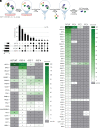
OGT’s N-terminal TPRs are critical for protein–protein interactions. (A) mKate2-2×FLAG-tagged OGT variants were immunoprecipitated from native cell iCre-dTAG MEF lysates, and subsequent mass spectrometry was performed to identify protein interactors. (B) Intersections of OGT interactor hits are illustrated by an UpSet plot; n = 113 proteins were classified as hits across all four experimental conditions. (C) Most OGT-WT hits do not interact with the OGT truncations. (D) OGT-8 hits illustrate importance of OGT’s N-terminal TPRs for stable interactions with known protein partners. In C and D, known OGT interactors are bolded, and known OGT substrates are italicized; denotes proteins for which expression data not obtained; * denotes proteins with elevated expression in “hit” cell line.

References

    1. Hardiville S., Hart G. W., Nutrient regulation of signaling, transcription, and cell physiology by O- GlcNAcylation. Cell Metab. 20, 208–213 (2014). - PMC - PubMed
    1. Decourcelle A., Leprince D., Dehennaut V., Regulation of polycomb repression by O- GlcNAcylation: Linking nutrition to epigenetic reprogramming in embryonic development and cancer. Front. Endocrinol. (Lausanne) 10, 117 (2019). - PMC - PubMed
    1. Zachara N. E., et al. , Dynamic O-GlcNAc modification of nucleocytoplasmic proteins in response to stress. A survival response of mammalian cells. J. Biol. Chem. 279, 30133–30142 (2004). - PubMed
    1. Martinez M. R., Dias T. B., Natov P. S., Zachara N. E., Stress-induced O-GlcNAcylation: An adaptive process of injured cells. Biochem. Soc. Trans. 45, 237–249 (2017). - PMC - PubMed
    1. Zhang F., et al. , O-GlcNAc modification is an endogenous inhibitor of the proteasome. Cell 115, 715–725 (2003). - PubMed

LinkOut - more resources