. 2025 May:71:533-550.
doi: 10.1016/j.jare.2024.05.017. Epub 2024 May 19.

Design of a targeted dual drug delivery system for boosting the efficacy of photoimmunotherapy against melanoma proliferation and metastasis

Affiliations

Design of a targeted dual drug delivery system for boosting the efficacy of photoimmunotherapy against melanoma proliferation and metastasis

Yi Chen et al. J Adv Res. 2025 May.

Abstract

Introduction: The combination of a photosensitizer and indoleamine-2,3 dioxygenase (IDO) inhibitor provides a promising photoimmunotherapy (PIT) strategy for melanoma treatment. A dual drug delivery system offers a potential approach for optimizing the inhibitory effects of PIT on melanoma proliferation and metastasis.

Objective: To develop a dual drug delivery system based on PIT and to study its efficacy in inhibiting melanoma proliferation and metastasis.

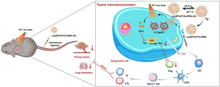
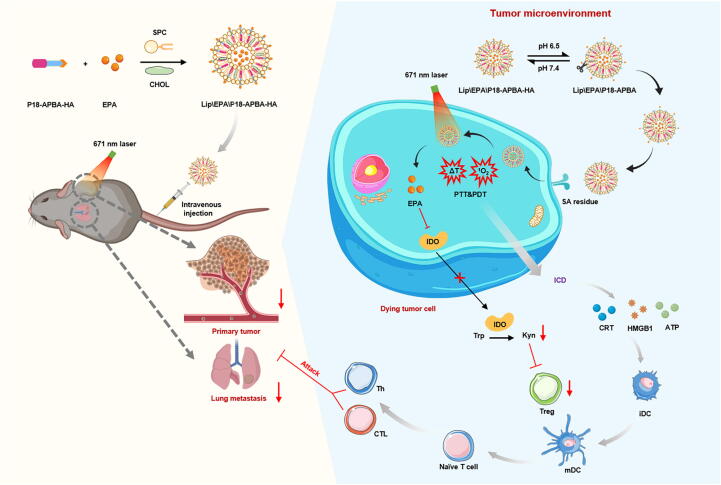
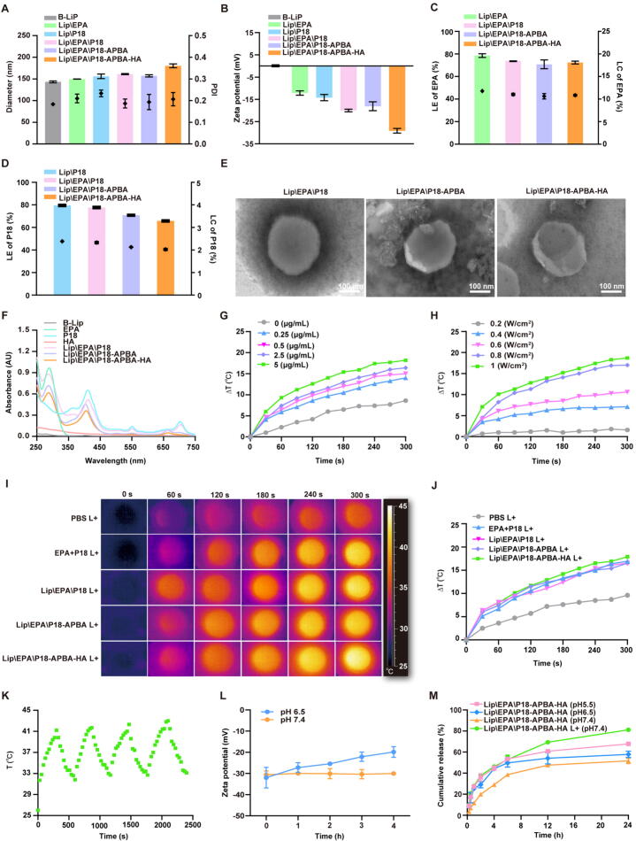
Methods: We constructed a multifunctional nano-porphyrin material (P18-APBA-HA) using the photosensitizer-purpurin 18 (P18), hyaluronic acid (HA), and 4-(aminomethyl) phenylboronic acid (APBA). The resulting P18-APBA-HA was inserted into a phospholipid membrane and the IDO inhibitor epacadostat (EPA) was loaded into the internal phase to prepare a dual drug delivery system (Lip\EPA\P18-APBA-HA). Moreover, we also investigated its physicochemical properties, targeting, anti-tumor immunity, and anti-tumor proliferation and metastasis effects.

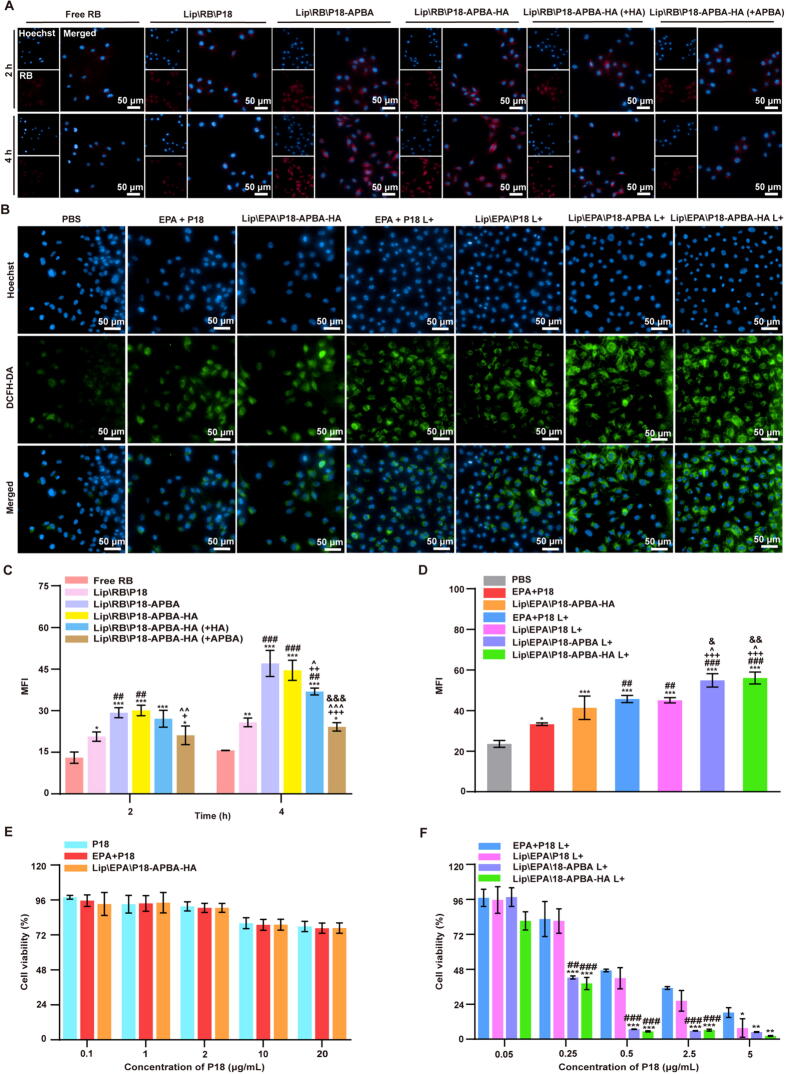
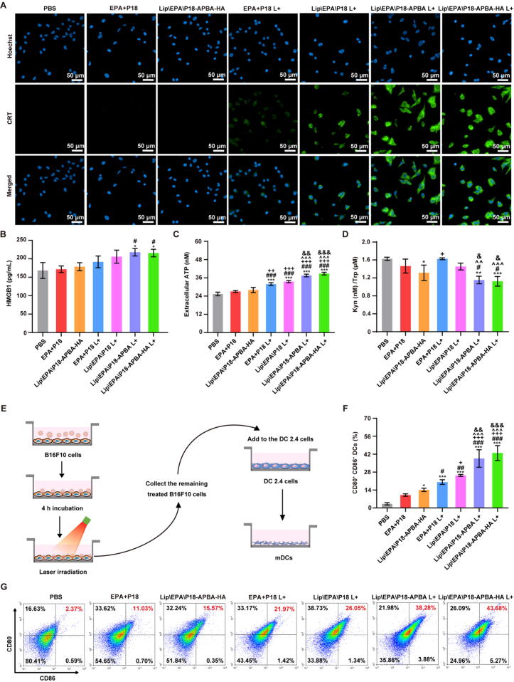
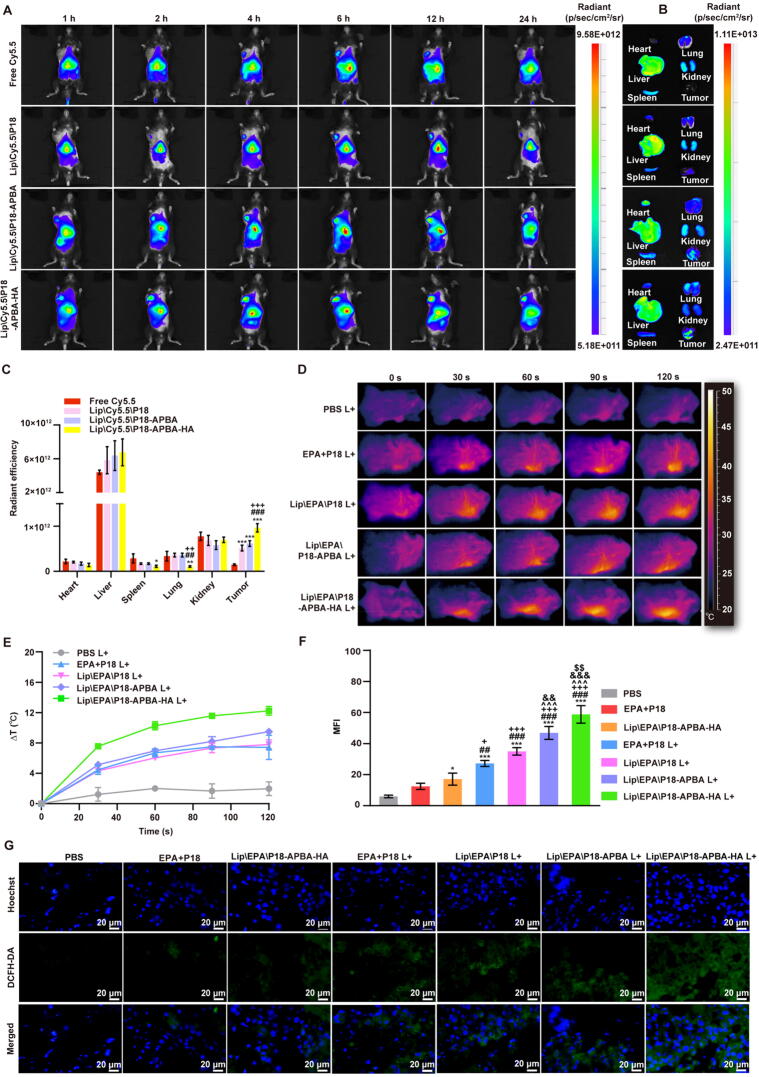
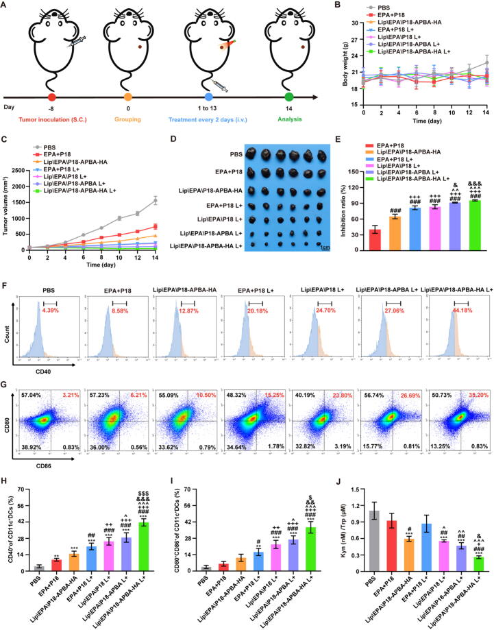
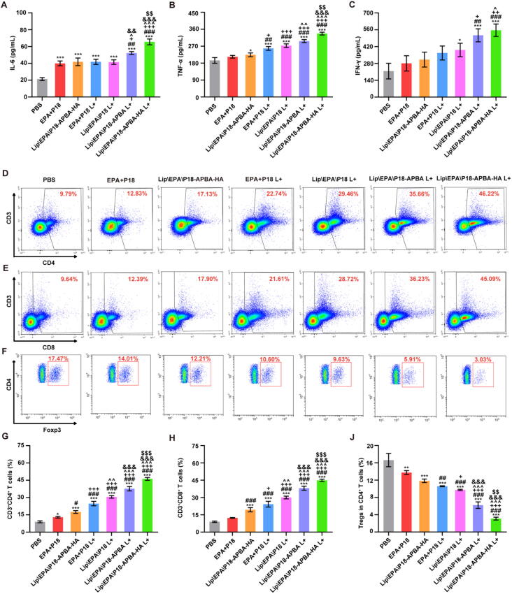
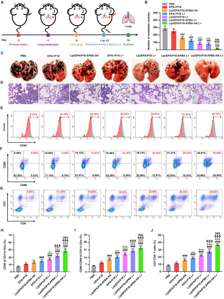
Results: The designed system utilized the pH sensitivity of borate ester to realize an enhanced-targeting strategy to facilitate the drug distribution in tumor lesions and efficient receptor-mediated cellular endocytosis. The intracellular release of EPA from Lip\EPA\P18-APBA-HA was triggered by thermal radiation, thereby inhibiting IDO activity in the tumor microenvironment, and promoting activation of the immune response. Intravenous administration of Lip\EPA\P18-APBA-HA effectively induced anti-tumor immunity by promoting dendritic cell maturation, cytotoxic T cell activation, and regulatory T cell suppression, and regulating cytokine secretion, to inhibit the proliferation of melanoma and lung metastasis.

Conclusion: The proposed nano-drug delivery system holds promise as offers a promising strategy to enhance the inhibitory effects of the combination of EPA and P18 on melanoma proliferation and metastasis.

Keywords: 3 dioxygenase inhibitor; Indoleamine-2; Melanoma; Photoimmunotherapy; Purpurin 18; pH-triggered delivery.


Conflict of interest statement

Declaration of competing interest The authors declare that they have no known competing financial interests or personal relationships that could have appeared to influence the work reported in this paper.

Figures

None
Graphical abstract
Scheme 1
Scheme 1
Schematic illustration of Lip\EPA\P18-APBA-HA preparation and its mechanism for inhibiting primary tumors and lung metastasis based on PIT. After intravenous administration, Lip\EPA\P18-APBA-HA aggregated at the tumor region, aided by its long circulation and EPR effect. The weakly acidic TME and the SA residues on the tumor cell surface will cause cleavage of the iso-PBA ester bond of P18-APBA-HA, and the HA will be stripped off, thereby exposing the PBA group. The PBA group could then be specifically recognized by the SA residues on the surface of the melanoma cells, thus enhancing their specific uptake of the nanoparticles. Following laser irradiation, the heat and ROS generated by P18 resulted in tumor cell death and the release of large amounts of TAAs and EPA. The released TAAs activated the immune response and stimulated the formation of effector T cells, while the EPA released in the TME increased the viability of effector T cells by inhibiting IDO activity and the immune evasion of tumor cells.
Fig. 1
Fig. 1
Preparation and characterization of liposomes. (A) Liposome diameter (bars) and PDI (dots) and (B) zeta potential. (C, D) LE (bars) and LC (dots) of liposomes of EPA and P18. (E) Representative TEM images of Lip\EPA\P18, Lip\EPA\P18-APBA, and Lip\EPA\P18-APBA-HA (scale bar = 100 nm). (F) UV spectra of EPA, P18, HA, and liposomes. (G) Temperature changes of different concentrations of P18 (0–5 μg/mL) under 0.8 W/cm2 laser irradiation. (H) Temperature changes of P18 under laser irradiation at different powers (0.2–1 W/cm2). (I, J) Infrared thermal images and temperature changes under 0.8 W/cm2 laser irradiation of PBS, EPA + P18, Lip\EPA\P18, Lip\EPA\P18-APBA, and Lip\EPA\P18-APBA-HA. (K) Temperature change curves of Lip\EPA\P18-APBA-HA solution under four cycles of irradiation. (L) Zeta potentials of Lip\EPA\P18-APBA-HA incubated at different pH values. (M) EPA release profiles of Lip\EPA\P18-APBA-HA at different pH values. L+ indicates liposomes exposed to 671 nm laser irradiation at 0.8 W/cm2 for 5 min before dialysis. Data presented as mean ± SD (n = 3).
Fig. 2
Fig. 2
In vitro cellular uptake, ROS generation, and cytotoxic effect. (A) B16F10 cells were incubated with free RB and RB-loaded liposomes for 2 and 4 h, respectively, and captured using a high-content imaging system (scale bar = 50 μm). (B) High-content images of ROS in B16F10 cells treated with different formulations with/without laser irradiation (scale bar = 50 μm). (C) Mean fluorescence intensity (MFI) of RB in B16F10 cells after incubation with free RB and RB-loaded liposomes for 2 and 4 h, respectively. *p < 0.05, **p < 0.01, ***p < 0.001 vs. free RB; ##p < 0.01, ###p < 0.001 vs. Lip\RB\P18; +p < 0.05, ++p < 0.01, ++p < 0.001 vs. Lip\RB\P18-APBA; ^p < 0.05, ^^p < 0.01, ^^^p < 0.001 vs. Lip\RB\P18-APBA-HA; &&&p < 0.001 vs. Lip\RB\P18-APBA-HA (+HA). (D) MFI of intracellular DCF in cells treated with different formulations with/without laser irradiation. *p < 0.05, ***p < 0.001 vs. PBS; ##p < 0.01, ###p < 0.001 vs. EPA + P18; +++p < 0.001 vs. Lip\EPA\P18-APBA-HA; ^p < 0.05 vs. EPA + P18 L+; &p < 0.05, &&p < 0.01 vs. Lip\EPA\P18 L+ . (E, F) Cytotoxic effects of P18, EPA + P18, Lip\EPA\P18, Lip\EPA\P18-APBA, and Lip\EPA\P18-APBA-HA with/without laser irradiation in B16F10 cells. *p < 0.05, **p < 0.01, ***p < 0.001 vs. EPA + P18 L+; ##p < 0.01, ###p < 0.001 vs. Lip\EPA\P18 L+. Data presented as mean ± SD (n = 3).
Fig. 3
Fig. 3
In vitro enhancement of anti-tumor immunity under culture conditions. (A) High-content images showing the exposure of CRT on B16F10 cells after treatment with different formulations (scale bar = 50 µm). (B) Extracellular HMGB1 and (C) ATP levels following different treatments. (D) Quantification of Try/Kyn ratio in cell supernatants. (E) Schematic illustration of DC activation. B16F10 cells were treated with different formulations and co-cultured with DC2.4 cells, stained for various surface markers (CD80 and CD86), and then examined by FCM. (F, G) FCM quantification of CD80 and CD86 expression on DCs in vitro. *p < 0.05, **p < 0.01, ***p < 0.001 vs. PBS; #p < 0.05, ##p < 0.01, ###p < 0.001 vs. EPA + P18; +p < 0.05, ++p < 0.01, +++p < 0.001 vs. Lip\EPA\P18-APBA-HA; ^^p < 0.01, ^^^p < 0.001 vs. EPA + P18 L+; &p < 0.05, &&p < 0.01, &&&p < 0.001 vs. Lip\EPA\P18 L+. Data presented as mean ± SD (n = 3).
Fig. 4
Fig. 4
In vivo biodistribution, PTT performance, and PDT efficiency of Lip\EPA\P18-APBA-HA. (A) Biodistribution of Cy5.5 in B16F10 tumor-bearing mice at 1, 2, 4, 6, 12, and 24 h post intravenous injection of free Cy5.5, Lip\Cy5.5\P18, Lip\Cy5.5\P18-APBA, and Lip\Cy5.5\P18-APBA-HA, respectively. (B, C) Ex vivo imaging and MFI quantification of transplanted heart, liver, spleen, lung, kidney, and tumor from mice at 24 h. *p < 0.05, **p < 0.01, ***p < 0.001 vs. free Cy5.5; ##p < 0.01, ###p < 0.001 vs. Lip\Cy5.5\P18; ++p < 0.01, +++p < 0.001 vs. Lip\Cy5.5\P18-APBA. (D, E) Infrared thermal images and temperature change for mice following intravenous administration of different formulations followed by laser irradiation. (F, G) ROS detection in B16F10 tumor observed by CLSM (scale bar = 20 µm) and MFI quantification. *p < 0.05, ***p < 0.001 vs. PBS; ##p < 0.01, ###p < 0.001 vs. EPA + P18; +p < 0.05, +++p < 0.001 vs. Lip\EPA\P18-APBA-HA; ^^^p < 0.001 vs. EPA + P18 L+; &&p < 0.01, &&&p < 0.001 vs. Lip\EPA\P18 L+; $$p < 0.01 vs. Lip\EPA\P18-APBA L+. Data presented as mean ± SD (n = 3).
Fig. 5
Fig. 5
In vivo therapeutic efficiencies of formulations against primary tumors and anti-tumor immunity. (A) Treatment schedule of anti-primary tumor experiments. (B) Change in body weight of B16F10 tumor-bearing mice (n = 6). (C) Primary tumor volume changes after various treatments (n = 6). (D) Images of excised tumors on day 14 (scale bar = 1 cm) (n = 6). (E) The inhibition ratio of tumor weight on day 14. (F-I) FCM analysis and percentage of mDCs (CD40+, CD80+ CD86+) in TDLNs (n = 3). (J) Quantification of intratumoral Try/Kyn ratio in mice after different treatments (n = 3). **p < 0.01, ***p < 0.001 vs. PBS; #p < 0.05, ##p < 0.01, ###p < 0.001 vs. EPA + P18; +p < 0.05, ++p < 0.01, +++p < 0.001 vs. Lip\EPA\P18-APBA-HA; ^p < 0.05, ^^p < 0.01 ^^^p < 0.001 vs. EPA + P18 L+; &p < 0.05, &&p < 0.01, &&&p < 0.001 vs. Lip\EPA\P18 L+; $p < 0.05, $$$p < 0.001 vs. Lip\EPA\P18-APBA L+. Data presented as mean ± SD.
Fig. 6
Fig. 6
In vivo anti-tumor immunity. Serum levels of (A) IL-6, (B) TNF-α, and (C) IFN-γ in mice after different treatments (n = 3). FCM analysis and percentages of (D, G) Th cells (CD3+ CD4+), (E, H) CTLs (CD3+ CD8+), and (F, J) Tregs (CD4+ Foxp3+) in primary tumors (n = 3). *p < 0.05, **p < 0.01, ***p < 0.001 vs. PBS; #p < 0.05, ##p < 0.01, ###p < 0.001 vs. EPA + P18; +p < 0.05, ++p < 0.01, +++p < 0.001 vs. Lip\EPA\P18-APBA-HA; ^p < 0.05, ^^p < 0.01, ^^^p < 0.001 vs. EPA + P18 L+; &&p < 0.01, &&&p < 0.001 vs. Lip\EPA\P18 L+; $$p < 0.01, $$$p < 0.001 vs. Lip\EPA\P18-APBA L+. Data presented as mean ± SD.
Fig. 7
Fig. 7
In vivo anti-tumor lung metastasis efficacy. (A) Schematic illustration of anti-metastasis experiment. (B) Number of metastatic nodules in the lung (n = 6). (C, D) Images of metastatic nodules and H&E staining images in the lung (n = 6) (scale bar = 50 µm). FCM analysis and percentages of (E-I) mDCs (CD40+, CD80+ CD86+) and (G, J) CTLs (CD3+ CD8+) in the lung (n = 6). **p < 0.01, ***p < 0.001 vs. PBS; ###p < 0.001 vs. EPA + P18; +p < 0.05, ++p < 0.01, +++p < 0.001 vs. Lip\EPA\P18-APBA-HA; ^p < 0.05, ^^p < 0.01, ^^^p < 0.001 vs. EPA + P18 L+; &&&p < 0.001 vs. Lip\EPA\P18 L+; $$$p < 0.001 vs. Lip\EPA\P18-APBA L+. Data presented as mean ± SD.

Similar articles

Cited by

References

    1. Deboever N., Feldman H.A., Hofstetter W.L., Mehran R.J., Rajaram R., Rice D.C., et al. The role of surgery in the treatment of melanoma pulmonary metastases in the modern era. J Surg Res. 2022;277:125–130. doi: 10.1016/j.jss.2022.04.021. - DOI - PubMed
    1. Shain A.H., Bastian B.C. From melanocytes to melanomas. Nat Rev Cancer. 2016;16:345–358. doi: 10.1038/nrc.2016.37. - DOI - PubMed
    1. Phoon Y.P., Tannenbaum C., Diaz-Montero C.M. Immunobiology of melanoma. Clin Plast Surg. 2021;48:561–576. doi: 10.1016/j.cps.2021.06.005. - DOI - PubMed
    1. Song M., Liu C., Chen S., Zhang W. Nanocarrier-based drug delivery for melanoma therapeutics. Int J Mol Sci. 2021;22:1873. doi: 10.3390/ijms22041873. - DOI - PMC - PubMed
    1. Zou J., Li L., Yang Z., Chen X. Phototherapy meets immunotherapy: a win–win strategy to fight against cancer. Nanophotonics. 2021;10:3229. doi: 10.1515/nanoph-2021-0209. - DOI

MeSH terms