. 2024 May 20;14(1):11497.
doi: 10.1038/s41598-024-62262-1.

Upregulation of the AMPK-FOXO1-PDK4 pathway is a primary mechanism of pyruvate dehydrogenase activity reduction in tafazzin-deficient cells

Affiliations

Upregulation of the AMPK-FOXO1-PDK4 pathway is a primary mechanism of pyruvate dehydrogenase activity reduction in tafazzin-deficient cells

Zhuqing Liang et al. Sci Rep. .

Erratum in

Abstract

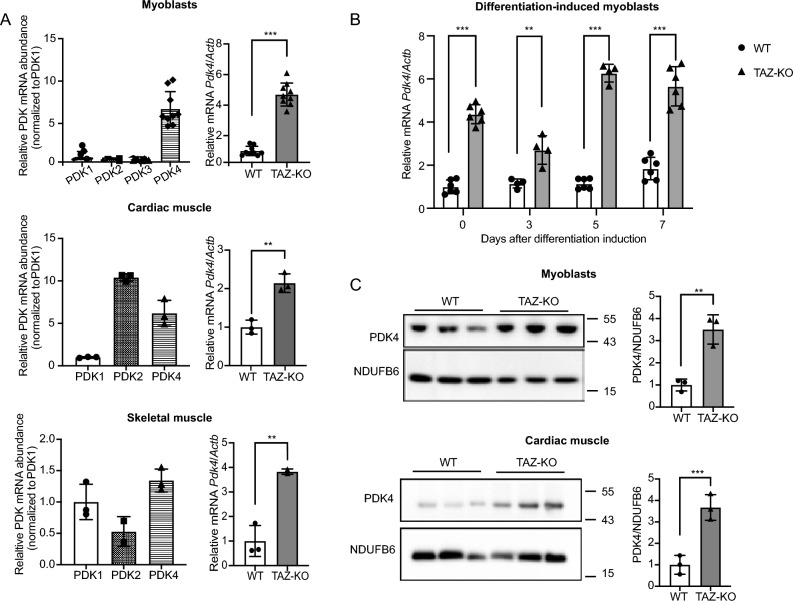
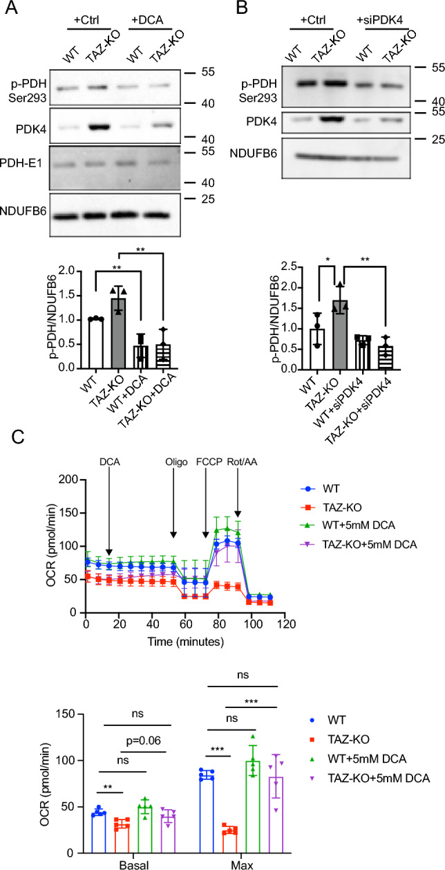
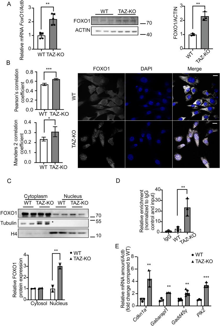
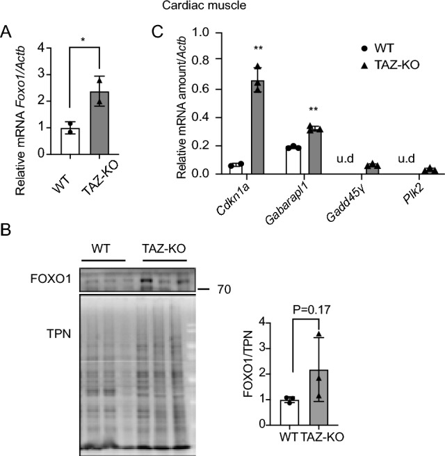
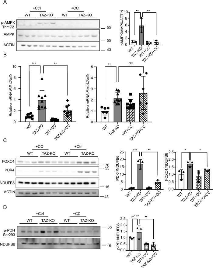
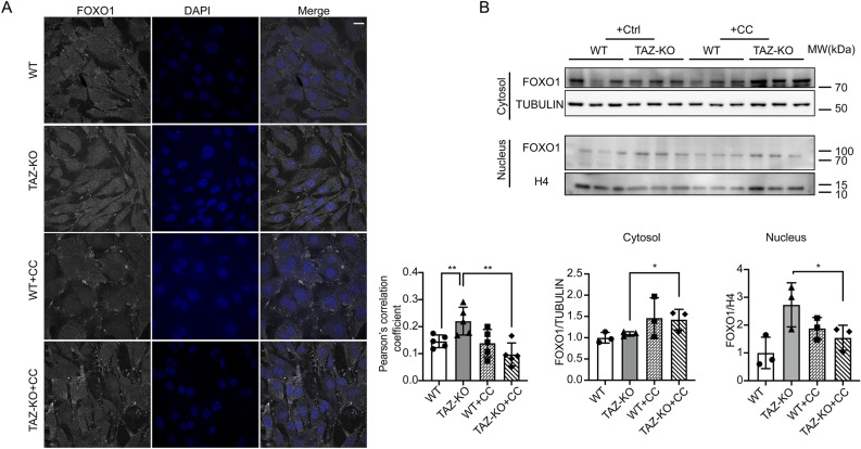
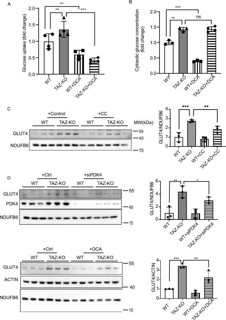
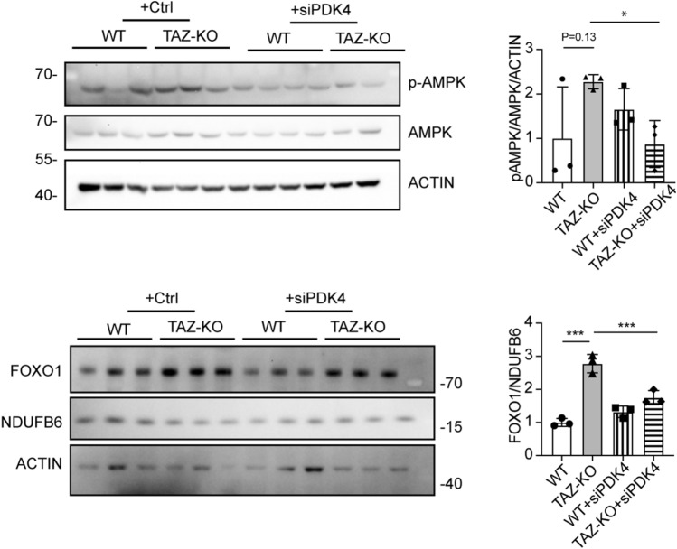
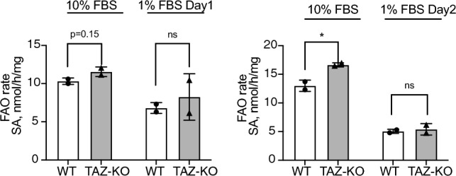
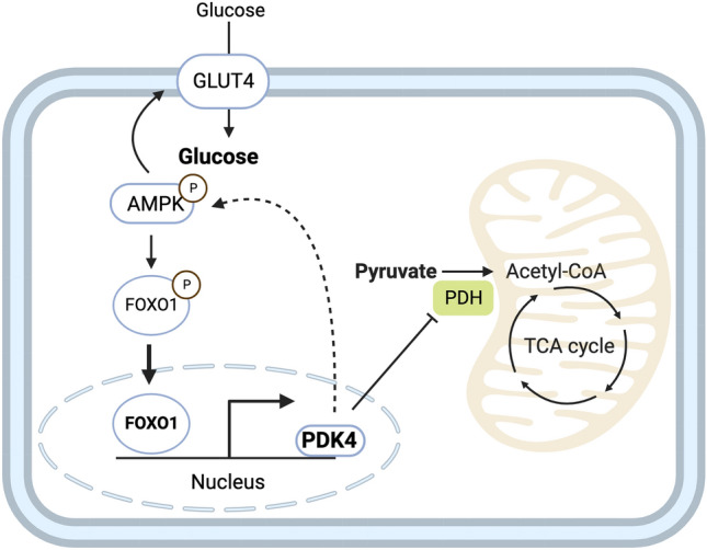
Barth syndrome (BTHS) is a rare disorder caused by mutations in the TAFAZZIN gene. Previous studies from both patients and model systems have established metabolic dysregulation as a core component of BTHS pathology. In particular, features such as lactic acidosis, pyruvate dehydrogenase (PDH) deficiency, and aberrant fatty acid and glucose oxidation have been identified. However, the lack of a mechanistic understanding of what causes these conditions in the context of BTHS remains a significant knowledge gap, and this has hindered the development of effective therapeutic strategies for treating the associated metabolic problems. In the current study, we utilized tafazzin-knockout C2C12 mouse myoblasts (TAZ-KO) and cardiac and skeletal muscle tissue from tafazzin-knockout mice to identify an upstream mechanism underlying impaired PDH activity in BTHS. This mechanism centers around robust upregulation of pyruvate dehydrogenase kinase 4 (PDK4), resulting from hyperactivation of AMP-activated protein kinase (AMPK) and subsequent transcriptional upregulation by forkhead box protein O1 (FOXO1). Upregulation of PDK4 in tafazzin-deficient cells causes direct phospho-inhibition of PDH activity accompanied by increased glucose uptake and elevated intracellular glucose concentration. Collectively, our findings provide a novel mechanistic framework whereby impaired tafazzin function ultimately results in robust PDK4 upregulation, leading to impaired PDH activity and likely linked to dysregulated metabolic substrate utilization. This mechanism may underlie previously reported findings of BTHS-associated metabolic dysregulation.


Conflict of interest statement

The authors declare no competing interests.

Figures

Figure 1
Figure 1
PDK4 is upregulated in tafazzin-deficient cells. PDK4 and other isoforms levels were evaluated using qPCR (A) (B) or Western blot (WB) analysis (C) of whole-cell lysates extracted from WT and TAZ-KO myoblasts, differentiation-induced myotubes, and mouse muscle tissue. The figures in panel (C) are representative images with each lane representing one biological replicate. The mRNA expression levels were normalized to the internal control Actb, and protein levels were normalized to NDUFB6. Densitometry was quantified using Image J and statistical significance was determined with an unpaired, two-tailed Student's t-test. The data points represent mean ± S.D. (error bars) for each individual biological replicate of each group. * 0.05 < p < 0.1, ** 0.01 < p < 0.05, *** 0.001 < p < 0.01.
Figure 2
Figure 2
Inhibiting PDK4 decreases PDH phosphorylation in TAZ-KO cells. Myoblasts were treated with either 5 mM DCA for 24 h (A) or PDK4-targeted siRNA (B). Phosphorylation of PDH on Ser293 (p-PDH) was assessed via WB. Image J software was used for quantitative analysis of p-PDH (bottom panels). (C) Oxygen consumption rates (OCR) of myoblasts subjected to 5 mM DCA treatments. n = 5. The data points represent mean ± S.D. (error bars) for each individual biological replicate of each group. * 0.05 < p < 0.1, ** 0.01 < p < 0.05, *** 0.001 < p < 0.01.
Figure 3
Figure 3
Upregulation of FOXO1 activates PDK4 transcription in TAZ-KO cells. (A) Total mRNA and whole-cell lysate were extracted from both WT and TAZ-KO myoblasts, and the mRNA and protein levels of FOXO1 were measured using real-time qPCR and WB analysis, respectively. Data are presented as fold-change relative to WT, and each lane represents an individual biological replicate. (B) Right: The subcellular localization of FOXO1 was detected through immunofluorescence (IF) confocal microscopy, with representative figures showing the relative distribution of FOXO1 in WT and TAZ-KO myoblasts. Left: Quantification of colocalization between FOXO1 and DAPI based on Pearson's and Mander's 2 colocalization analysis,. The scale bar equals 20 µm. (C) Nuclear protein fractionation was performed, followed by WB analysis using tubulin and histone 4 (H4) as internal controls for cytoplasmic and nuclear fractions, respectively. * indicates PDK4 band residue after stripping the membrane. (D) Chromatin immunoprecipitation/qPCR was performed in myoblasts with either an IgG control or anti-FOXO1 antibody. The Pdk4 promoter was detected using the specific primers listed in “Experimental procedures”. (E) Real-time qPCR was used to measure mRNA of the FOXO1 targets Cdkn11 (Cyclin Dependent Kinase Inhibitor 1A), Gabarapl1 (Gamma-aminobutyric acid receptor-associated protein-like 1), Gadd45γ (Growth Arrest and DNA Damage Inducible γ), and Plk2 (Polo Like Kinase 2), using Actb as the internal control. Data points represent mean ± S.D. (error bars) for each individual biological replicate of each group. * 0.05 < p < 0.1, ** 0.01 < p < 0.05, *** 0.001 < p < 0.01.
Figure 4
Figure 4
TAZ-KO cardiac muscle shows enrichment of nuclear FOXO1. Total mRNA and whole-cell lysate were extracted from both WT and TAZ-KO mouse cardiac tissues, and FOXO1 mRNA (A) and protein (B) were measured using real-time qPCR and WB analysis, respectively. A total protein stain was used for WB analysis normalization (TPN). Data are presented as fold-change relative to WT, with each lane representing an individual biological replicate. (C) Real-time qPCR was used to measure the mRNAs of four FOXO1 substrates (Cdkn11, Gabarapl1, Gadd45γ, and Plk2) using Actb as the internal control. Data points represent mean ± S.D. (error bars) for each individual biological replicate of each group. * 0.05 < p < 0.1, ** 0.01 < p < 0.05, *** 0.001 < p < 0.01.
Figure 5
Figure 5
AMPK-mediated upregulation of FOXO1 increases PDK4 in TAZ-KO cells. (A) Phosphorylation of AMPK residue Thr172 (p-AMPK) was assayed in WT and TAZ-KO myoblasts treated with the AMPK inhibitor compound C (CC) or vehicle. 10 μg of total protein was loaded for each sample, and ACTIN was used as an internal control. Representative images and quantitative analysis are shown. (B) Total mRNA was extracted from myoblasts, and Pdk4 and Foxo1 mRNA levels were measured via real-time qPCR following treatment with CC (10 μM, 16 h) or vehicle. (C) Protein expression of FOXO1 and PDK4 in myoblasts was measured via WB analysis following treatment with CC (10 μM, 16 h) or vehicle. Representative images and corresponding quantitative analyses are shown. (D) In myoblasts, p-PDH was measured by WB analysis following treatment with CC (10 μM, 16 h) or vehicle. Representative images and corresponding quantitative analyses are shown. Data points represent mean ± S.D. (error bars) for each individual biological replicate of each group. * 0.05 < p < 0.1, ** 0.01 < p < 0.05, *** 0.001 < p < 0.01.
Figure 6
Figure 6
AMPK activation promotes FOXO1 nuclear translocation in TAZ-KO cells. (A) IF staining was used to show the localization of FOXO1 in myoblasts following treatment with CC (5 μM, 16 h) or vehicle. Scale bar = 10 μm. Pearson's colocalization analysis between DAPI and FOXO1 (bottom); n = 5. (B) FOXO1 protein levels were assayed in nuclear and cytoplasmic cellular fractions following treatment with CC (5 μM, 16 h) or vehicle. Tubulin and histone H3 were used as internal controls for cytoplasmic and nuclear fractions, respectively. Corresponding quantitative analyses are also presented (bottom right). Data points represent mean ± S.D. (error bars) for each individual biological replicate of each group. * 0.05 < p < 0.1, ** 0.01 < p < 0.05, *** 0.001 < p < 0.01.
Figure 7
Figure 7
Glucose utilization is dysregulated in TAZ-KO cells. (A) Myoblasts were incubated with 1 mM 2DG. The glucose uptake rate was determined by measuring the amount of 2DG taken up by the cells, with values normalized to total protein in each sample before analysis. (B) Cytosolic glucose concentration was measured according to “Experimental procedures”. GLUT4 protein expression was measured by WB in myoblasts treated with either 10 μM CC (C) or 5 mM DCA for 16 h or PDK4-targeted siRNA for 24 h (D). NDUFB6 and ACTIN were used as internal controls. The data points represent means ± S.D. (error bars) for each individual biological replicate of each group. * 0.05 < p < 0.1, ** 0.01 < p < 0.05, *** 0.001 < p < 0.01.
Figure 8
Figure 8
PDK4 upregulation in TAZ-KO cells exacerbates AMPK activation and FOXO1 expression. Phosphorylation of AMPK residue Thr172 (p-AMPK) and FOXO1 protein levels were assayed in WT and TAZ-KO myoblasts treated with PDK4-targeted siRNA. The data points represent means ± S.D. (error bars) for each individual biological replicate of each group. * 0.05 < p < 0.1, *** 0.001 < p < 0.01.
Figure 9
Figure 9
Fatty acid oxidation is not significantly changed in TAZ-KO cells. WT and TAZ-KO cells were incubated with tritium-labeled oleate to allow for import and subsequent oxidation. FAO rate was calculated based on the amount of tritium-labeled H+ released back into the medium as a result of oleate oxidation; n = 4. Data points represent means ± S.D. (error bars) for each individual biological replicate of each group. * 0.05 < p < 0.1, ** 0.01 < p < 0.05, *** 0.001 < p < 0.01.
Figure 10
Figure 10
Proposed mechanism underlying metabolic perturbation in TAZ-KO cells. Activation of AMPK in TAZ-KO cells leads to enrichment of FOXO1 in the nucleus, where it acts to increase transcription of Pdk4. Upregulation of PDK4 results in altered cellular metabolism by inactivating PDH and applying positive feedback to activate AMPK. This mechanism represents a potential link between tafazzin-deficiency and the metabolic alterations observed in BTHS.

Update of

References

    1. Cade WT, et al. Blunted fat oxidation upon submaximal exercise is partially compensated by enhanced glucose metabolism in children, adolescents, and young adults with Barth syndrome. J. Inherit. Metab. Dis. 2019;42:480–493. doi: 10.1002/jimd.12094. - DOI - PMC - PubMed
    1. Cade WT, et al. Substrate metabolism during basal and hyperinsulinemic conditions in adolescents and young-adults with Barth syndrome. J. Inherit. Metab. Dis. 2013;36:91–101. doi: 10.1007/s10545-012-9486-x. - DOI - PMC - PubMed
    1. Carling D, et al. Bypassing the glucose/fatty acid cycle: AMP-activated protein kinase. Biochem. Soc. Trans. 2003;31:1157–1160. doi: 10.1042/bst0311157. - DOI - PubMed
    1. Raja V, Joshi AS, Li G, Maddipati KR, Greenberg ML. Loss of cardiolipin leads to perturbation of acetyl-CoA synthesis. J. Biol. Chem. 2017;292:1092–1102. doi: 10.1074/jbc.M116.753624. - DOI - PMC - PubMed
    1. Li Y, et al. Cardiolipin-induced activation of pyruvate dehydrogenase links mitochondrial lipid biosynthesis to TCA cycle function. J. Biol. Chem. 2019;294:11568–11578. doi: 10.1074/jbc.RA119.009037. - DOI - PMC - PubMed

MeSH terms