. 2024 Jul;25(7):2878-2895.
doi: 10.1038/s44319-024-00158-x. Epub 2024 May 20.

Adipocyte HSL is required for maintaining circulating vitamin A and RBP4 levels during fasting

Affiliations

Adipocyte HSL is required for maintaining circulating vitamin A and RBP4 levels during fasting

Julia S Steinhoff et al. EMBO Rep. 2024 Jul.

Abstract

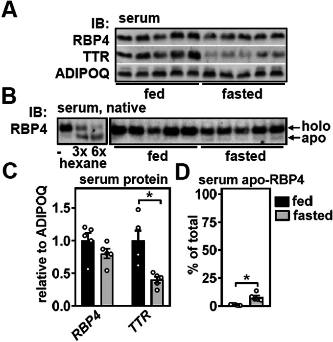
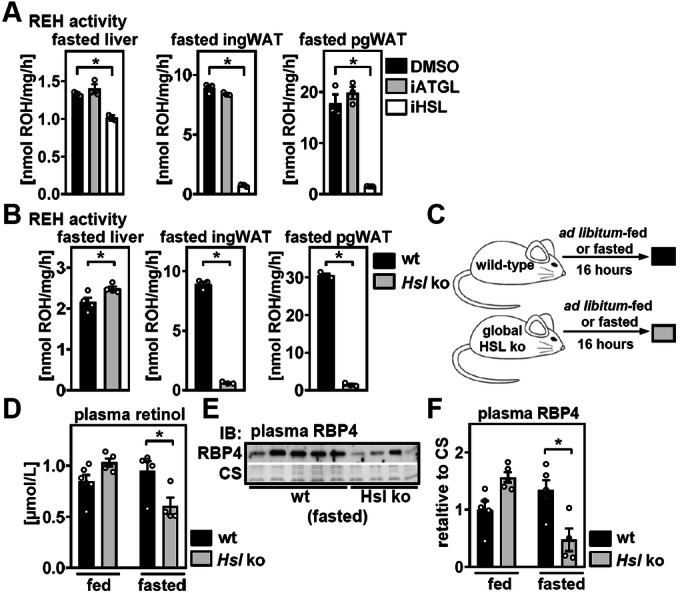
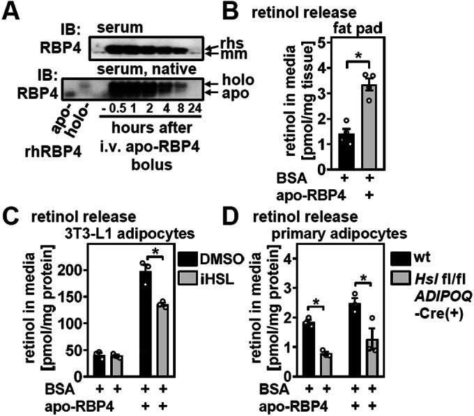
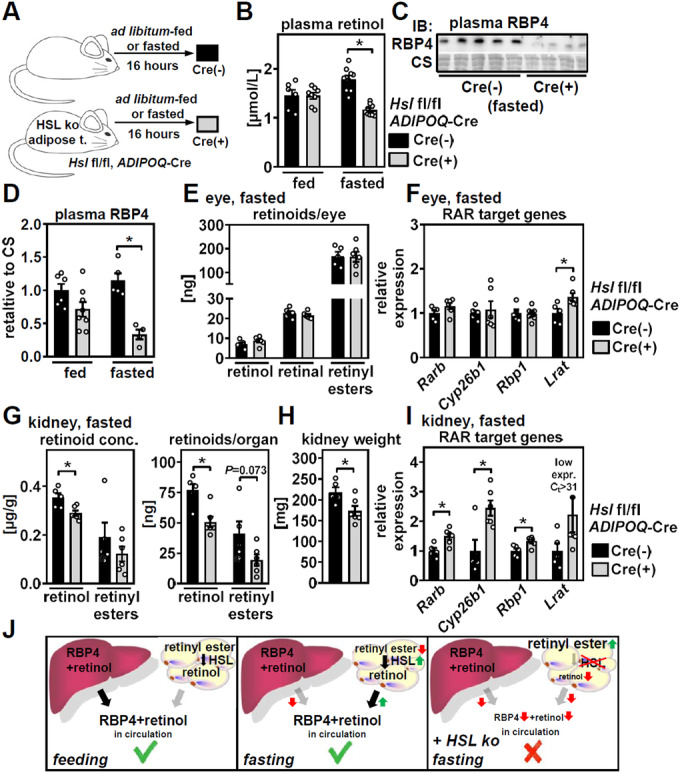
Vitamin A (retinol) is distributed via the blood bound to its specific carrier protein, retinol-binding protein 4 (RBP4). Retinol-loaded RBP4 is secreted into the circulation exclusively from hepatocytes, thereby mobilizing hepatic retinoid stores that represent the major vitamin A reserves in the body. The relevance of extrahepatic retinoid stores for circulating retinol and RBP4 levels that are usually kept within narrow physiological limits is unknown. Here, we show that fasting affects retinoid mobilization in a tissue-specific manner, and that hormone-sensitive lipase (HSL) in adipose tissue is required to maintain serum concentrations of retinol and RBP4 during fasting in mice. We found that extracellular retinol-free apo-RBP4 induces retinol release by adipocytes in an HSL-dependent manner. Consistently, global or adipocyte-specific HSL deficiency leads to an accumulation of retinoids in adipose tissue and a drop of serum retinol and RBP4 during fasting, which affects retinoid-responsive gene expression in eye and kidney and lowers renal retinoid content. These findings establish a novel crosstalk between liver and adipose tissue retinoid stores for the maintenance of systemic vitamin A homeostasis during fasting.

Keywords: Fasting; HSL; RBP4; Retinol; Vitamin A.


Conflict of interest statement

The authors declare no competing interests.

Figures

Figure 1
Figure 1. Fasting induces RBP4 and lowers TTR in the liver.
(A) Male C57BL/6J mice were fed ad libitum or fasted for 24 h as depicted. (B) Blood glucose and non-esterified fatty acids (NEFA) in fed and fasted mice were determined. (C) Serum retinol concentrations were analyzed by HPLC. (D) Hepatic expression of indicated genes was measured by qPCR. (E) Retinyl ester hydrolase (REH) activity of liver lysates from fed and fasted mice was analyzed ex vivo. (F) Protein abundance of RBP4 and TTR in livers of fed and fasted mice, GAPDH served as loading control. (G) The densitometric analysis of proteins shown in (F). Data information: Data are represented as individual data points of n = 5, 5 (BD) or n = 4, 4 (EG) biological replicates and mean ± s.e.m. and *P < 0.05 vs. ad libitum-fed mice using an unpaired two-tailed Student’s t test. Source data are available online for this figure.
Figure 2
Figure 2. Fasting does not affect total RBP4 but reduces TTR and increases apo-RBP4 in the circulation.
(A) Serum RBP4 and TTR levels of fed and fasted mice were analyzed by immunoblotting, and serum ADIPOQ served as loading control. (B) Abundance of apo- and holo-RBP4 in serum of fed and fasted mice was analyzed by SDS-free native immunoblotting (right panel). Repeated hexane-extractions of serum was used as a control for visualization of each isoform (left panel). (C) Densitometric analysis of blots shown in (A). (D) Densitometric analysis of blots shown in (B). Data information: Data are represented as individual data points of n = 5, 5 (AD) biological replicates and mean ± s.e.m. and *P < 0.05 vs. ad libitum-fed mice using an unpaired two-tailed Student’s t test. Source data are available online for this figure.
Figure 3
Figure 3. Expression and secretion of RBP4 in primary hepatocytes is regulated by cAMP and FOXO1.
Primary hepatocytes were isolated from mouse liver and treated with vehicle or (A) the synthetic PPARα agonist WY14643, (B) all-trans retinoic acid (atRA), (C) dexamethasone (Dex), 8-Br-cAMP, and (D) glucagon as indicated for 24 h. mRNA expression of appropriate control genes and Rbp4 were determined by qPCR. (E) Hepatocytes were incubated with increasing concentrations of 8-Br-cAMP and RBP4 protein in cell lysates and media analyzed by immunoblotting, RAN protein and Ponceau membrane staining served as loading controls, respectively. (F) Densitometric analysis of blots shown in (E). (G) Primary hepatocytes were treated with non-targeting (Control) or Foxo1 siRNA and FOXO1 protein analyzed by immunoblotting, ACTB served as loading control. (H) mRNA expression of indicated genes in hepatocytes described in (G) was analyzed by qPCR. (I) Primary hepatocytes were treated with insulin as indicated for 24 h and mRNA expression of Igfbp1 and Rbp4 determined by qPCR. Data information: In (AH) Data are representative for three independent experiments and performed in triplicates (AF), or quadruplicates (H). (I) Data are representative of two independent experiments performed with six replicates. Data are represented as individual data points and mean ± s.e.m. and *P < 0.05 vs. vehicle- or siControl-treated hepatocytes using an unpaired two-tailed Student’s t test (A, D, I) or one-way ANOVA with Dunnett’s correction for multiple testing (B, C, F, H). Source data are available online for this figure.
Figure 4
Figure 4. Fasting reduces retinoid levels in white adipose tissue but not in liver or lung.
Mice were fed ad libitum or fasted for 24 h and tissue retinol and retinyl esters in (A) liver, (B) lung, (C) inguinal-, and (D) perigonadal white adipose tissue (ing/pgWAT) analyzed by HPLC. Retinoids are normalized to tissue weight (left panel) or shown as µg per total organ (right panel). Data information: Data are represented as individual data points of n = 5, 5 biological replicates and mean ± s.e.m. and *P < 0.05 vs. ad libitum-fed mice using an unpaired two-tailed Student’s t test. Source data are available online for this figure.
Figure 5
Figure 5. HSL is required for mobilization of WAT retinoids and to maintain circulating RBP4 and retinol concentrations during fasting.
(A) Retinyl ester hydrolase (REH) activity of liver and white adipose tissue (WAT) lysates of 16 h-fasted wild-type mice (wt) in the presence of DMSO vehicle, the ATGL inhibitor Atglistatin, or the HSL inhibitor NNC-0076-0000-0079 was analyzed ex vivo. Wild-type (wt) mice or mice with global Hsl deletion were fasted for 16 h and (B) retinyl ester hydrolase (REH) activity of indicated tissues analyzed ex vivo. (C) Wt mice or mice with global Hsl deletion were fasted as indicated and (D) plasma retinol analyzed by HPLC. (E) Plasma RBP4 was determined by immunoblotting. Coomassie staining (CS) served as loading control. (F) Densitometric analysis of plasma RBP4 in ad libitum-fed and 16 h-fasted mice. Data information: Data are representative for three independent experiments, performed in triplicates (A). (B) Data show biological replicates with n = 4, 4 (liver) and n = 3, 3 (ing and pgWAT), in (D), data show biological replicates with n = 6, 5, 5, 4, and in (F), data show biological replicates with n = 5, 5, 5, 4. Data are presented as mean ± s.e.m. and *P < 0.05 vs. DMSO vehicle or wt mice using a one-way ANOVA with Dunnett’s correction for multiple testing (A), an unpaired two-tailed Student’s t test (B), or a two-way ANOVA with Sidak’s correction for multiple testing (D, F). Source data are available online for this figure.
Figure 6
Figure 6. Retinol-free apo-RBP4 induces HSL-dependent retinol release from adipocytes.
(A) Serum RBP4 was analyzed by immunoblotting (top) or native PAGE (bottom) at indicated times after i.v. injection of 100 µg of recombinant human RBP4 (rhsRBP4) into the mouse tail vein. Apo- and holo-rhsRBP4 were used as controls for native PAGE. (B) Murine inguinal fat pads were incubated with 4% BSA and 2 µM apo-RBP4 for 2 h as indicated and retinol content in the supernatant analyzed by HPLC. (C) Differentiated 3T3-L1 or (D) in vitro-differentiated primary adipocytes from wild-type (wt) and adipose tissue-specific Hsl knockout mice were loaded with 20 µM retinol overnight. The next day, 3T3-L1 adipocytes were incubated with the HSL inhibitor NNC-0076-0000-0079 and all adipocytes with 4% BSA and 2 µM apo-RBP4 for 2 h as indicated and retinol in the adipocyte supernatant analyzed by HPLC. Data information: In (A), data are representative for two independent experiments. In (B), data show n = 4, 4 biological replicates and in (C, D), data show n = 3 biological replicates for each condition. Data are represented as individual data points and mean ± s.e.m. and *P < 0.05 vs. fat pads/adipocytes incubated with BSA or BSA+apo-RBP4 using an unpaired two-tailed Student’s t test (B) or a two-way ANOVA with Sidak’s correction for multiple testing (C, D). Source data are available online for this figure.
Figure 7
Figure 7. Adipose tissue-specific HSL deletion lowers circulating retinol and RBP4 and modulates retinoid content and all-trans retinoic acid (atRA)-responsive gene expression in the kidney during fasting.
(A) ADIPOQ-Cre(-) and Cre(+) mice with floxed Hsl alleles were treated as depicted and (B) plasma retinol analyzed by HPLC. (C) Plasma RBP4 in 16 h-fasted ADIPOQ-Cre(−) and Cre(+) mice was determined by immunoblotting. Coomassie staining (CS) served as loading control. (D) Densitometric analysis of plasma RBP4 in ad libitum-fed and 16 h-fasted mice. ADIPOQ-Cre(−) and Cre(+) mice were fasted for 16 h and retinoid content (E) of the eye and (G) of the kidney determined by HPLC and gene expression (F) of the eye and (I) of the kidney analyzed by qPCR. (H) Kidney weights of 16 h-fasted ADIPOQ-Cre(−) and Cre(+) mice were determined. Data information: Data in (B, DI) are represented as individual data points of n = 6, 8, 9, 9 (B), n = 5, 4 (C), n = 6, 8, 5, 4 (D), and n = 5, 6 (EI) of biological replicates and mean ± s.e.m. and *P < 0.05 vs. Cre(−) mice using a two-way ANOVA with Sidak’s correction for multiple testing (B, D) or an unpaired two-tailed Student’s t test (FI). Source data are available for this figure. (J) Summary, simplified for better illustration: in the fed state, the liver secretes holo-RBP4. In the fasted state, hepatic holo-RBP4 secretion is reduced, whereas HSL in adipose tissue is activated, providing retinol for release and thereby depleting adipose tissue retinyl ester stores. Subjecting mice that lack HSL in adipose tissue to fasting impairs retinyl ester mobilization, reduces retinol release, and lowers circulating retinol and RBP4 which, in turn, modulates retinoid-sensitive gene expression and retinoid content in other organs. Source data are available online for this figure.
Figure EV1
Figure EV1. Refeeding mice reduces RBP4 protein abundance in liver.
(A) Mice were fasted or re-fed as depicted. (B) Blood glucose and serum NEFA of fasted and re-fed mice were determined. (C) Hepatic abundance of RBP4 was determined by immunoblotting, GAPDH served as loading control. (D) Densitometric analysis of the blots shown in (C). Data information: Data are represented as individual data points of n = 4, 4 (BD) biological replicates and mean ± s.e.m. and *P < 0.05 vs. fasted mice using an unpaired two-tailed Student’s t test.
Figure EV2
Figure EV2. RBP4 accumulation and secretion in primary hepatocytes is regulated by cAMP signaling but not by all-trans retinoic acid (atRA).
(A) Primary hepatocytes were incubated with increasing concentrations of atRA and RBP4 protein in cell lysates and media analyzed by immunoblotting, RAN protein and Ponceau membrane staining served as loading controls, respectively. (B) Densitometric analysis of blots shown in (A). (C) Hepatocytes were incubated with 0.5 mM of 8-Br-cAMP for 24 h and RBP4 protein in cell lysates and media analyzed by immunoblotting, RAN protein and Ponceau membrane staining served as loading controls, respectively. 10 µM of the proteasome inhibitor MG132 was added to vehicle and 8-Br-cAMP-treated hepatocytes for the last 4 h before harvesting. (D) Densitometric analysis of blots shown in (C). Data information: Data are represented as individual data points of n = 3 for each condition and mean ± s.e.m. and *P < 0.05 vs. vehicle treatment using an unpaired two-tailed Student’s t test (D).
Figure EV3
Figure EV3. Global Hsl knockout increases retinyl ester content in WAT but not liver and reduces expression of RBP4 in WAT.
(A) HSL protein abundance in liver (left panel) and perigonadal white adipose tissue (pgWAT) (right panel) of wild-type (wt) and Hsl knockout (ko) mice was determined by immunoblotting. GAPDH protein served as loading control. (B) Tissue retinol and retinyl esters in liver and pgWAT were analyzed by HPLC. Retinoids are shown as n/µmol per total organ. (C) Wt and Hsl ko mice were fed and fasted as depicted and (D) abundance of RBP4 protein in livers was determined by immunoblotting. GAPDH served as loading control. (E) Densitometric analysis of blots shown in (D). (F) mRNA expression of Rbp4 and (G) that of canonical PPAR target genes in pgWAT was determined by qPCR. (H) Hepatic abundance of RBP4 was determined by immunoblotting, GAPDH served as loading control. (I) Densitometric analysis of the blots shown in (H). Data information: Data are represented as individual data points of n = 3, 3 (A), n = 6, 6 (B), n = 3, 3, 4, 4 (D, E), n = 6, 5, 6, 6 (F), n = 4, 4 (G), and n = 3, 3, 4, 4 (H, I) biological replicates and mean ± s.e.m., *P < 0.05 vs. wt mice using an unpaired two-tailed Student’s t test (B, G) or a two-way ANOVA with Sidak’s correction for multiple testing (E, F, I).
Figure EV4
Figure EV4. Global deletion of HSL does not impair hepatic retinol mobilization upon feeding Vitamin A-deficient diet (VAD).
(A) Mice of indicated genotype were fed normal chow or VAD and fasted for 16 h or not prior plasma and tissue collection as depicted. (B) Plasma retinol in ad libitum-fed mice on VAD for 9 weeks was determined by HPLC. (C) Plasma retinol in fasted mice on VAD for 9 weeks was determined by HPLC. (D) Retinol (left panel) and retinyl ester content (right panel) of liver after feeding normal chow or VAD in fasted mice was determined by HPLC. Data information: Data are represented as individual data points of n = 5, 5 (B), n = 8, 6 (C), and n = 4 for each group (D) biological replicates and mean ± s.e.m., *P < 0.05 vs. wt mice using an unpaired two-tailed Student’s t test (C).
Figure EV5
Figure EV5. Adipose tissue-specific Hsl knockout does not affect mRNA and protein expression of hepatic RBP4 but reduces RBP4 levels in WAT.
(A) HSL protein abundance in liver (left panel) and perigonadal white adipose tissue (pgWAT, right panel) of ADIPOQ-Cre(−) and Cre(+) mice with floxed Hsl alleles was determined by immunoblotting. ACTB protein served as loading control. (B) ADIPOQ-Cre(-) and Cre(+) mice with floxed Hsl alleles were fasted as depicted and (C) hepatic mRNA expression of Rbp4 determined by qPCR. (D) Abundance of RBP4 protein in livers of mice described in (B) was determined by immunoblotting. ACTB served as loading control. (E) Densitometric analysis of blots shown in (D). (F) mRNA expression of Rbp4 and (G) that of canonical PPAR target genes in pgWAT of fasted mice was determined by qPCR. (H) Abundance of RBP4 in pgWAT of fasted mice was determined by immunoblotting, GAPDH served as loading control. (I) Densitometric analysis of the blots shown in (H). Data information: Data are represented as individual data points of n = 3, 3, 6, 6 (C), n = 3, 3, 4, 4 (E), n = 4, 3 (F), n = 5, 6 (G), n = 6, 5 (I) biological replicates and mean ± s.e.m., *P < 0.05 vs. Cre(−) mice using an unpaired two-tailed Student’s t test (F, G, I).

References

    1. Alapatt P, Guo F, Komanetsky SM, Wang S, Cai J, Sargsyan A, Rodríguez Díaz E, Bacon BT, Aryal P, Graham TE. Liver retinol transporter and receptor for serum retinol-binding protein (RBP4) J Biol Chem. 2013;288:1250–1265. doi: 10.1074/jbc.M112.369132. - DOI - PMC - PubMed
    1. Altarejos JY, Montminy M. CREB and the CRTC co-activators: sensors for hormonal and metabolic signals. Nat Rev Mol Cell Biol. 2011;12:141–151. doi: 10.1038/nrm3072. - DOI - PMC - PubMed
    1. Badman MK, Pissios P, Kennedy AR, Koukos G, Flier JS, Maratos-Flier E. Hepatic fibroblast growth factor 21 is regulated by PPARalpha and is a key mediator of hepatic lipid metabolism in ketotic states. Cell Metab. 2007;5:426–437. doi: 10.1016/j.cmet.2007.05.002. - DOI - PubMed
    1. Batten ML, Imanishi Y, Maeda T, Tu DC, Moise AR, Bronson D, Possin D, Van Gelder RN, Baehr W, Palczewski K. Lecithin-retinol acyltransferase is essential for accumulation of all-trans-retinyl esters in the eye and in the liver. J Biol Chem. 2004;279:10422–10432. doi: 10.1074/jbc.M312410200. - DOI - PMC - PubMed
    1. Bellovino D, Morimoto T, Tosetti F, Gaetani S. Retinol binding protein and transthyretin are secreted as a complex formed in the endoplasmic reticulum in HepG2 human hepatocarcinoma cells. Exp Cell Res. 1996;222:77–83. doi: 10.1006/excr.1996.0010. - DOI - PubMed