. 2024 May 20;7(1):606.
doi: 10.1038/s42003-024-06307-1.

MDM2 amplification in rod-shaped chromosomes provides clues to early stages of circularized gene amplification in liposarcoma

Affiliations

MDM2 amplification in rod-shaped chromosomes provides clues to early stages of circularized gene amplification in liposarcoma

Saskia Sydow et al. Commun Biol. .

Abstract

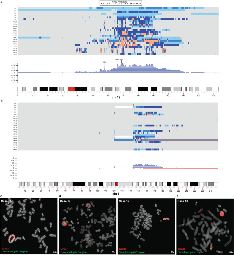
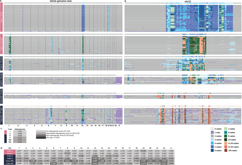
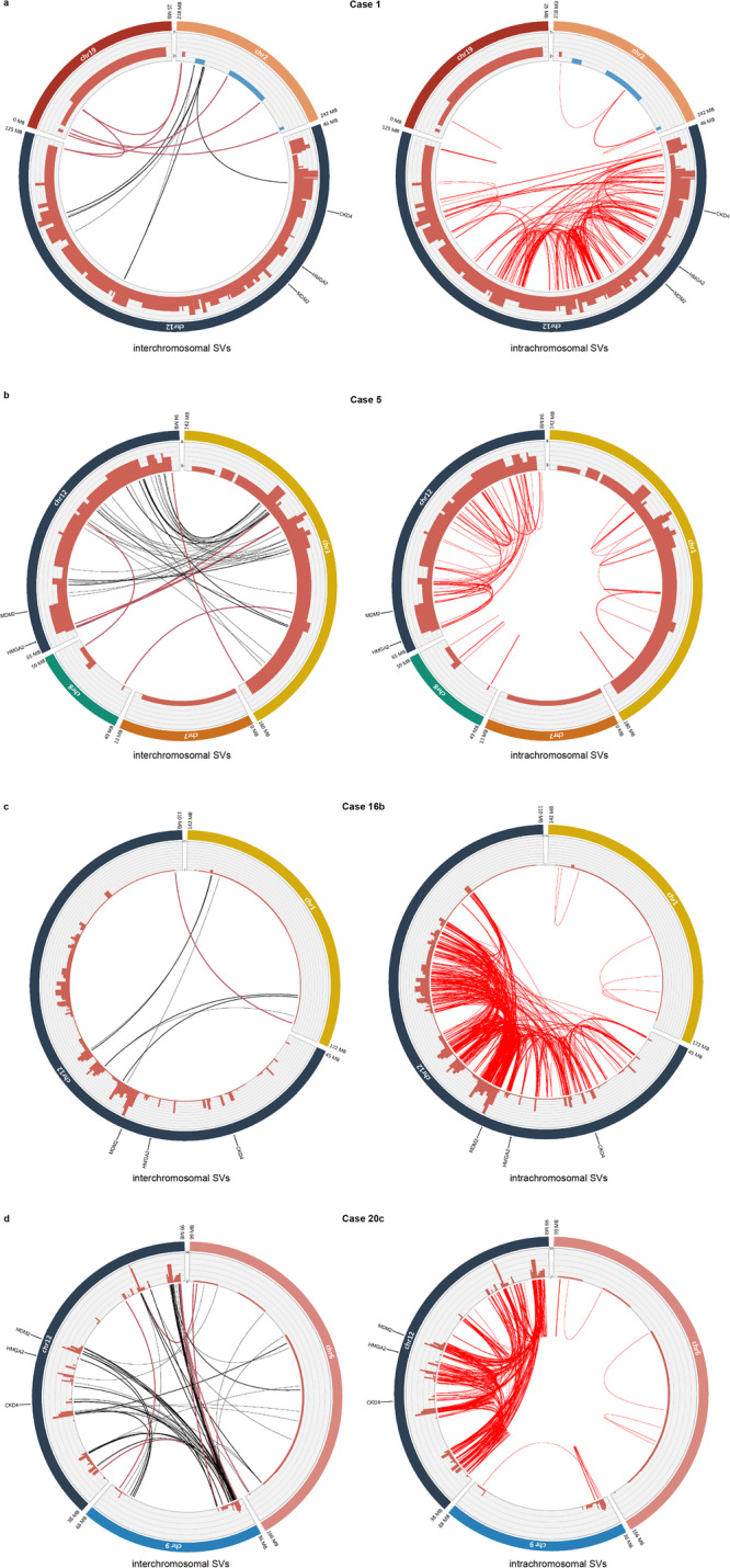
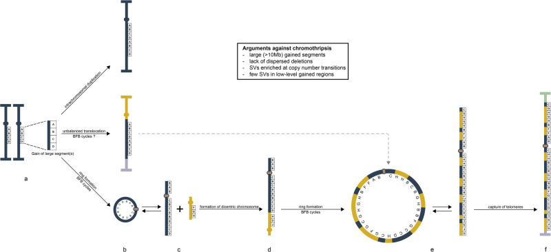
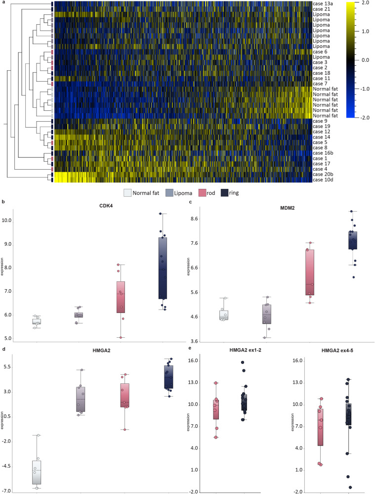
Well-differentiated liposarcoma (WDLS) displays amplification of genes on chromosome 12 (Chr12) in supernumerary ring or giant marker chromosomes. These structures have been suggested to develop through chromothripsis, followed by circularization and breakage-fusion-bridge (BFB) cycles. To test this hypothesis, we compared WDLSs with Chr12 amplification in rod-shaped chromosomes with WDLSs with rings. Both types of amplicons share the same spectrum of structural variants (SVs), show higher SV frequencies in Chr12 than in co-amplified segments, have SVs that fuse the telomeric ends of co-amplified chromosomes, and lack interspersed deletions. Combined with the finding of cells with transient rod-shaped structures in tumors with ring chromosomes, this suggests a stepwise process starting with the gain of Chr12 material that, after remodeling which does not fit with classical chromothripsis, forms a dicentric structure with other chromosomes. Depending on if and when telomeres from other chromosomes are captured, circularized or linear gain of 12q sequences will predominate.


Conflict of interest statement

The authors declare no competing interests.

Figures

Fig. 1
Fig. 1. Copy number (CN) variation in lipomatous tumors with 12q gain.
Distribution and frequency plots of CN gain in (a) Chr12 and (b) Chr1 from bulk DNA. Fluorescence in situ hybridization on metaphase spreads from c Case 10c, d Case 17 and e Case 18 shows that also low-level gained segments (green signals; MDM2 signals in red) are included in the ring chromosomes, as well as in the two normal homologues. Also, Case 17 (d) highlights the interchangeability between ring and rod-shaped chromosomes.
Fig. 2
Fig. 2. Single cell copy number (CN) variation in lipomatous tumors with 12q gain.
Single cell whole genome sequencing with 1 Mb bins showing distribution of (a) genome-wide CN changes in individual cells and (b) with 40 kb bins showing distribution of CN gain in Chr12 in individual cells. Heterogeneity scores (c) genome-wide and (d) chromosome-specific based on 1 Mb bins.
Fig. 3
Fig. 3. Structural variants in lipomatous tumors with 12q gain.
a Case 1; b Case 5; c Case 16b; d Case 20c. Inter- (black lines) and intrachromosomal (red lines) structural variants (SVs) detected at whole-genome sequencing in lipomatous tumors with 12q-gain. Copy number state is displayed in terracotta red bars. Important interchromosomal SVs are highlighted in terracotta red.
Fig. 4
Fig. 4. A schematic view of the mechanisms behind gain and amplification of MDM2 in lipomatous tumors.
a The initial event is duplication of one or more large (often >10 Mb) segments from chromosome arm 12q (oval circles represent centromeres; horizontal bars represent telomeres). b These duplicated segments can, after more or less extensive reorganization, either be (top) inserted in one of the two homologs of Chr12, as exemplified by Case 2, or (middle) translocated to one or more other chromosomes, as exemplified by Case 3; in both scenarios the gained 12q segments end up in a chromosome with intact telomeres, providing relative mitotic stability. The third option (bottom) involves circularization of the gained segments in a ring chromosome, which subsequently develops a neocentromere (beige oval circle); the structural variants, as well as the copy number data in Case 16, suggest that gain of 12q precedes co-amplification with other chromosomes. Due to mitotic instability, the ring chromosome will experience extensive copy number alterations, resulting in amplification of some segments. c The ring will occasionally break up to form a rod-shaped structure (see Fig. 1d) and fuse with material from other chromosomes, notably Chr1. d The consistent finding of structural variants fusing the most telomeric sequences of the gained sequences from Chr12 and co-amplified chromosomes, as seen in Cases 5, 16, 20, and 21, strongly suggests that the newly formed rod-shaped chromosome is dicentric. e After breakage-fusion-bridge cycles, the dicentric chromosome will break and form a new ring chromosome. f After further cycles of breaking up and recircularization, the amplified structure will eventually capture telomeres from other chromosomes and become relatively stable.
Fig. 5
Fig. 5. Gene expression profiles in lipomatous tumors with 12q gain at RNA-sequencing (RNA-seq) and quantitative real-time PCR (qRT-PCR).
a Unsupervised heatmap showing clustering of cases from Group A and B compared to control tissue/tumors (normal fat/lipoma). Box plots at RNA-seq showing expression levels of (b) CDK4, c MDM2, d HMGA2, and at qRT-PCR (e, left) HMGA2 ex1-2; (e, right) HMGA2 ex4-5. Normal fat (n = 6), Lipoma (n = 8), rod-shaped (n = 7, Cases 1–7), ring (n = 13, Cases 8–21).

References

    1. Santarius T, Shipley J, Brewer D, Stratton MR, Cooper CS. A census of amplified and overexpressed human cancer genes. Nat. Rev. Cancer. 2010;10:59–64. doi: 10.1038/nrc2771. - DOI - PubMed
    1. Wahl GM. The importance of circular DNA in mammalian gene amplification. Cancer Res. 1989;49:1333–1340. - PubMed
    1. Shimizu N. Extrachromosomal double minutes and chromosomal homogeneously staining regions as probes for chromosome research. Cytogenet Genome Res. 2009;124:312–326. doi: 10.1159/000218135. - DOI - PubMed
    1. Verhaak RGW, Bafna V, Mischel PS. Extrachromosomal oncogene amplification in tumour pathogenesis and evolution. Nat. Rev. Cancer. 2019;19:283–288. doi: 10.1038/s41568-019-0128-6. - DOI - PMC - PubMed
    1. Shoshani O, et al. Chromothripsis drives the evolution of gene amplification in cancer. Nature. 2021;591:137–141. doi: 10.1038/s41586-020-03064-z. - DOI - PMC - PubMed

Publication types

Substances