. 2024 May 20;22(1):271.
doi: 10.1186/s12951-024-02538-w.

Bio-nanoparticles loaded with synovial-derived exosomes ameliorate osteoarthritis progression by modifying the oxidative microenvironment

Affiliations

Bio-nanoparticles loaded with synovial-derived exosomes ameliorate osteoarthritis progression by modifying the oxidative microenvironment

Haifei Cao et al. J Nanobiotechnology. .

Abstract

Background and aims: Osteoarthritis (OA) is a prevalent degenerative joint disorder, marked by the progressive degeneration of joint cartilage, synovial inflammation, and subchondral bone hyperplasia. The synovial tissue plays a pivotal role in cartilage regulation. Exosomes (EXOs), small membrane-bound vesicles released by cells into the extracellular space, are crucial in mediating intercellular communication and facilitating the exchange of information between tissues. Our study aimed to devise a hydrogel microsphere infused with SOD3-enriched exosomes (S-EXOs) to protect cartilage and introduce a novel, effective approach for OA treatment.

Materials and methods: We analyzed single-cell sequencing data from 4247 cells obtained from the GEO database. Techniques such as PCR, Western Blot, immunofluorescence (IF), and assays to measure oxidative stress levels were employed to validate the cartilage-protective properties of the identified key protein, SOD3. In vivo, OA mice received intra-articular injections of S-EXOs bearing hydrogel microspheres, and the effectiveness was assessed using safranine O (S.O) staining and IF.

Results: Single-cell sequencing data analysis suggested that the synovium influences cartilage via the exocrine release of SOD3. Our findings revealed that purified S-EXOs enhanced antioxidant capacity of chondrocytes, and maintained extracellular matrix metabolism stability. The S-EXO group showed a significant reduction in mitoROS and ROS levels by 164.2% (P < 0.0001) and 142.7% (P < 0.0001), respectively, compared to the IL-1β group. Furthermore, the S-EXO group exhibited increased COL II and ACAN levels, with increments of 2.1-fold (P < 0.0001) and 3.1-fold (P < 0.0001), respectively, over the IL-1β group. Additionally, the S-EXO group showed a decrease in MMP13 and ADAMTS5 protein expression by 42.3% (P < 0.0001) and 44.4% (P < 0.0001), respectively. It was found that S-EXO-containing hydrogel microspheres could effectively deliver SOD3 to cartilage and significantly mitigate OA progression. The OARSI score in the S-EXO microsphere group markedly decreased (P < 0.0001) compared to the OA group.

Conclusion: The study demonstrated that the S-EXOs secreted by synovial fibroblasts exert a protective effect on chondrocytes, and microspheres laden with S-EXOs offer a promising therapeutic alternative for OA treatment.

Keywords: Exosomes; Microspheres; Osteoarthritis; SOD3.


Conflict of interest statement

The authors declare no competing interests.

Figures

Scheme 1
Scheme 1
The process of our study. (a) Analysis of single-cell sequencing data. (b) Preparation of S-EXOs. (c) Preparation of hydrogel microspheres loaded with S-EXOs. (d) Intra-articular injection of hydrogel microspheres loaded with S-EXOs for OA treatment
Fig. 1
Fig. 1
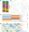
The analysis of single-cell sequencing. (a-b) Percentage distribution of the 9 different subsets according to either early OA and severe OA. (c) Heatmap of significant cellular functions of the synovial fibroblast in early OA and severe OA. (d) Heatmap of significant cellular functions for each of the 9 synovial fibroblast subsets
Fig. 2
Fig. 2
The analysis of antioxidant function of synovial fibroblasts from early OA and severe OA. Biological process bubble diagrams of samples from cluster 0 (a) and cluster 3 (b) based on GO analysis. (c) The difference of SOD3 between early OA and severe OA based on whole-cell population analysis. (d) The network diagram of cluster 3. The difference of SOD3 between early OA and severe OA based on cluster 0 (e) and cluster 3 (f) population analysis. (g) RT-PCR analysis for the mRNA expression of Sod3, n = 4. (h) Elisa analysis for the secretion of SOD3, n = 4
Fig. 3
Fig. 3
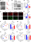
S-EXOs enhanced the antioxidant capacity of chondrocytes in inflammatory conditions induced by IL-1β. (a) Morphology of EXOs and S-EXOs captured by TEM and the particle size distribution detected by NTA. Scale bar = 200 nm, n = 3. (b) Western blot analysis of the EXOs and S-EXOs specific protein markers, n = 3. (c-e) The effects of EXOs and S-EXOs on the amount of mitoROS (Red) and intracellular ROS (Green) in chondrocytes, n = 3. (f) DPPH, (g) ABTS, (h) PTIO and (i) hydroxyl radical-scavenging ability of the EXOs and S-EXOs detected via UV − vis spectroscopy, n = 4
Fig. 4
Fig. 4
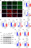
The effects of EXOs and S-EXOs on ECM metabolism of chondrocytes in inflammatory micro-environment induced by IL-1β. (a-b) IF analysis for the expression of ACAN. (c-d) IF analysis for the expression of MMP13. Scale bar = 100 μm, n = 3. (e-h) RT-PCR analysis for the mRNA expression of Col2a1, Acan, Mmp13 and Adamts5, n = 4. (i-m) The protein levels of COL II, ACAN, MMP13 and ADAMTS5, n = 3
Fig. 5
Fig. 5
Characterization of EXOs bearing microspheres. (a) The gross view and diameter distribution of microspheres. Scale bar = 100 μm. (b) The representative SEM and EDS images of dispersed microspheres. Scale bar = 20 μm. (c) The rate of remaining weight, n = 3. (d) The loading efficiency curves for the EXOs-loaded microspheres, n = 3. (e) Release profiles of EXOs from the EXOs-loaded microspheres, n = 3. (f) The quantitative analysis of Live&dead cells cultured on various microspheres, n = 3. (g) The cell proliferation analysis of the cells cultured on various microspheres using CCK-8, n = 3
Fig. 6
Fig. 6
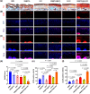
EXOs-loaded microspheres alleviated the progress of OA by delivering SOD3. (a) Representative images of cartilage stained by Safranin O-fast green (S.O.). (b&e) IF analysis for the expression of SOD3. (c&f) IF analysis for the expression of ACAN. (d) OARSI scores. Scale bar = 100 μm, n = 3

References

    1. Giorgino R, Albano D, Fusco S, Peretti GM, Mangiavini L, Messina C. Knee osteoarthritis: Epidemiology, Pathogenesis, and mesenchymal stem cells: what else is New? An update. Int J Mol Sci. 2023;24(7). - PMC - PubMed
    1. Tong L, Yu H, Huang X, Shen J, Xiao G, Chen L, et al. Current understanding of osteoarthritis pathogenesis and relevant new approaches. Bone Res. 2022;10(1):60. doi: 10.1038/s41413-022-00226-9. - DOI - PMC - PubMed
    1. Stewart HL, Kawcak CE. The importance of subchondral bone in the pathophysiology of Osteoarthritis. Front Vet Sci. 2018;5:178. doi: 10.3389/fvets.2018.00178. - DOI - PMC - PubMed
    1. Donell S. Subchondral bone remodelling in osteoarthritis. EFORT Open Rev. 2019;4(6):221–9. doi: 10.1302/2058-5241.4.180102. - DOI - PMC - PubMed
    1. Hu Y, Chen X, Wang S, Jing Y, Su J. Subchondral bone microenvironment in osteoarthritis and pain. Bone Res. 2021;9(1):20. doi: 10.1038/s41413-021-00147-z. - DOI - PMC - PubMed

Substances