. 2024 Oct 1;196(2):788-809.
doi: 10.1093/plphys/kiae287.

Extracellular vesicles of Norway spruce contain precursors and enzymes for lignin formation and salicylic acid

Affiliations

Extracellular vesicles of Norway spruce contain precursors and enzymes for lignin formation and salicylic acid

Santeri Kankaanpää et al. Plant Physiol. .

Abstract

Lignin is a phenolic polymer in plants that rigidifies the cell walls of water-conducting tracheary elements and support-providing fibers and stone cells. Different mechanisms have been suggested for the transport of lignin precursors to the site of lignification in the cell wall. Extracellular vesicle (EV)-enriched samples isolated from a lignin-forming cell suspension culture of Norway spruce (Picea abies L. Karst.) contained both phenolic metabolites and enzymes related to lignin biosynthesis. Metabolomic analysis revealed mono-, di-, and oligolignols in the EV isolates, as well as carbohydrates and amino acids. In addition, salicylic acid (SA) and some proteins involved in SA signaling were detected in the EV-enriched samples. A proteomic analysis detected several laccases, peroxidases, β-glucosidases, putative dirigent proteins, and cell wall-modifying enzymes, such as glycosyl hydrolases, transglucosylase/hydrolases, and expansins in EVs. Our findings suggest that EVs are involved in transporting enzymes required for lignin polymerization in Norway spruce, and radical coupling of monolignols can occur in these vesicles.


Conflict of interest statement

Conflict of interest statement. None declared.

Figures

Figure 1.
Figure 1.
Tissue culture model and extracellular vesicles (EVs) isolated from the culture medium of tissue-cultured cells of Norway spruce. A) Setup for experiments using an extracellular lignin-forming Norway spruce cell culture (according to Laitinen et al. 2017 with some modifications). Spruce cells were transferred at Day 0 from the solid maintenance medium to the liquid nutrient medium supplemented either with an H2O2 scavenger, KI (5 mm; non-lignin-forming conditions), or the corresponding volume of water (H2O; lignin-forming conditions). At Days 7, 10, and 14, 4 replicate cultures of each treatment were harvested, and the media were collected for the isolation of extracellular vesicles (EVs) (see Materials and methods). B) Transmission electron micrographs of the EVs pelleted with ultracentrifugation from the spruce cell culture media. Common characteristics of EVs are seen in the EM images with moon-shaped particles with membrane characteristics shown in the close-ups. i) Close-up of the EVs marked with a rectangular shape in the big image. A scale bar (100 nm) shown is for all close-up images. C) Histogram of the size distribution of EVs as measured from the transmission electron microscopy images using ImageJ (n = 100).
Figure 2.
Figure 2.
Metabolomic analysis of compounds in the extracellular vesicle (EV) samples and in the EV-depleted culture media of Norway spruce cultured cells. Principal component analysis (PCA) plot of the gas chromatography–mass spectrometry (GC–MS)-detected compounds in A) the EV samples and in B) the culture media at 3 different time points (7, 10, and 14 d) for 2 treatments, namely, lignin-forming and non-lignin-forming (KI-treatment). Ellipses and rectangles frame biological repeats and are added for clarity. The value between the brackets is the percentage of the variation explained by the respective principal component (PC). PCA plot of the ultra-performance liquid chromatography–mass spectrometry (UPLC–MS)-detected compounds in C) the EV samples and in D) the EV-depleted culture media in 3 different time points (7, 10, and 14 d) in 2 treatments [lignin-forming and non-lignin-forming (KI-treatment)]. Ellipses and rectangles frame biological repeats and are added for clarity. The value between the brackets is the percentage of the variation explained by the respective principal component (PC).
Figure 3.
Figure 3.
Accumulation of the phenolic metabolites in the extracellular vesicle (EV) samples as observed in the metabolic analysis. A) Example of the phenolic metabolites detected only in the EV samples, and not in the medium samples, by gas chromatography–mass spectrometry (GC–MS). B) Accumulation of coniferyl alcohol and coniferaldehyde in the EV samples as detected by GC–MS in lignin-forming and non-lignin-forming (KI-treatment) conditions (mean ± SD, n = 4). C) Accumulation of G(8-8)G hexoside (34, pinoresinol hexoside) in the EV samples as observed by ultra-performance liquid chromatography–mass spectrometry (UPLC–MS) in lignin-forming and non-lignin-forming conditions (KI-treatment). The boxplots show the median (horizontal lines), 25th to 75th percentiles (edges of the boxes), and minimum to maximum values (edges of the whiskers) (n = 4).
Figure 4.
Figure 4.
Compounds detected by ultra-performance liquid chromatography–mass spectrometry (UPLC–MS) analysis in the extracellular vesicle (EV) samples and in the EV-depleted culture media of Norway spruce cultured cells. A) Heat maps showing compounds detected in the EV samples and in the culture media. The structural characterizations were based on the MS/MS-fragmentation spectra. B) Structural formulae of di- and oligolignols detected in the EV samples of Norway spruce.
Figure 5.
Figure 5.
Extracted ion chromatograms (EIC) and electron impact (EI) spectra of salicylic acid (SA) and presence of SA in the extracellular vesicle (EV) samples of Norway spruce. A) EIC of m/z 267.0869 of a SA reference standard (top), a pooled sample of all EV samples (middle), and a pooled sample of all EV-depleted culture medium samples (bottom). B) Deconvoluted EI spectrum of the peak eluting at 16.38 min in a pooled sample of all EV samples. C) Deconvoluted EI spectrum of the peak eluting at 16.39 min in the chromatogram of the SA reference standard. D) EI spectrum of the 2 trimethylsilyl (TMS) derivative of SA in the National Institute of Standards and Technology (NIST) ‘20 spectral library. E) Accumulation of SA in the EV samples as detected by gas chromatography–mass spectrometry (GC–MS) in lignin-forming and non-lignin-forming (KI-treatment) conditions (mean ± SD, n = 4).
Figure 6.
Figure 6.
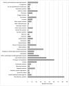
Proteomic analysis of the extracellular vesicle (EV) samples of Norway spruce. Distribution of proteins (n = 557) detected in the EV samples in groups with annotated functions.
Figure 7.
Figure 7.
Proteins detected in extracellular vesicle (EV)-enriched samples of Norway spruce, and their annotated functions. The median and the average abundancy of unique peptides per protein across all samples are also shown. Proteins shown are listed based on their reported presence in EVs or their putative role in lignin biosynthesis. A wider list of proteins detected is given in Supplementary Table S3.
Figure 8.
Figure 8.
Hypothetical pathways for the export of oxidative enzymes (squares) and lignin mono-, di-, and oligomers (hexagons with varying linkages) to the apoplastic space in tissue-cultured cells of Norway spruce during lignin formation. A) Conventional protein secretion exports oxidative enzymes that are synthesized in the endoplasmic reticulum (ER), modified in the Golgi apparatus, sorted in the trans-Golgi network (TGN), and transported in Golgi vesicles to the plasma membrane (PM), where the vesicles fuse with the PM to release oxidative enzymes. In addition, monolignols are synthesized via the phenylpropanoid and monolignol biosynthesis pathways (waved arrow indicates the pathways and intermediates are represented with hexagons), with some enzymes associated with the ER and transported through the PM via diffusion and/or a transporter-mediated transport. Monolignol oxidation and polymerization occur in the apoplast (cell wall, and the culture medium in case of cultured cells). B) Double membrane-containing exocyst-positive organelles (EXPOs) form in the cytoplasm, thereby sealing the oxidative enzymes inside. Monolignols diffuse along the concentration gradient or are actively transported to the vesicles where they are radicalized by the oxidative enzymes and begin to polymerize. The outer membranes of EXPOs fuse with the PM, releasing the inner vesicle (now called an extracellular vesicle, EV) into the apoplast. C) Multivesicular bodies (MVBs) originate from the TGN and contain vesicles (intraluminal vesicles, ILVs) that form by the inward budding of the MVB membrane. Oxidative enzymes get trapped inside the vesicles. Monolignols could follow the oxidative enzymes into the vesicles in a similar way as described above in B). MVB fuses with the PM releasing ILVs into the apoplast. EVs secreted in this way are called exosomes. In B) and C), the monolignol accumulation in the EVs could continue in the apoplast, with the monolignols secreted directly through the PM. D) Microvesicle-type EVs are produced by blebbing of the PM into the apoplast and seal proteins inside. These EVs accumulate monolignols only in the apoplast; thus, the original monolignol transport through PM would be based on diffusion and/or transporter-mediated transport. In B) to D), the EVs finally release their contents to the apoplast by a yet unknown unloading mechanism.

Similar articles

Cited by

References

    1. Alejandro S, Lee Y, Tohge T, Sudre D, Osorio S, Park J, Bovet L, Lee Y, Geldner N, Fernie AR, et al. . AtABCG29 is a monolignol transporter involved in lignin biosynthesis. Curr Biol. 2012:22(13):1207–1212. 10.1016/j.cub.2012.04.064 - DOI - PubMed
    1. Bachurski D, Schuldner M, Nguyen PH, Malz A, Reiners KS, Grenzi PC, Babatz F, Schauss AC, Hansen HP, Hallek M, et al. . Extracellular vesicle measurements with nanoparticle tracking analysis—an accuracy and repeatability comparison between NanoSight NS300 and ZetaView. J Extracell Vesicles. 2019:8(1):1596016. 10.1080/20013078.2019.1596016 - DOI - PMC - PubMed
    1. Benedetti M, Verrascina I, Pontiggia D, Locci F, Mattei B, De Lorenzo G, Cervone F. Four Arabidopsis berberine bridge enzyme-like proteins are specific oxidases that inactivate the elicitor-active oligogalacturonides. Plant J. 2018:94(2):260–273. 10.1111/tpj.13852 - DOI - PubMed
    1. Berthet S, Demont-Caulet N, Pollet B, Bidzinski P, Cézard L, Le Bris P, Borrega N, Hervé J, Blondet E, Balzergue S, et al. . Disruption of LACCASE4 and 17 results in tissue-specific alterations to lignification of Arabidopsis thaliana stems. Plant Cell. 2011:23(3):1124–1137. 10.1105/tpc.110.082792 - DOI - PMC - PubMed
    1. Blaschek L, Murozuka E, Serk H, Ménard D, Pesquet E. Different combinations of laccase paralogs nonredundantly control the amount and composition of lignin in specific cell types and cell wall layers in Arabidopsis. Plant Cell. 2023:35(2):889–909. 10.1093/plcell/koac344 - DOI - PMC - PubMed

LinkOut - more resources