

VEGF-C and 5-Fluorouracil Improve Bleb Survival in a Rabbit Glaucoma Surgery Trabeculectomy Model
- PMID: 38771570
- PMCID: PMC11114614
- DOI: 10.1167/iovs.65.5.32
VEGF-C and 5-Fluorouracil Improve Bleb Survival in a Rabbit Glaucoma Surgery Trabeculectomy Model
Abstract
Purpose: To evaluate VEGF-C-induced lymphoproliferation in conjunction with 5-fluorouracil (5-FU) antimetabolite treatment in a rabbit glaucoma filtration surgery (GFS) model.
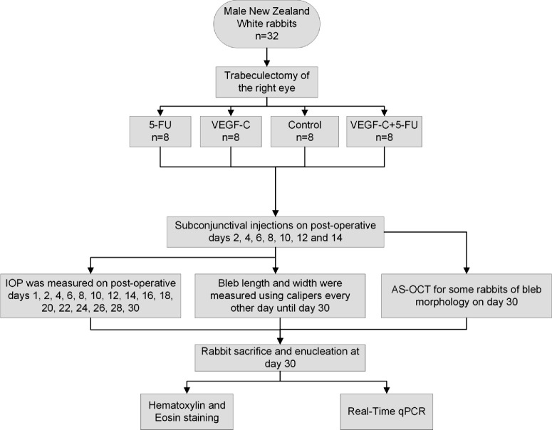
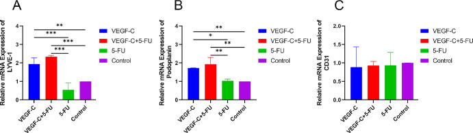
Methods: Thirty-two rabbits underwent GFS and were assigned to four groups (n = 8 each) defined by subconjunctival drug treatment: (a) VEGF-C combined with 5-FU, (b) 5-FU, (c) VEGF-C, (d) and control. Bleb survival, bleb measurements, and IOP were evaluated over 30 days. At the end, histology and anterior segment OCT were performed on some eyes. mRNA was isolated from the remaining eyes for RT-PCR evaluation of vessel-specific markers (lymphatics, podoplanin and LYVE-1; and blood vessels, CD31).
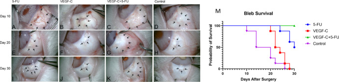
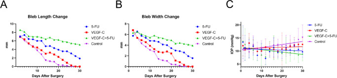
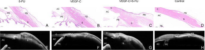
Results: Qualitatively and quantitatively, VEGF-C combined with 5-FU resulted in blebs which were posteriorly longer and wider than the other conditions: vs. 5-FU (P = 0.043 for longer, P = 0.046 for wider), vs. VEGF-C (P < 0.001, P < 0.001) and vs. control (P < 0.001, P < 0.001). After 30 days, the VEGF-C combined with 5-FU condition resulted in longer bleb survival compared with 5-FU (P = 0.025), VEGF-C (P < 0.001), and control (P < 0.001). Only the VEGF-C combined with 5-FU condition showed a negative correlation between IOP and time that was statistically significant (r = -0.533; P = 0.034). Anterior segment OCT and histology demonstrated larger blebs for the VEGF-C combined with 5-FU condition. Only conditions including VEGF-C led to increased expression of lymphatic markers (LYVE-1, P < 0.001-0.008 and podoplanin, P = 0.002-0.011). Expression of CD31 was not different between the groups (P = 0.978).
Conclusions: Adding VEGF-C lymphoproliferation to standard antimetabolite treatment improved rabbit GFS success and may suggest a future strategy to improve human GFSs.
Conflict of interest statement
Disclosure:
Figures
Similar articles
-
Subconjunctival Lymphatics Respond to VEGFC and Anti-Metabolites in Rabbit and Mouse Eyes.Invest Ophthalmol Vis Sci. 2022 Sep 1;63(10):16. doi: 10.1167/iovs.63.10.16. Invest Ophthalmol Vis Sci. 2022. PMID: 36166215 Free PMC article.
-
Comparison of Efficacy and Safety of Bleb Needle Revision With and Without 5-Fluorouracil for Failing Trabeculectomy Bleb.J Glaucoma. 2019 May;28(5):386-391. doi: 10.1097/IJG.0000000000001226. J Glaucoma. 2019. PMID: 30839411
-
Comparison of needle revision with subconjunctival bevacizumab and 5-fluorouracil injection of failed trabeculectomy blebs.J Ocul Pharmacol Ther. 2012 Oct;28(5):542-6. doi: 10.1089/jop.2012.0035. Epub 2012 Jun 25. J Ocul Pharmacol Ther. 2012. PMID: 22731246 Clinical Trial.
-
Postoperative use of bevacizumab as an antifibrotic agent in glaucoma filtration surgery in the rabbit.Invest Ophthalmol Vis Sci. 2009 Jul;50(7):3233-7. doi: 10.1167/iovs.08-2441. Epub 2009 Jan 31. Invest Ophthalmol Vis Sci. 2009. PMID: 19182254
-
Updates on the utility of anterior segment optical coherence tomography in the assessment of filtration blebs after glaucoma surgery.Acta Ophthalmol. 2022 Feb;100(1):e29-e37. doi: 10.1111/aos.14881. Epub 2021 May 4. Acta Ophthalmol. 2022. PMID: 33942540 Review.
Cited by
-
Mechanisms of fibrosis formation following glaucoma filtration surgery.Int J Ophthalmol. 2025 Aug 18;18(8):1579-1586. doi: 10.18240/ijo.2025.08.21. eCollection 2025. Int J Ophthalmol. 2025. PMID: 40827283 Free PMC article. Review.
References
-
- Jonas JB, Aung T, Bourne RR, et al. .. Glaucoma. Lancet. 2017; 390(10108): 2183–2193. - PubMed
-
- Mathew DJ, Buys YM.. Minimally invasive glaucoma surgery: a critical appraisal of the literature. Annu Rev Vis Sci. 2020; 6: 47–89. - PubMed
-
- Yu DY, Morgan WH, Sun X, et al. .. The critical role of the conjunctiva in glaucoma filtration surgery. Prog Retin Eye Res. 2009; 28(5): 303–328. - PubMed
-
- Morgan WH, Yu DY.. XEN-45 gelatin microfistula for uveitic glaucoma. Clin Exp Ophthalmol. 2018; 46(4): 323–324. - PubMed
MeSH terms
LinkOut - more resources
Full Text Sources
Medical
Miscellaneous
