. 2024 May 21;9(13):e177588.
doi: 10.1172/jci.insight.177588.

ADAM17 variant causes hair loss via ubiquitin ligase TRIM47-mediated degradation

Affiliations

ADAM17 variant causes hair loss via ubiquitin ligase TRIM47-mediated degradation

Xiaoxiao Wang et al. JCI Insight. .

Abstract

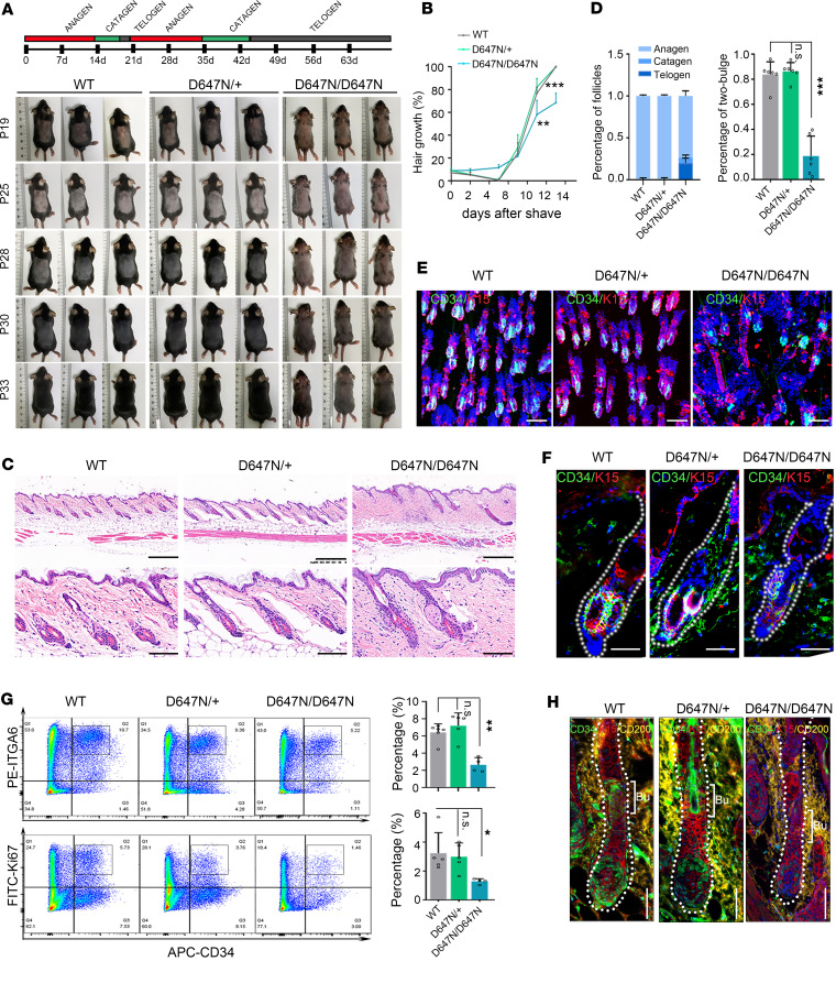
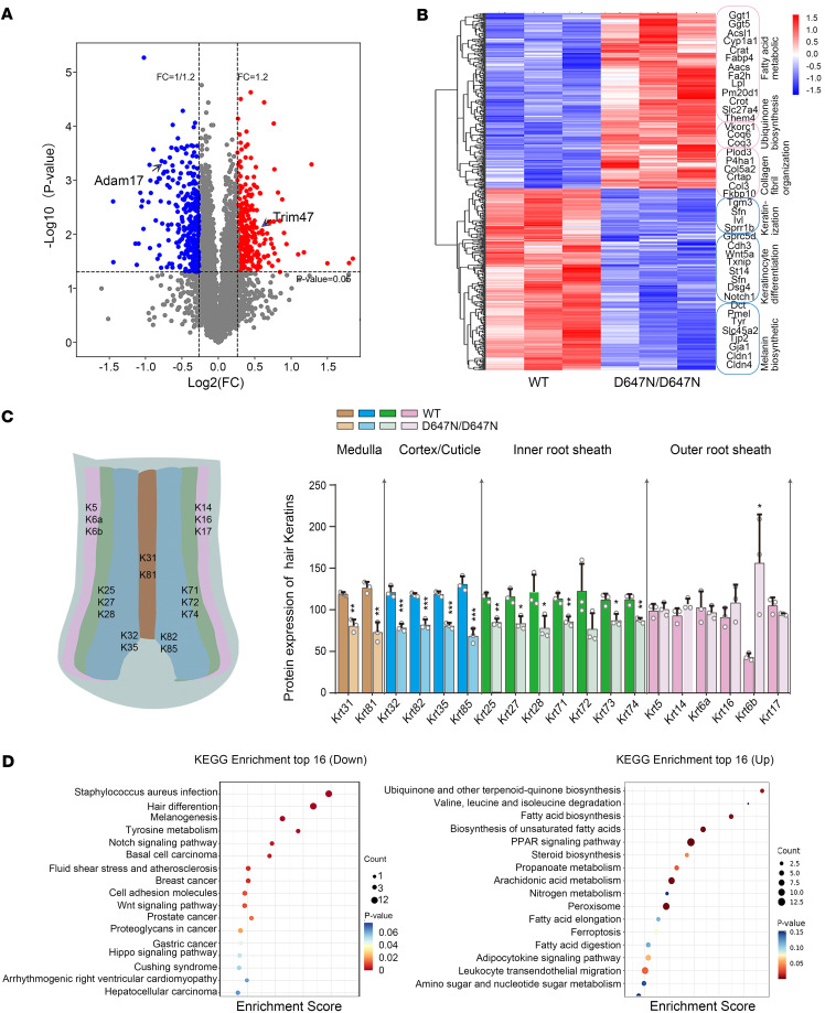
Hypotrichosis is a genetic disorder characterized by a diffuse and progressive loss of scalp and/or body hair. Nonetheless, the causative genes for several affected individuals remain elusive, and the underlying mechanisms have yet to be fully elucidated. Here, we discovered a dominant variant in a disintegrin and a metalloproteinase domain 17 (ADAM17) gene caused hypotrichosis with woolly hair. Adam17 (p.D647N) knockin mice mimicked the hair abnormality in patients. ADAM17 (p.D647N) mutation led to hair follicle stem cell (HFSC) exhaustion and caused abnormal hair follicles, ultimately resulting in alopecia. Mechanistic studies revealed that ADAM17 binds directly to E3 ubiquitin ligase tripartite motif-containing protein 47 (TRIM47). ADAM17 variant enhanced the association between ADAM17 and TRIM47, leading to an increase in ubiquitination and subsequent degradation of ADAM17 protein. Furthermore, reduced ADAM17 protein expression affected the Notch signaling pathway, impairing the activation, proliferation, and differentiation of HFSCs during hair follicle regeneration. Overexpression of Notch intracellular domain rescued the reduced proliferation ability caused by Adam17 variant in primary fibroblast cells.

Keywords: Dermatology; Genetic diseases; Genetic variation; Genetics; Ubiquitin-proteosome system.


Figures

Figure 1
Figure 1. ADAM17 variant leads to ADWH.
(A) Representative clinical pictures of patients with ADWH. (B) Hematoxylin and eosin (HE) staining of scalp tissues from patients and the normal controls. Scale bars, 125 μm. (C) The scanning electron microscopy (SEM) analysis revealed that the patient’s hair shaft displayed a nonuniform and abnormal cross-sectional shape. Furthermore, there was a substantial peeling of hair cuticle, which normally functions as the hair shaft’s outermost protective layer. Scale bars, 50 μm. (D) Pedigree of the family for patients. Hollow boxes represented the healthy men, while solid boxes represented the affected men. Hollow circles represented the healthy women, while solid circles represented the affected women in the family tree. The proband was identified with a black arrow. (E) A genome-wide linkage analysis provided evidence of linkage to chromosome 2, with a maximum LOD score of 3.18. (F) Gene sequencing revealed the heterozygous ADAM17 p.Asp647Asn (c.1939G>A) variant in patients. The arrows indicate the variant. (G) ADAM17 was predominantly expressed in the hair cortex, inner root sheath (IRS), and outer root sheath (ORS) of human hair follicle. Scale bar, 500 μm. (H) Schematic overview of ADAM17 protein and its domains. The p.D647N mutation was localized in CANDIS domain (indicated by a red arrow). SP, signal peptide; PD, pro-domain; Catalytic, catalytic metalloprotease domain; DD, disintegrin domain; MPD, membrane proximal domain; CANDIS, conserved ADAM17 seventeen dynamic interaction sequence; TM, transmembrane domain; CD, cytoplasmic domain. (I) Aspartic residue at position 647 is located within the conserved ADAM17 dynamic interaction sequence (CANDIS) domain of ADAM17, which is highly conserved.
Figure 2
Figure 2. Adam17 (p.D647N) variant causes abnormal hair follicle morphology and hair loss in mice.
(A) Hair coats and vibrissa hairs of wild-type (WT), Adam17D647N/+, and Adam17D647N/D647N mice at postnatal day (P) 19, P35, and 6 months (M). (B) Pelage hairs were observed under an optical microscope. Left and middle panel: compared with wild-type hairs, all 4 hair types of Adam17D647N/D647N pelage hairs showed waviness. Scale bars, 2 mm. Right panel: Adam17D647N/D647N mice displayed a significantly reduced proportion of primary and secondary hairs and a significant increase in the proportion of zigzag hairs. (n = 4 biological replicates.) (C) The scanning electron microscopy (SEM) analysis unveiled a peeling of the hair cuticle in the hair of Adam17D647N/D647N mice. Scale bar, 10 μm. (D) HE staining revealed notable structural abnormalities of hair follicles (HFs) in Adam17D647N/D647N mice at P28. Upper panel scale bars, 250 μm; lower panel scale bars, 50 μm. (E) HE staining of longitudinal sections revealed obvious structural abnormalities of HFs in Adam17D647N/D647N mice during the first anagen (P7) and second anagen (P28). Scale bars, 250 μm. (F) Immunofluorescence staining revealed a substantial decrease in IRS markers within the HFs of Adam17D647N/D647N mice, suggesting significant morphological abnormalities of the IRS. Scale bar, 100 μm. (G) A schematic illustration of HF layers and markers expressed in HF. (H) Immunoblot analysis of HF layer-specific markers in dorsal skin. (I) Transmission electron microscopy (TEM) of HFs at approximately 500 μm depth. Left panel scale bar, 5 μm; right panel scale bar, 2 μm. ORS, outer root sheath; IRS, inner root sheath; Cl, companion layer; He, Henle’s layer; Hu, Huxley’s layer; Cu, cuticle; Co, cortex; Me, medulla. All experiments were repeated 3 times. Data were expressed as mean ± SD; *P < 0.05; **P < 0.01; ***P < 0.001; 1-way ANOVA (B).
Figure 3
Figure 3. Adam17 (p.D647N) variant affects homeostasis of the HFSC niche in hair follicles.
(A) Phenotypes of wild-type (WT), Adam17D647N/+, and Adam17D647N/D647N mice after shaving back skin during telogen (P19) and monitoring initiation of the next hair cycle. Adam17 mutation impeded hair regeneration. (B) Statistical data on the proportion of the skin with pigmentation in mice after hair shaving. (n = 6 biological replicates.) (C) HE staining of back skin during second telogen (P63). Upper panel scale bars, 300 μm; lower panel scale bars, 50 μm. (DF) WT hair follicles (HFs) possessed a 2-bulge architecture, whereas Adam17D647N/D647N HFs usually had only 1. (D) Statistical data on the proportion of 3 HF types. (n = 6 biological replicates.) (E) Immunofluorescence was performed on whole-mount back skin hair follicles at the second telogen stage using HFSC markers K15 and CD34. Scale bar, 100 μm. (F) Skin sections underwent immunofluorescence staining using antibodies specific to HFSC markers. Scale bars, 40 μm. (G) Fluorescence-activated cell sorting (FACS) analyses of HFSC populations sorted by high α6-integrin and CD34. Right upper panel: Quantification of CD34-positive/α6-high cells (indicated by the black square brackets) among epithelial cells in second telogen mice. (n = 4–6 biological replicates.) Right lower panel: Quantification of CD34-positive/Ki67-positive cells among epithelial cells in second telogen mice. (n = 4–6 biological replicates.) (H) HFSC differentiation was blocked in Adam17D647N/D647N mice. Scale bars, 40 μm. CD200, the marker of secondary hair germ. Bu, hair bulge. All experiments were repeated 3 times. Data were expressed as mean ± SD; *P < 0.05; **P < 0.01; ***P < 0.001; 1-way ANOVA (B and D); Mann-Whitney test (G: right upper panel); Kruskal-Wallis test (G: right lower panel).
Figure 4
Figure 4. ADAM17 (p.D647N) variant decreases its protein stability owing to enhanced auto-ubiquitination.
(A) Immunohistochemical staining showed that the expression of ADAM17 in patients’ hair follicles (HFs) was significantly lower than that in normal controls. Scale bar, 300 μm (n = 8 biological replicates of normal controls; n = 8 technical replicates of patient III:1). (B) ADAM17 protein level in the scalp tissues of patients was lower than that in the controls. (n = 6 biological replicates of controls; n = 6 technical replicates of patient III:1.) (C) No significant alteration detected in the mRNA levels of ADAM17. (n = 4 biological replicates of controls; n = 4 technical replicates of patient III:1.) (D) Adam17 protein level in HFs of Adam17D647N/D674N mice was lower than that in wild-type (WT) mice. Left scale bar, 100 μm; right scale bar, 50 μm. (n = 28–32 technical replicates.) (E) Adam17 protein level in skin tissues was significantly reduced in Adam17D647N/D674N mice compared with the WT mice. (n = 3 biological replicates.) (F) No significant changes were observed in the mRNA levels of Adam17 between ADAM17D647N/D674N and WT mice. (n = 6 biological replicates.) (G) Cycloheximide (CHX) chase analysis showed that ADAM17 (p.D674N) mutation induced rapid degradation of ADAM17 in HaCaT cells. (H) ADAM17 (p.D647N) mutation resulted in heightened degradation of ADAM17 though proteasome pathway in HaCaT cells. (I) ADAM17 (p.D647N) mutation had no bearing on the degradation of ADAM17 through the autophagy pathway in HaCaT cells. (J) ADAM17 (p.D647N) mutation resulted in heightened ubiquitination and subsequent degradation of ADAM17 via the proteasomal pathway. All experiments were repeated 3 times. Results were expressed as mean ± SD; *P < 0.05; **P < 0.01; ***P < 0.001; unpaired 2-tailed t test (B and C); 1-way ANOVA (E and F); Mann-Whitney test (A).
Figure 5
Figure 5. TRIM47 is identified as a specific E3 ubiquitin ligase of ADAM17.
(A) Liquid chromatography-tandem mass spectrometry (LC-MS/MS) analysis of wild-type and mutant ADAM17 binding proteins. (B) Peptide spectrum match score ratios for the proteins identified by mass spectrometry. (C) ADAM17 interacts with TRIM47 in HaCaT cells, and their association was enhanced by the ADAM17 (p.D647N) mutation. (D) The direct interaction between ADAM17 and TRIM47 was validated by pulldown assay. (E) Endogenous Adam17 binds to Trim47 in the epidermal tissue lysates obtained from mice, and Adam17 (p.D647N) mutation enhances their association. (F) Confocal immunofluorescence revealed a colocalization (yellow) of ADAM17 (red) and TRIM47 (green) in HaCaT cells, and ADAM17 (p.D647N) mutation enhanced their colocalization. Scale bars, 8 μm (first 3 images); 1 μm (fourth image). (G) Confocal immunofluorescence revealed a colocalization (yellow) of Adam17 (red) and Trim47 (green) in primary cultured mouse skin fibroblasts. Scale bars, 10 μm (first 3 images); 1 μm (fourth image). (H) Adam17 and Trim47 colocalized in both IRS and ORS of hair follicles. The white arrows indicated the colocalization (yellow) of Trim47 (green) and Adam17 (red). Scale bars, 40 μm (first 4 images); 20 μm (fifth image). (I) Knockdown of TRIM47 impeded the proteasomal degradation of ADAM17. Left panel: Representative immunoblot images of the ADAM17 and TRIM47 protein levels during CHX chase assays. Right panel: quantification of immunoblotting results corresponding to the left panel. (n = 3 biological replicates.) (J) The 3-dimensional structure of ADAM17/TRIM47 complex in stereo. Right upper panel: essential amino acids of ADAM17 (blue) that polar contacted TRIM47 (green) were depicted. Right lower panel: residues of TRIM47 (green) which polar contacted to ADAM17 (blue) were depicted. All experiments were repeated 3 times. Results were expressed as mean ± SD; *P < 0.05; unpaired 2-tailed t test (I).
Figure 6
Figure 6. ADAM17 variant inhibits hair follicle development through the Notch signaling pathway.
(A) Log2 fold-change in the normalized counts of Proteomics-Seq reads of differentially expressed proteins (DEPs) in wild-type and Adam17D647N/D647N mice. (B) Heatmap of DEPs in the indicated mice, annotated for selected proteins. (C) DEP analysis revealed a substantial decrease of hair shaft and IRS markers in Adam17D647N/D647N mice. (n = 3 biological replicates.) (D) Pathway enrichment analysis of DEPs revealed marked upregulation of ubiquinone and other terpenoid-quinone biosynthesis pathways and downregulation of Notch signaling pathways. Results were expressed as mean ± SD; *P < 0.05; **P < 0.01; ***P < 0.001; unpaired 2-tailed t test (C).
Figure 7
Figure 7. ADAM17 (p.D647N) variant affects Notch signaling, resulting in hair follicle malformation.
(A) Effects of ADAM17 mutation on protein levels of key molecules involved in Notch signaling observed in the skin biopsy of the patient. (B) Adam17 mutation inhibited Notch signaling in the skin tissues of mice. (C) Effects of Adam17 mutation on mRNA levels of key molecules involved in Notch signaling observed in the skin tissues of mice. (n = 6 biological replicates.) (D) Immunohistochemical staining showed that Adam17 mutation led to downregulation of ADAM17 and Notch intracellular domain (NICD) expression but not full-length Notch1 in Adam17D647N/D647N mice. Scale bar, 250 μm. (E) Immunofluorescence showed that Adam17 mutation led to downregulation of ADAM17 and NICD expression in hair follicle of Adam17D647N/D647N mice. Scale bar, 80 μm. (F) Cellular component separation assay showed that NICD protein level was decreased in nucleus of ADAM17-mutant HaCaT cells. Lamin B2 and α-tubulin were used as the nuclear (Nue) and cytosolic (Cyto) protein makers, respectively. (G and H) Overexpressing NICD significantly rescued the proliferation activity of primary fibroblasts derived from Adam17D647N/D647N mice. (n = 3 biological replicates.) (I) Schematic diagram of Notch signaling pathway. Following sequentially cleaving by ADAM10 and γ-secretase, Notch released its NICD. NICD then translocated to nucleus, and, in collaboration with RBPJ and Mastermind, activated the transcription of target genes, including members of the Hes and Hey families. All experiments were repeated 3 times. Results were expressed as mean ± SD; *P < 0.05; **P < 0.01; ***P < 0.001; 1-way ANOVA and Kruskal-Wallis test (C); Brown-Forsythe and Welch ANOVA tests (G).

Similar articles

References

    1. Chen CL, et al. Functional complexity of hair follicle stem cell niche and therapeutic targeting of niche dysfunction for hair regeneration. J Biomed Sci. 2020;27(1):43. doi: 10.1186/s12929-020-0624-8. - DOI - PMC - PubMed
    1. Singam V, et al. Association of alopecia areata with hospitalization for mental health disorders in US adults. J Am Acad Dermatol. 2019;80(3):792–794. doi: 10.1016/j.jaad.2018.07.044. - DOI - PubMed
    1. Prendke M, et al. Quality of life in children and adolescents with alopecia areata-a systematic review. J Eur Acad Dermatol Venereol. doi: 10.1111/jdv.18848. [published online January 5, 2023]. - DOI - PubMed
    1. Toussi A, et al. Psychosocial and psychiatric comorbidities and health-related quality of life in alopecia areata: a systematic review. J Am Acad Dermatol. 2021;85(1):162–175. doi: 10.1016/j.jaad.2020.06.047. - DOI - PMC - PubMed
    1. Wasif N, et al. Novel mutations in the keratin-74 (KRT74) gene underlie autosomal dominant woolly hair/hypotrichosis in Pakistani families. Hum Genet. 2011;129(4):419–424. doi: 10.1007/s00439-010-0938-9. - DOI - PubMed

MeSH terms