Pseudopregnant mice generated from Piwil1 deficiency sterile mice
- PMID: 38771805
- PMCID: PMC11108164
- DOI: 10.1371/journal.pone.0296414
Pseudopregnant mice generated from Piwil1 deficiency sterile mice
Abstract
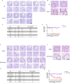
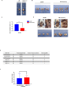
Vasectomized mice play a key role in the production of transgenic mice. However, vasectomy can cause great physical and psychological suffering to mice. Therefore, there is an urgent need to find a suitable replacement for vasectomized mice in the production of transgenic mice. In this study, we generated C57BL/6J mice (Piwil1 D633A-INS99, Piwil1mt/mt) with a 99-base insertion in the Miwi (Piwil1) gene using CRISPR/Cas9 technology and showed that Piwil1mt/+ heterozygous mice were normally fertile and that homozygous Piwil1mt/mt males were sterile and females were fertile. Transplantation of normal fertilized eggs into wild pseudopregnant females following mating with Piwil1mt/mt males produced no Piwil1mt/mt genotype offspring, and the number of offspring did not differ significantly from that of pseudopregnant mice following mating and breeding with ligated males. The CRISPR‒Cas9 system is available for generating Miwi-modified mice, and provides a powerful resource to replace ligated males in assisted reproduction research.
Copyright: © 2024 Xie et al. This is an open access article distributed under the terms of the Creative Commons Attribution License, which permits unrestricted use, distribution, and reproduction in any medium, provided the original author and source are credited.
Conflict of interest statement
The authors have declared that no competing interests exist.
Figures


Similar articles
-
Replacement of surgical vasectomy through the use of wild-type sterile hybrids.Lab Anim (NY). 2021 Feb;50(2):49-52. doi: 10.1038/s41684-020-00692-w. Epub 2021 Jan 4. Lab Anim (NY). 2021. PMID: 33398200 Free PMC article.
-
Naturally sterile Mus spretus hybrids are suitable for the generation of pseudopregnant embryo transfer recipients.Lab Anim (NY). 2024 Jul;53(7):181-185. doi: 10.1038/s41684-024-01393-4. Epub 2024 Jun 17. Lab Anim (NY). 2024. PMID: 38886565 Free PMC article.
-
Direct comparison of vasectomized males and genetically sterile Gapdhs knockout males for the induction of pseudopregnancy in mice.Lab Anim. 2018 Aug;52(4):365-372. doi: 10.1177/0023677217748282. Epub 2017 Dec 25. Lab Anim. 2018. PMID: 29277131
-
Noncanonical functions of PIWIL1/piRNAs in animal male germ cells and human diseases†.Biol Reprod. 2022 Jul 25;107(1):101-108. doi: 10.1093/biolre/ioac073. Biol Reprod. 2022. PMID: 35403682 Review.
-
Transgenic animals in male reproduction research.Exp Clin Endocrinol. 1994;102(6):419-33. doi: 10.1055/s-0029-1211314. Exp Clin Endocrinol. 1994. PMID: 7890018 Review.
References
MeSH terms
Substances
LinkOut - more resources
Full Text Sources
Molecular Biology Databases
Research Materials

