. 2024 May 28;121(22):e2402890121.
doi: 10.1073/pnas.2402890121. Epub 2024 May 21.

Matriglycan maintains t-tubule structural integrity in cardiac muscle

Affiliations

Matriglycan maintains t-tubule structural integrity in cardiac muscle

Jeffrey M Hord et al. Proc Natl Acad Sci U S A. .

Abstract

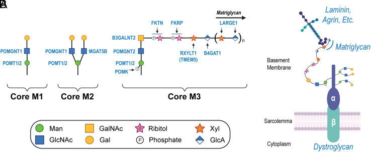
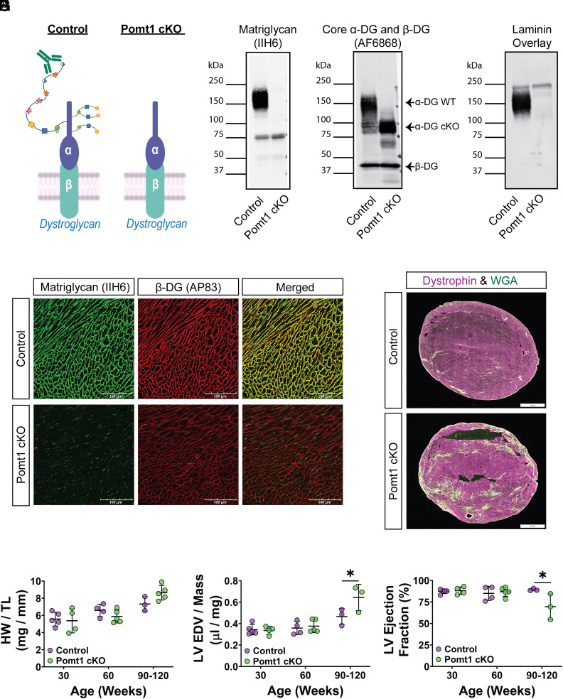
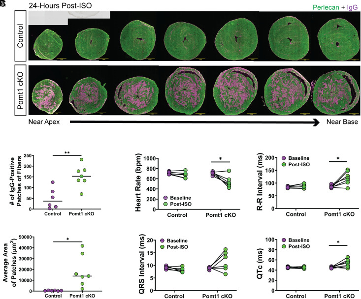
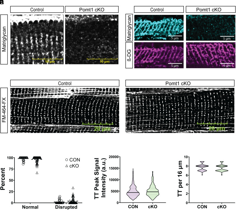
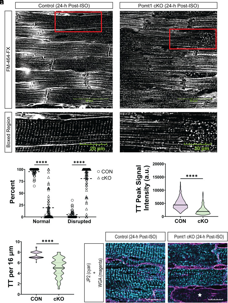
Maintaining the structure of cardiac membranes and membrane organelles is essential for heart function. A critical cardiac membrane organelle is the transverse tubule system (called the t-tubule system) which is an invagination of the surface membrane. A unique structural characteristic of the cardiac muscle t-tubule system is the extension of the extracellular matrix (ECM) from the surface membrane into the t-tubule lumen. However, the importance of the ECM extending into the cardiac t-tubule lumen is not well understood. Dystroglycan (DG) is an ECM receptor in the surface membrane of many cells, and it is also expressed in t-tubules in cardiac muscle. Extensive posttranslational processing and O-glycosylation are required for DG to bind ECM proteins and the binding is mediated by a glycan structure known as matriglycan. Genetic disruption resulting in defective O-glycosylation of DG results in muscular dystrophy with cardiorespiratory pathophysiology. Here, we show that DG is essential for maintaining cardiac t-tubule structural integrity. Mice with defects in O-glycosylation of DG developed normal t-tubules but were susceptible to stress-induced t-tubule loss or severing that contributed to cardiac dysfunction and disease progression. Finally, we observed similar stress-induced cardiac t-tubule disruption in a cohort of mice that solely lacked matriglycan. Collectively, our data indicate that DG in t-tubules anchors the luminal ECM to the t-tubule membrane via the polysaccharide matriglycan, which is critical to transmitting structural strength of the ECM to the t-tubules and provides resistance to mechanical stress, ultimately preventing disruptions in cardiac t-tubule integrity.

Keywords: O-mannosylation; cardiac muscle; dystroglycan; matriglycan; t-tubule.


Conflict of interest statement

Competing interests statement:The authors declare no competing interest.

Figures

Fig. 1.
Fig. 1.
Biosynthesis of core M glycans on α-DG. (A) The α-DG O-mannosyl glycosylation pathway is illustrated, highlighting the synthesis of core M1, M2, and M3 glycans. (B) Illustration depicts O-mannosylated DG situated at the sarcolemma and its interaction with ECM ligands. Abbreviations: Man, mannose; GlcNAc, N-acetyl-glucosamine; Gal, galactose; GalNAc, N-acetyl-galactosamine; Xyl, xylose; GlcA, glucuronic acid; POMT1/2, protein O-mannosyltransferases 1 and 2; POMGNT1, protein O-linked mannose N-acetyl-glucosaminyltransferase 1; POMGNT2, protein O-linked mannose N-acetyl-glucosaminyltransferase 2; MGAT5B, mannosyl α1,6-glycoprotein β1,6,-N-acetyl-glucosaminyltransferase; POMGNT2, protein O-linked mannose N-acetyl-glucosaminyltransferase 2; B3GALNT2, β1,3-N-acetylgalactosaminyltransferase 2; POMK, protein O-mannose kinase; FKTN, Fukutin; FKRP, Fukutin-related protein; RXYLT1, ribitol xylosyltransferase 1; TMEM5, transmembrane protein 5; B4GAT1, β1,4-glucuronyltransferase 1; LARGE1, like-acetyl-glucosaminyltransferase 1.
Fig. 2.
Fig. 2.
Lack of core M glycans on α-DG in cardiac muscle leads to progressive cardiomyopathy. (A) Alpha-DG core M modification in control and Pomt1 cKO. (BD) Immunoblots on ventricles to detect B, matriglycan; C, core α-DG/β-DG; and D, laminin binding. Two sets of pooled samples per group (control n = 13; cKO n = 11). (E) Immunofluorescence on ventricles to detect matriglycan and β-DG (n = 12 controls; n = 12 cKO mice). (Scale bar, 100 μm.) (F) Immunofluorescence on ventricles from 120-wk-old mice to detect fibrotic accumulation. (Scale bar, 1 mm.) (G) Heart weight per tibial length (HW/TL); (H) left ventricular end diastolic volume per left ventricular mass (LV EDV/Mass); and (I) LV ejection fraction in 30-, 60-, and 90-120-wk-old mice. Mice of both sexes were utilized. Number of mice for HW/TL and echocardiography: controls = 5; cKO = 4 at 30-wk; controls = 4, cKO = 5 at 60-wk; controls = 3, cKO = 5 for HW/TL at 90 to 120-wk; and controls = 3, cKO = 3 for echocardiography at 90 to 120-wk. *P < 0.05. Unpaired t tests with the Holm–Sidak post hoc test performed on age-matched groups. Data expressed as mean ± SD.
Fig. 3.
Fig. 3.
Myocardial stress leads to contractile-induced damage and disrupts normal electrophysiology in Pomt1 cKO mice. ISO (10 mg/kg body weight) was administered to promote an acute bout of increased cardiac workload in control and Pomt1 cKO mice. Mice were killed 24-h post-injection. (A) Cardiac cross-sections through the ventricles to assess cardiomyofiber damage as detected by intramyocyte IgG. (Scale bar, 1 mm scale.) (B) Quantification of the number of IgG-positive fiber patches (Upper) and average area of the patches (Bottom) in control and Pomt1 cKO hearts. Image analysis was performed on hearts of mice from both sexes (n = 6 controls; n = 7 cKO). *P < 0.05; **P = 0.008. Unpaired t tests with the Holm–Sidak post hoc test were performed. Data expressed as mean ± SD. (C) Electrophysiological analysis of control and Pomt1 cKO mice at baseline and 24-h post-ISO to determine heart rate, R-R interval, QRS interval, and corrected QT intervals (QTc). Experiments were performed with mice of both sexes. Controls, n = 6; Pomt1 cKO, n = 7 for baseline and post-ISO. *P < 0.05. Paired t tests with the Holm–Sidak post hoc test were implemented. Data expressed as mean ± SD.
Fig. 4.
Fig. 4.
The t-tubule network develops in the absence of O-mannosylated α-DG. (A and B) Immunofluorescence of cryosections of ventricles from control and Pomt1 cKO mice to detect matriglycan (A and B) and β-DG (B). Ten µm cryosections were used in A. (Scale bar, 10 µm.) Sixteen µm cryosections were used in B. (Scale bar, 5 µm.) (C) FM 464-FX fluorescence of whole left ventricles to detect plasma membranes, including t-tubule membranes. (Scale bar, 20 µm.) (D) Quantification of the percent of myofibers with normal or disrupted t-tubule staining within a 90× magnification field. (E) Line scan analysis to determine the peak signal intensity of FM 464-FX labeled t-tubules (TT). (F) Number of t-tubules observed within 16 µm regions. Image analysis was performed on hearts of mice from both sexes (n = 4 controls; n = 5 cKO). Unpaired t tests with the Holm–Sidak post hoc test were performed. Data are expressed as mean ± SD.
Fig. 5.
Fig. 5.
Pomt1 cKO ventricular myocytes exhibit detubulation and disruption of the cardiac dyad in response to contractile stress. (A) Labeling of control and Pomt1 cKO whole left ventricles stained with FM 464-FX 24-h after a single bolus of ISO (10 mg/kg body weight). (Scale bar, 50 µm.) Bottom images show the boxed region (red rectangular box) from the Upper images. (Scale bar, 20 µm.) (B) Percentage of myofibers that showed either normal or disrupted patterns of t-tubules. (C) Line scan analysis to determine the peak signal intensity of FM 464-FX labeled t-tubules (TT). (D) Number of t-tubules observed within a 16 µm region. Image analysis was performed on hearts of mice from both sexes (n = 4 controls; n = 5 cKO). Unpaired t tests with the Holm–Sidak post hoc test were performed. Data expressed as mean ± SD. ****P < 0.0001. (E) Immunofluorescence to detect JP2 and WGA-AlexaFluor 594 in hearts from mice subjected to a single bolus of ISO. (Scale bar, 10 µm.) The white star in the Pomt1 cKO image indicates a cardiomyofiber that lacked JP2 immunodetection.

References

    1. Naba A., et al. , The extracellular matrix: Tools and insights for the "omics" era. Matrix Biol. 49, 10–24 (2016). - PMC - PubMed
    1. Klietsch R., Ervasti J. M., Arnold W., Campbell K. P., Jorgensen A. O., Dystrophin-glycoprotein complex and laminin colocalize to the sarcolemma and transverse tubules of cardiac muscle. Circ. Res. 72, 349–360 (1993). - PubMed
    1. Crossman D. J., et al. , Increased collagen within the transverse tubules in human heart failure. Cardiovasc. Res. 113, 879–891 (2017). - PubMed
    1. Cheah J. X., Nieuwenhuis T. O., Halushka M. K., An expanded proteome of cardiac t-tubules. Cardiovasc. Pathol. 42, 15–20 (2019). - PMC - PubMed
    1. Sheikh M. O., Halmo S. M., Wells L., Recent advancements in understanding mammalian O-mannosylation. Glycobiology 27, 806–819 (2017). - PMC - PubMed

LinkOut - more resources