. 2024 May 28;121(22):e2402764121.
doi: 10.1073/pnas.2402764121. Epub 2024 May 21.

Type VII secretion system extracellular protein B targets STING to evade host anti- Staphylococcus aureus immunity

Affiliations

Type VII secretion system extracellular protein B targets STING to evade host anti- Staphylococcus aureus immunity

Lin Gao et al. Proc Natl Acad Sci U S A. .

Abstract

Staphylococcus aureus (S. aureus) can evade antibiotics and host immune defenses by persisting within infected cells. Here, we demonstrate that in infected host cells, S. aureus type VII secretion system (T7SS) extracellular protein B (EsxB) interacts with the stimulator of interferon genes (STING) protein and suppresses the inflammatory defense mechanism of macrophages during early infection. The binding of EsxB with STING disrupts the K48-linked ubiquitination of EsxB at lysine 33, thereby preventing EsxB degradation. Furthermore, EsxB-STING binding appears to interrupt the interaction of 2 vital regulatory proteins with STING: aspartate-histidine-histidine-cysteine domain-containing protein 3 (DHHC3) and TNF receptor-associated factor 6. This persistent dual suppression of STING interactions deregulates intracellular proinflammatory pathways in macrophages, inhibiting STING's palmitoylation at cysteine 91 and its K63-linked ubiquitination at lysine 83. These findings uncover an immune-evasion mechanism by S. aureus T7SS during intracellular macrophage infection, which has implications for developing effective immunomodulators to combat S. aureus infections.

Keywords: EsxB; STING; Staphylococcus aureus; inflammation; macrophages.


Conflict of interest statement

Competing interests statement:The authors declare no competing interest.

Figures
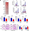
Fig. 1.
Fig. 1.
STING activates proinflammatory responses in macrophages. (A) Heat map of down-regulated mRNAs in STING knockout (STING−/−) vs. WT peritoneal macrophages (PMs) infected with the USA300 strain for 6 h (n = 3). (B) qPCR analysis of Tnf, Il1b, Il6, and Il12p40 mRNA from WT or STING−/− PMs infected with USA300 (multiplicity of infection [MOI] = 25) for the indicated times. (C) ELISA quantification of the TNF-α, IL-1β, IL-6, and IL-12 levels in homogenized lung-tissue samples (1 mL, phosphate-buffered saline) infected with the USA300 strain for 6 h. STING[flox/flox] (Flox) or STING[flox/flox, Lyz2-Cre] (STING CKO) mice were infected via the intratracheal administration of the test strains (approximately 2 × 10⁸ colony-forming units [CFU] per mouse, n = 8). (D and E) Histopathological examination of lung-tissue sections stained with hematoxylin and eosin. The infection procedure is the same as in D. Scale bars, 1,000 μm (Top) and 200 μm (Bottom); the boxed areas at the top are enlarged below. (F) CFU quantification of the bacterial titers in lung-tissue homogenates from D. The two-way ANOVA multiple comparisons test (B) or two-tailed unpaired Student t test (C, E, and F) was used for statistical analysis; *P < 0.05, **P < 0.01, ***P < 0.001, and ****P < 0.0001. The bars show the mean and SD of n = 3 (B), n = 8 (C and F), or n = 5 (E). The data shown are a representative result of 3 independent biological experiments.
Fig. 2.
Fig. 2.
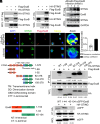
EsxB interacts with STING. (A and B) Immunoblots (IBs) of whole cell lysates (WCLs) or immunoprecipitation (IP) products of WCLs of HEK293T cells transfected with plasmids expressing Flag-tagged or HA-tagged EsxB and Flag-tagged or HA-tagged STING. (C) MH-S cells infected with ΔEsxB+Flag-EsxB for 1 h (MOI = 100). After the elimination of extracellular bacteria by gentamicin (100 μg/mL), the endogenous interaction of EsxB with STING was tested for 4 h. (D) Confocal microscopy of MH-S cells infected with the ΔEsxB+Flag-EsxB strain or USA300 strain. The infection procedure is the same as in C; (scale bar, 10 µm.) The colocalization ratio of EsxB and STING in 30 ΔEsxB+Flag-EsxB-infected macrophages was analyzed. (E) Truncation of STING. (F) IBs of WCLs and IP products of WCLs of HEK293T cells transfected with plasmids expressing Flag-tagged EsxB and HA-tagged truncated STING; WT, full-length STING. (G) Truncation of EsxB. (H) IBs of WCLs and IP products of WCLs of HEK293T cells transfected with plasmids expressing HA-tagged STING and eGFP-tagged truncated EsxB. The values below the blots indicate densitometry quantification (A, B, F, and H) or densitometry quantification relative to GAPDH (C). The data shown are a representative result of 3 independent biological experiments.
Fig. 3.
Fig. 3.
EsxB inhibits host proinflammatory responses. (A) qPCR analysis of Tnf, Il1b, Il6, and Il12p40 mRNA from MH-S cells infected with the USA300, ΔEsxB, or ΔEsxB+Flag-EsxB strain (MOI = 25) for the indicated times. (B) ELISA quantification of the TNF-α, IL-1β, IL-6, and IL-12 levels in homogenized samples of lung-tissue (1 mL, phosphate-buffered saline) infected with the USA300, ΔEsxB, or ΔEsxB+Flag-EsxB strain for 24 h. C57/BL mice were infected by the intratracheal administration of the test strains (approximately 2 × 10⁸ CFUs per mouse). (C) CFU quantification of the bacterial titers in lung-tissue homogenates. (D and E) Histopathological examination of lung-tissue sections stained with hematoxylin and eosin at 24 h after infection. Scale bars, 1,000 μm (Top) and 200 μm (Bottom); the boxed areas at the Top are enlarged below. The two-way ANOVA multiple comparisons test (A) or two-tailed Student unpaired t test (BD) was used for statistical analysis: *P < 0.05, **P < 0.01, ***P < 0.001, and ****P < 0.0001. The bars show the mean and SD of n = 3 (A), n = 8 (B and C), or n = 5 (D). The data shown are a representative result of 3 independent biological experiments.
Fig. 4.
Fig. 4.
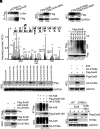
EsxB targets STING to evade degradation. (A) IBs of IP products of WCLs of HEK293T cells transfected with Flag-tagged EsxB-expressing plasmids. (B) Analysis of the phosphorylation sites of EsxB through tandem mass spectrometry. (C and D) IBs of WCLs of HEK293T cells transfected with plasmids expressing Flag-tagged WT EsxB, EsxB S84A (serine 84 replaced with alanine), or EsxB S84D (serine 84 replaced with aspartate). (E) IBs of IP products of WCLs from HEK293T cells transfected with plasmids expressing Flag-tagged EsxB and Myc-tagged ubiquitin or different mutant ubiquitins. (F) IBs of WCLs of HEK293T cells transfected with plasmids expressing Flag-tagged EsxB 84S (all serine residues, except for serine 84, are replaced) or different mutant EsxB 84S (lysine is replaced with arginine one by one). (G) IBs of WCLs of HEK293T cells transfected with plasmids expressing Flag-tagged EsxB or HA-tagged STING; Bortezomib (BTZ): a proteasome inhibitor. (H) IBs of WCLs and IP products of WCLs of HEK293T cells transfected with plasmids expressing Flag-tagged EsxB 84S K33R (lysine 33 replaced with arginine, and all serine residues are replaced, except for serine 84) and HA-tagged STING. (I) IBs of WCLs and IP products of WCLs of HEK293T cells transfected with plasmids expressing Flag-tagged EsxB 33K (all lysine residues replaced with arginine, except for lysine 33), HA-tagged STING, and HA-tagged K48 ubiquitin. (J) WT or STING−/− PMs infected with ΔEsxB+Flag-EsxB or ΔEsxB+Flag-EsxB S84A strain (MOI = 100) for 1 h. The EsxB or EsxB S84A levels were tested for 4 h after eliminating extracellular bacteria by gentamicin (100 μg/mL). The values below the blots indicate densitometry quantification. The data shown are representative results of 3 independent biological experiments.
Fig. 5.
Fig. 5.
EsxB inhibits palmitoylation of STING. (A) IBs of WCLs and IP products of WCLs of PMs infected with the USA300 or ΔEsxB strain for the indicated times (MOI = 25). (B) IBs of WCLs and IP products of WCLs of STING knockout PMs (transfected with STING, STING C88S, or STING C91S mRNA) infected with the USA300 or ΔEsxB strain for the indicated times (MOI = 25). (C and D) IBs of WCLs and IP products of WCLs of PMs infected with the USA300 or ΔEsxB strain for the indicated times (MOI = 25). HAM, hydroxylamine. The values below the blots indicate densitometry quantification relative to GAPDH. The data shown are a representative result of 3 independent biological experiments.
Fig. 6.
Fig. 6.
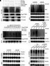
EsxB inhibits K63 ubiquitination of STING. (A) IBs of WCLs and IP products of WCLs of HEK293T cells transfected with plasmids expressing Flag-tagged STING, HA-tagged EsxB, and HA-tagged WT (Ub), K48, K63 ubiquitin. (B) IBs or IP products of the lysates of PMs infected with the USA300 or ΔEsxB strain for the indicated times (MOI = 25). (C) IBs of WCLs and IP products of WCLs of HEK293T cells transfected with plasmids expressing HA-tagged EsxB, HA-tagged K63 ubiquitin, and Flag-tagged STING or its mutants (a specific lysine is replaced with arginine). (D) IBs of WCLs and IP products of WCLs of STING knockout PMs (transfected with STING or STING K83R mRNA) infected with the USA300 or ΔEsxB strain for the indicated times (MOI = 25). (E and F) IBs or IP products of the lysates of PMs infected with the USA300 or ΔEsxB strain for the indicated times (MOI = 25). The values below the blots indicate densitometry quantification (AD and F) or densitometry quantification relative to GAPDH (E). The data shown are a representative result of 3 independent biological experiments.
Fig. 7.
Fig. 7.
EsxB inhibits the polymerization of STING. (A) Nonreduced and reduced IBs of the lysates of PMs infected with the USA300 or ΔEsxB strain for the indicated times (MOI = 25). (B) Nonreduced and reduced IBs of the lysates of STING knockout PMs (transfected with STING or STING K83R/C91S mRNA) infected with the USA300 or ΔEsxB strain for the indicated times (MOI = 25). (C) qPCR analysis of Tnf, Il1b, Il6, and Il12p40 mRNA from STING knockout PMs (transfected with STING or STING K83R/C91S mRNA) infected with the USA300 or ΔEsxB strain for the indicated times (MOI = 25). The two-way ANOVA multiple comparisons test (C) was used for statistical analysis; *P < 0.05, **P < 0.01, ***P < 0.001, and ****P < 0.0001. The bars show the mean and SD of n = 3 (C). The values below the blots indicate densitometry quantification relative to GAPDH (A and B); we used the dimer band value as the value of STING. The data shown are a representative result of 3 independent biological experiments.

References

    1. A. S. Lee et al., Methicillin-resistant Staphylococcus aureus. Nat. Rev. Dis. Primers 4, 18033 (2018), 10.1038/nrdp.2018.34. - DOI - PubMed
    1. Lakhundi S., Zhang K., Methicillin-resistant Staphylococcus aureus: Molecular characterization, evolution, and epidemiology. Clin. Microbiol. Rev. 31, e00020-18 (2018), 10.1128/cmr.00020-18. - DOI - PMC - PubMed
    1. Proctor R. A., Challenges for a Universal Staphylococcus aureus Vaccine. Clin. Infect. Dis. 54, 1179–1186 (2012), 10.1093/cid/cis033. - DOI - PubMed
    1. Pozzi C., et al. , Phagocyte subsets and lymphocyte clonal deletion behind ineffective immune response toStaphylococcus aureus. FEMS Microbiol. Rev. 39, 750–763 (2015), 10.1093/femsre/fuv024. - DOI - PubMed
    1. Ergun S. L., Li L., Structural insights into STING signaling. Trends Cell Biol. 30, 399–407 (2020), 10.1016/j.tcb.2020.01.010. - DOI - PubMed

MeSH terms

Substances