. 2024 Jun:104:105163.
doi: 10.1016/j.ebiom.2024.105163. Epub 2024 May 20.

Sialic acid blockade inhibits the metastatic spread of prostate cancer to bone

Affiliations

Sialic acid blockade inhibits the metastatic spread of prostate cancer to bone

Kirsty Hodgson et al. EBioMedicine. 2024 Jun.

Abstract

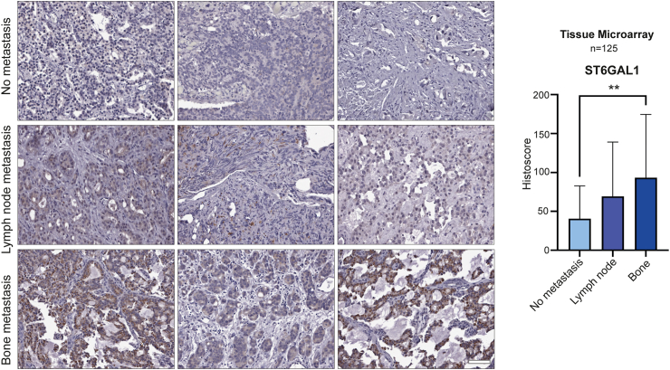
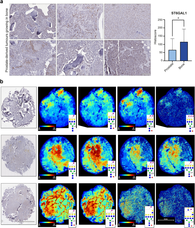
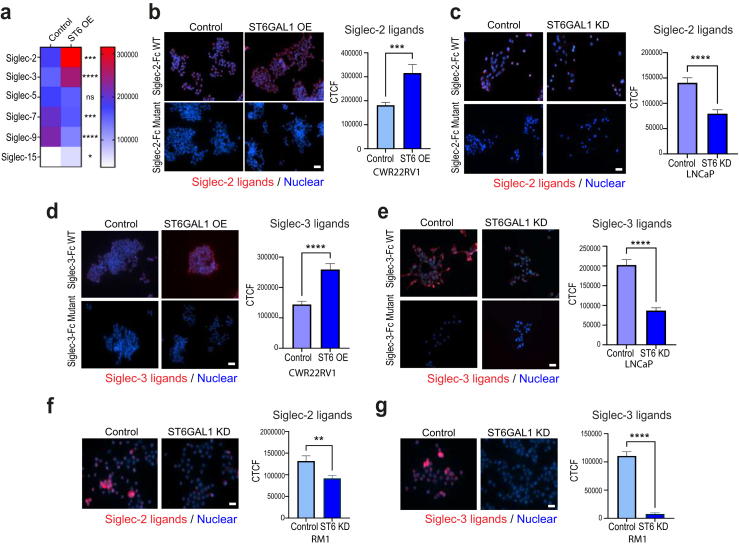
Background: Bone metastasis is a common consequence of advanced prostate cancer. Bisphosphonates can be used to manage symptoms, but there are currently no curative treatments available. Altered tumour cell glycosylation is a hallmark of cancer and is an important driver of a malignant phenotype. In prostate cancer, the sialyltransferase ST6GAL1 is upregulated, and studies show ST6GAL1-mediated aberrant sialylation of N-glycans promotes prostate tumour growth and disease progression.

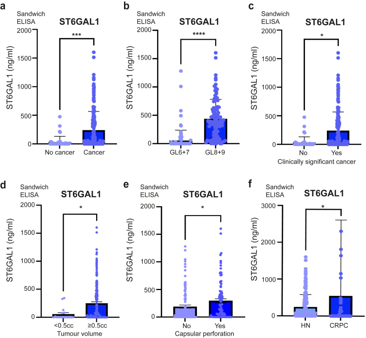
Methods: Here, we monitor ST6GAL1 in tumour and serum samples from men with aggressive prostate cancer and using in vitro and in vivo models we investigate the role of ST6GAL1 in prostate cancer bone metastasis.

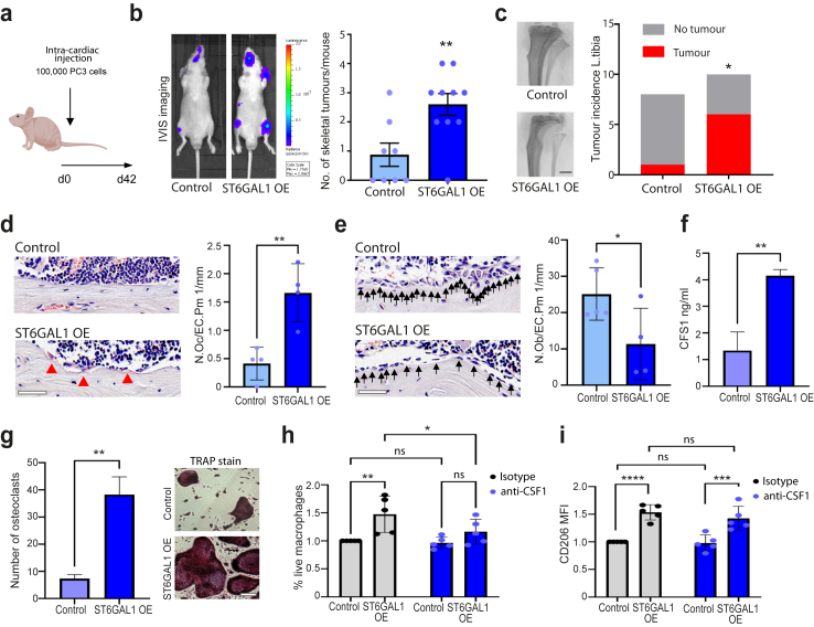
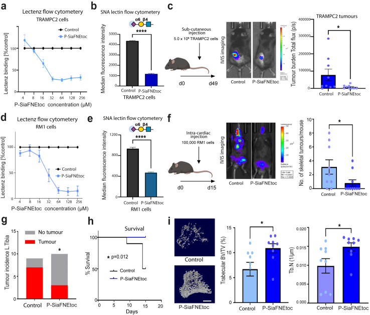
Findings: ST6GAL1 is upregulated in patients with prostate cancer with tumours that have spread to the bone and can promote prostate cancer bone metastasis in vivo. The mechanisms involved are multi-faceted and involve modification of the pre-metastatic niche towards bone resorption to promote the vicious cycle, promoting the development of M2 like macrophages, and the regulation of immunosuppressive sialoglycans. Furthermore, using syngeneic mouse models, we show that inhibiting sialylation can block the spread of prostate tumours to bone.

Interpretation: Our study identifies an important role for ST6GAL1 and α2-6 sialylated N-glycans in prostate cancer bone metastasis, provides proof-of-concept data to show that inhibiting sialylation can suppress the spread of prostate tumours to bone, and highlights sialic acid blockade as an exciting new strategy to develop new therapies for patients with advanced prostate cancer.

Funding: Prostate Cancer Research and the Mark Foundation For Cancer Research, the Medical Research Council and Prostate Cancer UK.

Keywords: Bone metastasis; Glycans; Prostate cancer; Sialic acid; Sialylation; Therapeutics.


Conflict of interest statement

Declaration of interests JM and ES are shareholders of GlycoScoreDx Ltd and have filed patent applications related to this work (GB patent numbers GB2,594,103, GB2,595,425 and US patent application 17/780,508). J.F.A.P., N.E., E.R. are shareholders of and employed by GlycoTherapeutics B.V.; C.B. and T.J.B. are shareholders of and scientific advisors of GlycoTherapeutics B.V.; S.J.M is a shareholder of and employed by Synvenio B.V.; J.F.A.P.; and T.J.B. are shareholders of Synvenio B.V.; Radboud University and Radboudumc have filed patent applications related to P-SiaFNEtoc (including patent number 11,639,364). MM has filed patents related to Siglec inhibitors (US patent applications 63/421,007 and 63/497,540). All other authors declare that there are no potential competing interests.

Figures

Fig. 1
Fig. 1
The sialyltransferase enzyme ST6GAL1 is upregulated in prostate cancer that has spread to the bone. (a) Immunohistochemistry analysis of a previously published 125 case tissue microarray (TMA) to compare ST6GAL1 levels in localised prostate cancer tumours and prostate cancer tissues presenting with metastasis to bone or lymph node (all biopsy samples were taken from the primary site). Further details for this cohort are provided in Supplementary Table S1. ST6GAL1 protein levels are 1.7-fold upregulated in patients with prostate cancer lymph node metastasis (unpaired t test, p = 0.1657) and 2.3-fold upregulated in patients with prostate cancer bone metastasis (unpaired t test, p = 0.0091). Scale bar is 100 μm.
Fig. 2
Fig. 2
ST6GAL1 is upregulated in prostate-derived tumours growing in bone. (a) Immunohistochemistry analysis of ST6GAL1 levels in 20 rapid autopsy samples from prostate-derived tumours growing in bone compared to 53 localised prostate cancer tissue samples biopsied at the primary site (taken from the TMA analysed in Fig. 1). ST6GAL1 levels were 1.74-fold higher in prostate-derived tumours growing in bone compared to tumours from the primary site (unpaired t test, p = 0.0103). Scale bar is 200 μm. (b) N-glycan MALDI imaging mass spectrometry analysis of all 20 bone metastases tissue samples. Three bone tumours with high immunostaining levels of ST6GAL1 are shown in the first image of row, with representative α2-6 sialylated N-glycan images that co-localize to ST6GAL1-stained regions. Scale bar for IHC and MALDI images is 3 mm.
Fig. 3
Fig. 3
ST6GAL1 is upregulated in the blood of men with aggressive prostate cancer. Analysis of ST6GAL1 protein levels in a cohort of 300 serum samples using pre-validated sandwich ELISA assays, including 40 men given a ‘no cancer’ diagnosis, 100 men with low grade prostate cancer (Gleason grade 6–7), 100 men with high grade prostate cancer (Gleason grade 8–9), and 60 men with metastatic CRPC. More information on this cohort is provided in Supplementary Table S1. (a) ST6GAL1 serum levels were 7.3-fold higher in men diagnosed with prostate cancer relative to men without prostate cancer (n = 240, unpaired t test, p < 0.0001). (b) Levels of serum ST6GAL1 levels were 6.7-fold higher in men with high grade prostate cancer compared to men with low grade disease (n = 200, unpaired t test, p < 0.0001). (c) Serum ST6GAL1 levels were 10.6-fold higher in men with clinically significant prostate cancer compared to non-clinically significant prostate cancer defined according to the PI-RADS v2.1 guidelines (n = 200, unpaired t test, p = 0.143). (d) ST6GAL1 serum levels were 4.4-fold higher in men with a prostate tumour volume ≥0.5 cc compared to those with a tumour volume <0.5 cc (where a tumour volume of ≥0.5 cc is defined as clinically significant), (n = 200 unpaired t test, p = 0.0174). (e) The level of serum ST6GAL1 was 1.55-fold higher in prostate tumours with capsular perforation compared to tumours not involving or extending beyond the prostate gland (n = 200, unpaired t test, p = 0.0394). (f) Levels of serum ST6GAL1 were 2.24-fold higher in men with metastatic CRPC (58/60 of whom had metastasis to bone) compared to men with hormone naïve (HN) disease (n = 260, unpaired t test, p = 0.046).
Fig. 4
Fig. 4
Upregulation of ST6GAL1 promotes the spread of prostate cancer to bone. (a) Luciferase tagged PC3 cells (control or ST6GAL1 overexpressing) were injected into BALB/c nude mice via intra cardiac injection. (b) Tumours were monitored over 6 weeks using in vivo bioluminescence imaging. Upregulation of ST6GAL1 significantly increased the number of skeletal tumours formed per mouse (n = 10, unpaired t test, p = 0.006). (c) Ex vivo micro-CT analysis was used to examine tumour induced bone destruction in left tibias and showed that overexpression of ST6GAL1 in PC3 cells significantly enhanced tumour incidence (representative images are shown) (n = 10, Chi-square test, p = 0.04). Scale bar is 1 mm. (d,e) Further bone histomorphometry demonstrated that in mice bearing ST6GAL1 overexpressing tumours there were significantly higher number of osteoclasts per mm medial bone surface (n = 4 unpaired t test, p = 0.0055) but lower number of osteoblasts per mm lateral bone surface in tibias without overt tumours, compared to mice bearing control tumours (n = 5 unpaired t test, p = 0.0452). (f) Sandwich ELISA analysis of CSF1 levels in conditioned media samples from PC3 prostate cancer cells with overexpression of ST6GAL1. CSF1 levels are significantly increased in conditioned media samples from PC3 cells with upregulation of ST6GAL1 (unpaired t test, p = 0.003). (g) Conditioned media samples from DU145 cells overexpressing ST6GAL1 promotes the differentiation of murine primary osteoclast pre-cursors into osteoclasts measured via TRAP staining (unpaired t test, p = 0.0005). Scale bar is 100 μm. (h,i) Primary monocytes from healthy controls were treated with 150 μl of concentrated conditioned media from PC3 cells (control or ST6GAL1 overexpressing) at days 0 and 3 in the presence of 5 μg/ml anti-MCSF or isotype control. Cells were harvested on day 6 and assessed for viability (h) and CD206 expression (i) using flow cytometry. (n = 5, two-way ANOVA, multiple comparisons, (h) cont. vs. OE isotype. p = 0.002. OE isotype vs. OE anti-MCSF p = 0.028. (i) cont. vs OE isotype. p < 0.0001. cont. vs OE isotype. p = 0.0002).
Fig. 5
Fig. 5
ST6GAL1 regulates immunosuppressive sialoglycans in prostate cancer cells. Siglec ligands were monitored in prostate cancer cells with immunocytochemistry using recently developed Siglec-Fc proteins. (a) Heatmap to illustrate Siglec ligands that are differentially expressed in CWR22RV1 prostate cancer cells with overexpression of ST6GAL1. (b) Upregulation of ST6GAL1 in CWR22RV1 cells significantly increases sialoglycans that engage Siglec-2 (CD-22) (unpaired t test, p = 0.0008). (c) Downregulation of ST6GAL1 in LNCaP prostate cancer cells significantly reduced the expression of sialoglycans that engage Siglec-2 (unpaired t test, p < 0.0001). (d) Upregulation of ST6GAL1 in CWR22RV1 cells significantly increases the levels of sialoglycans that engage Siglec-3 (CD33) (unpaired t test, p < 0.0001). (e) Downregulation of ST6GAL1 in LNCaP prostate cancer cells significantly reduced the expression of sialoglycans that engage Siglec-3 (unpaired t test, p < 0.0001). Scale bar is 50 μm. (f) Consistent with ST6GAL1 regulating ligands that are recognised by Siglec-2 and Siglec-3, we also detected downregulation of sialoglycans recognised by Siglec-2 (unpaired t test, p = 0.0036) and (g) Siglec-3 (unpaired t test, p = 0001) in murine RM1 prostate cancer cells depleted of ST6GAL1. Scale bar is 50 μm.
Fig. 6
Fig. 6
Sialic acid blockade can prevent/inhibit prostate cancer bone metastasis. (a) Inhibition of sialylation in TRAMPC2 cells using P-SiaFNEtoc detected using pan-specific Lectenz lectin flow cytometry. Cells were treated with a range of concentrations of P-SiaFNEtoc inhibitor from 2 μM to 512 μM for 72 h. The intensities were normalised to a DMSO control. (b) Detection of α2-6 linked sialylated N-glycans in TRAMPC2 cells using SNA lectin flow cytometry. TRAMPC2 cells treated with 64 μM P-SiaFNEtoc for 72 h had reduced levels of SNA binding indicating a reduction in α2-6 linked sialylation in these cells (unpaired t test, p = 0.0001). (c) Luciferase tagged TRAMPC2 cells (control or pre-treated with 64 μM P-SiaFNEtoc for 72 h) were injected into immunocompetent C57BL/6 mice via sub-cutaneous injection and tumours were monitored using in vivo bioluminescence imaging. Pre-treatment of TRAMPC2 cells with P-SiaFNEtoc (which removed sialylated glycans) significantly reduced tumour burden over 6 weeks (n = 10, Mann–Whitney test, p = 0.0233) thus suggesting that sialic acid blockade has the potential to inhibit the growth of prostate tumours. (d) Inhibition of sialylation in RM1 cells using P-SiaFNEtoc detected using pan-specific Lectenz lectin flow cytometry. Cells were treated with a range of concentrations of P-SiaFNEtoc inhibitor from 2 μM to 512 μM for 72 h. The intensities were normalised to a DMSO control. (e) Detection of α2-6 linked sialylated N-glycans in RM1 cells using SNA lectin flow cytometry. RM1 cells treated with 256 μM P-SiaFNEtoc for 72 h had reduced levels of SNA binding indicating a reduction in α2-6 linked sialylation in these cells (unpaired t test, p < 0.0001). (f) Luciferase tagged RM1 cells (control or pre-treated with 256 μM P-SiaFNEtoc for 72 h) were injected into immunocompetent C57BL/6 mice via intra cardiac injection. Tumours were monitored over 15 days using in vivo bioluminescence imaging. (g,h) Pre-treatment of RM1 cells with P-SiaFNEtoc (to remove sialylated glycans) significantly reduced the number of skeletal tumours formed (Mann–Whitney test, p = 0.0454), the incidence of tumour in left tibias (Chi-square test, p = 0.0455), and significantly increased survival time in mice (Log-rank test, p = 0.012). (i) Micro-CT analysis demonstrated that P-SiaFNEtoc significantly alleviated bone destruction in the trabecular bone of tibias and increased trabecular bone volume (BV/TV, p = 0.0211) and trabecular number (Tb. N, p = 0.035) (n = 9, unpaired t test, ∗p < 0.05). Representative images are shown. Scale bar is 200 μm.

References

    1. Siegel R.L., Miller K.D., Fuchs H.E., Jemal A. Cancer statistics, 2022. CA Cancer J Clin. 2022;72(1):7–33. - PubMed
    1. Kang J., La Manna F., Bonollo F., et al. Tumor microenvironment mechanisms and bone metastatic disease progression of prostate cancer. Cancer Lett. 2022;530:156–169. - PubMed
    1. Archer Goode E., Wang N., Munkley J. Prostate cancer bone metastases biology and clinical management (Review) Oncol Lett. 2023;25(4):163. - PMC - PubMed
    1. Nieder C., Haukland E., Pawinski A., Dalhaug A. Pathologic fracture and metastatic spinal cord compression in patients with prostate cancer and bone metastases. BMC Urol. 2010;10:23. - PMC - PubMed
    1. Pinho S.S., Reis C.A. Glycosylation in cancer: mechanisms and clinical implications. Nat Rev Cancer. 2015;15(9):540–555. - PubMed

MeSH terms