. 2024 Jun 6;111(6):1206-1221.
doi: 10.1016/j.ajhg.2024.04.019. Epub 2024 May 20.

Etiological involvement of KCND1 variants in an X-linked neurodevelopmental disorder with variable expressivity

Affiliations

Etiological involvement of KCND1 variants in an X-linked neurodevelopmental disorder with variable expressivity

Tassja Kalm et al. Am J Hum Genet. .

Abstract

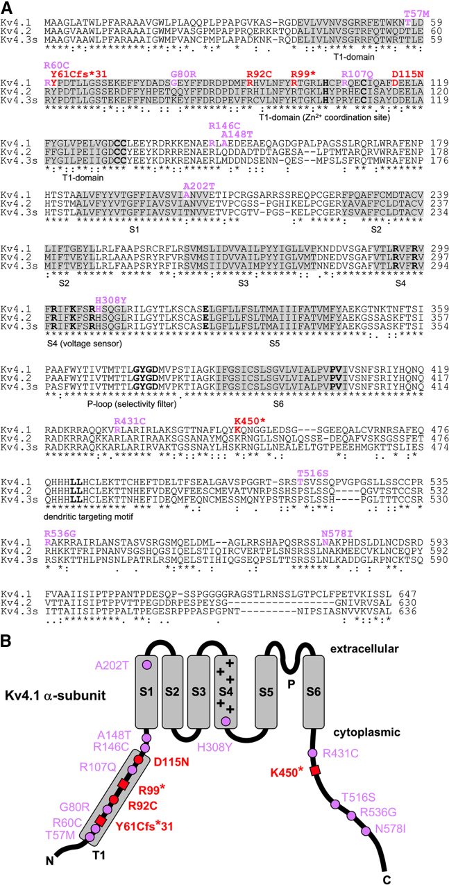
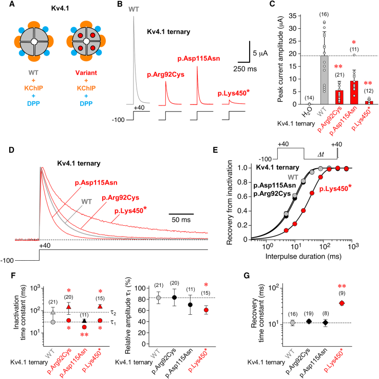
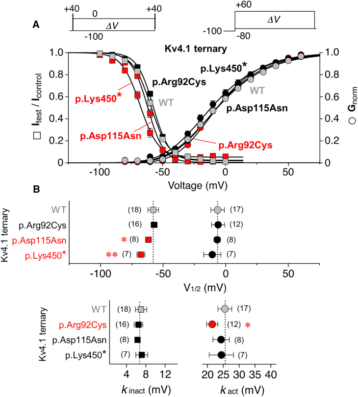
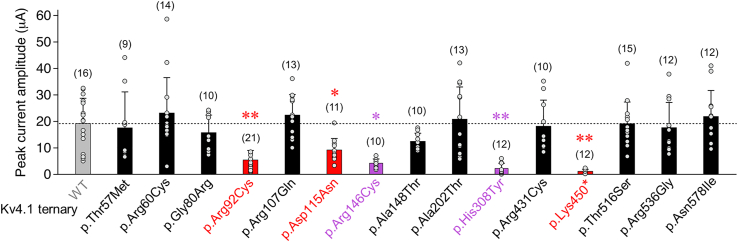
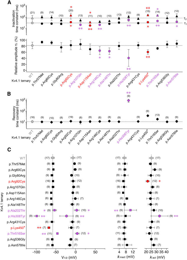
Utilizing trio whole-exome sequencing and a gene matching approach, we identified a cohort of 18 male individuals from 17 families with hemizygous variants in KCND1, including two de novo missense variants, three maternally inherited protein-truncating variants, and 12 maternally inherited missense variants. Affected subjects present with a neurodevelopmental disorder characterized by diverse neurological abnormalities, mostly delays in different developmental domains, but also distinct neuropsychiatric signs and epilepsy. Heterozygous carrier mothers are clinically unaffected. KCND1 encodes the α-subunit of Kv4.1 voltage-gated potassium channels. All variant-associated amino acid substitutions affect either the cytoplasmic N- or C-terminus of the channel protein except for two occurring in transmembrane segments 1 and 4. Kv4.1 channels were functionally characterized in the absence and presence of auxiliary β subunits. Variant-specific alterations of biophysical channel properties were diverse and varied in magnitude. Genetic data analysis in combination with our functional assessment shows that Kv4.1 channel dysfunction is involved in the pathogenesis of an X-linked neurodevelopmental disorder frequently associated with a variable neuropsychiatric clinical phenotype.


Conflict of interest statement

Declaration of interests The authors declare no competing interests.

Figures

Figure 1
Figure 1
KCND1 variant-associated amino acid substitutions (A) Alignment of human Kv4 subfamily members Kv4.1, Kv4.2, and Kv4.3s (short isoform). Kv4.1 amino acid residues affected by the reported KCND1 variants are shown in color, and respective changes are indicated above using single-letter code (red: initially studied KCND1 group 1 variants; purple: additional maternally inherited missense variants, group 2). Regions highlighted in gray indicate the tetramerization (T1) domain and transmembrane segments S1–S6, as indicated below. Sequence motifs critical for channel assembly trafficking and function are indicated by bold letters (HX5CX20CC motif in the T1-domain: Zn2+ coordination site; positively charged residues in S4: voltage sensor; potassium channel signature sequence GYGD in the pore loop [P]: selectivity filter; critical dynamic coupling residues, glutamate [E] in the S4-S5 linker, and proline-valine [PV] in the distal S6 segment: operation of the cytoplasmic gate;,,, C-terminal di-leucine motif: dendritic targeting12). Numbers on the right specify amino acid residue positions. Sequences were aligned using Clustal Omega software (http://www.clustal.org/omega/). Residues that are perfectly conserved () or exhibit either strong or weak similarity (: or . for a scoring of >0.5 or ≤0.5, respectively, in the Gonnet PAM 250 matrix) are indicated. Note that Kv4.1 amino acid residues in positions 57, 60, 61, 92, 99, 107, 115, 146, 308, 431, 450, 536, and 578 are fully conserved in all three Kv4 subfamily members. (B) Topology scheme of the Kv4.1 α-subunit (extracellular and cytoplasmic side indicated) with six transmembrane segments (S1–S6, gray boxes) and cytoplasmic N- and C-termini. Positively charged amino acid residues in S4 (+) mediate voltage sensing, and a re-entrant P loop between S5 and S6 harbors the selectivity filter sequence. The T1 domain (gray box) is located in the cytoplasmic N-terminus. Changes in amino acid sequence are indicated in single-letter code. The two amino acid substitutions and three truncation sites, respectively, associated with five KCND1 group 1 variants are depicted as red dots (DNVs R92C and D115N) and red squares (PTVs Y61Cfs31, R99, and K450). All other amino acid substitutions (12 maternally inherited missense variants) are depicted as purple dots. Note that all variant-associated amino acid substitutions, except for p.Ala202Thr (A202T in S1) and p.His308Tyr (H308Y in S4), reside within either the cytoplasmic N- or the cytoplasmic C-terminal domain of the protein and do not affect one of the indicated critical sequence motifs.
Figure 2
Figure 2
Functional expression and kinetic analysis of inactivation for KCND1 WT and group 1 variants in a ternary configuration (A) Kv4.1 wild-type channels (WT, gray quadrants) and variant channels with amino acid substitutions (quadrants with red dots) were functionally analyzed in a ternary configuration, i.e., with both KChIP (orange) and DPP (blue, see also Figure S1 and Video S1) in Xenopus laevis oocytes (see material and methods). (B) Macroscopic currents mediated by Kv4.1 WT, p.Arg92Cys, p.Asp115Asn, and p.Lys450 ternary channels. Currents were recorded under two-electrode voltage clamp using depolarizing voltage steps from −100 to +40 mV, as indicated (gray trace: WT; red traces: p.Arg92Cys, p.Asp115Asn, and p.Lys450). (C) Bar graph shows mean peak current amplitudes, including individual data points (number of observations indicated). Note that for all functionally tested group 1 variants (red bars) mean peak current amplitudes were smaller than for Kv4.1 WT (gray bar and horizontal dotted line). Water-injected oocytes served as control. (D) Normalized currents mediated by Kv4.1 WT (gray trace), p.Arg92Cys, p.Asp115Asn, and p.Lys450 (red traces) in a ternary configuration. Currents were elicited by extended voltage pulses, as indicated (2.5 s, +40 mV), to study the kinetics of macroscopic inactivation (i.e., current decay). (E) Recovery of ternary channels from inactivation was measured using a double-pulse protocol with long control and brief test pulses to +40 mV and variable interpulse durations at −100 mV (Δt, see inset). The normalized data (Itest/Icontrol) were plotted against the interpulse duration and described by a single-exponential function (gray symbols: Kv4.1 WT; black symbols: p.Arg92Cys and p.Asp115Asn; red symbols: p.Lys450; error bars are SEM and smaller than symbols). (F) Inactivation time constants obtained by fitting the current decay kinetics with a double-exponential function (τ1: circles, τ2: triangles) and the relative amplitude of the total decay accounted for by τ1. (G) Recovery time constants obtained by fitting the kinetics of recovery from inactivation with a single-exponential function. (F and G) Gray symbols and horizontal dotted lines: WT; black symbols: values obtained for variant ternary channels with no difference compared to Kv4.1 WT; red symbols: values obtained for variant ternary channels that significantly differ from Kv4.1 WT; number of observations indicated; all statistics based on one way ANOVA with Dunnett’s post-hoc testing; significant differences compared to Kv4.1 WT are indicated with p < 0.05 or ∗∗p < 0.0001.
Figure 3
Figure 3
Voltage dependence of activation and steady-state inactivation (A) Voltage dependences of peak conductance activation (circles) and steady-state inactivation (squares) are shown for Kv4.1 wild-type (WT), p.Arg92Cys, p.Asp115Asn, and p.Lys450 ternary channels. For the study of steady-state inactivation, brief control and test pulses to +40 mV were separated by a 10 s conditioning pulse (ΔV between −100 and 0 mV in 10 mV increments, see inset). Normalized data (Itest/Icontrol) were plotted against the conditioning pulse voltage. For the study of peak conductance activation, test pulses to voltages between −80 and +60 mV (ΔV in 10 mV increments) were applied from −100 mV (see inset). Normalized conductance values were plotted against the test pulse voltage. The data were fitted with appropriate Boltzmann functions., Gray symbols: Kv4.1 WT; black symbols: data which do not differ from Kv4.1 WT; red symbols: data that differ from Kv4.1 WT (error bars are SEM). (B) Voltages of half-maximal inactivation (squares) and half-maximal activation (circles) and corresponding slope factors (kinact and kact, respectively); gray symbols and vertical dotted lines: Kv4.1 WT; black symbols: V1/2 and k values of variant ternary channels with no difference compared to Kv4.1 WT; red symbols: V1/2 and k values of variant ternary channels that significantly differ from Kv4.1 WT; number of observations indicated; all statistics based on one way ANOVA with Dunnett’s post-hoc testing; significant differences compared to Kv4.1 WT are indicated with p < 0.05 or ∗∗p < 0.0001.
Figure 4
Figure 4
Functional expression of group 2 variants in a ternary configuration Mean peak current amplitudes at +40 mV including individual data points (number of observations indicated; gray bar and horizontal dotted line: Kv4.1 WT data from Figure 2C; red bars: variant ternary channel data from Figure 2C; black bars: Peak amplitude values for variant ternary channels with no difference compared to Kv4.1 WT; purple bars: peak amplitude values of variant ternary channels that significantly differ from Kv4.1 WT; all statistics based on one way ANOVA with Dunnett’s post-hoc testing; significant differences compared to Kv4.1 WT are indicated with p < 0.05 or ∗∗p < 0.0001.
Figure 5
Figure 5
Biophysical characterization of group 2 variants in a ternary configuration (A) Inactivation time constants obtained by fitting the Kv4.1-mediated current decay kinetics with a double-exponential function (τ1: circles, τ2: triangles) and the relative amplitude of the total decay accounted for by τ1 (number of observations indicated). (B) Recovery time constants obtained by fitting the kinetics of recovery from inactivation with a single-exponential function (number of observations indicated). (C) Voltages of half-maximal inactivation (squares) and half-maximal activation (circles) and corresponding slope factors (kinact and kact, respectively; number of observations indicated). (A–C) Gray symbols and dotted lines: Kv4.1 WT data from Figures 2F, 2G, and 3B; red symbols: variant ternary channel data from Figures 2F, 2G, and 3B that significantly differ from Kv4.1 WT when testing 15 variants, including p.His308Tyr; black symbols: values obtained for variant ternary channels with no difference compared to Kv4.1 WT (including part of the p.Asp115Asn and p.Lys450 data); purple symbols: values obtained for variant ternary channels (maternally inherited missense variants) that significantly differ from Kv4.1 WT; number of observations indicated; all statistics based on one way ANOVA with Dunnett’s post-hoc testing; significant differences compared to Kv4.1 WT are indicated with p < 0.05 or ∗∗p < 0.0001.

References

    1. Gutman G.A., Chandy K.G., Grissmer S., Lazdunski M., McKinnon D., Pardo L.A., Robertson G.A., Rudy B., Sanguinetti M.C., Stühmer W., Wang X. International Union of Pharmacology. LIII. Nomenclature and molecular relationships of voltage-gated potassium channels. Pharmacol. Rev. 2005;57:473–508. doi: 10.1124/pr.57.4.10. - DOI - PubMed
    1. Jerng H.H., Pfaffinger P.J., Covarrubias M. Molecular physiology and modulation of somatodendritic A-type potassium channels. Mol. Cell. Neurosci. 2004;27:343–369. doi: 10.1016/j.mcn.2004.06.011. - DOI - PubMed
    1. Kim J., Wei D.S., Hoffman D.A. Kv4 potassium channel subunits control action potential repolarization and frequency-dependent broadening in rat hippocampal CA1 pyramidal neurones. J. Physiol. 2005;569:41–57. doi: 10.1113/jphysiol.2005.095042. - DOI - PMC - PubMed
    1. Ramakers G.M.J., Storm J.F. A postsynaptic transient K+ current modulated by arachidonic acid regulates synaptic integration and threshold for LTP induction in hippocampal pyramidal cells. 2002;99:10144–10149. doi: 10.1073/pnas.152620399. - DOI - PMC - PubMed
    1. Hille B. 3 Edition. Sinauer Associates, Inc.; 2001. Ion Channels of Excitable Membranes.

Publication types

Substances

LinkOut - more resources