. 2024 May 20;12(5):e009013.
doi: 10.1136/jitc-2024-009013.

Developing a membrane-proximal CD33-targeting CAR T cell

Affiliations

Developing a membrane-proximal CD33-targeting CAR T cell

Ruby Freeman et al. J Immunother Cancer. .

Abstract

Background: CD33 is a tractable target in acute myeloid leukemia (AML) for chimeric antigen receptor (CAR) T cell therapy, but clinical success is lacking.

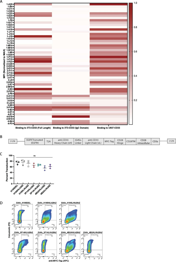
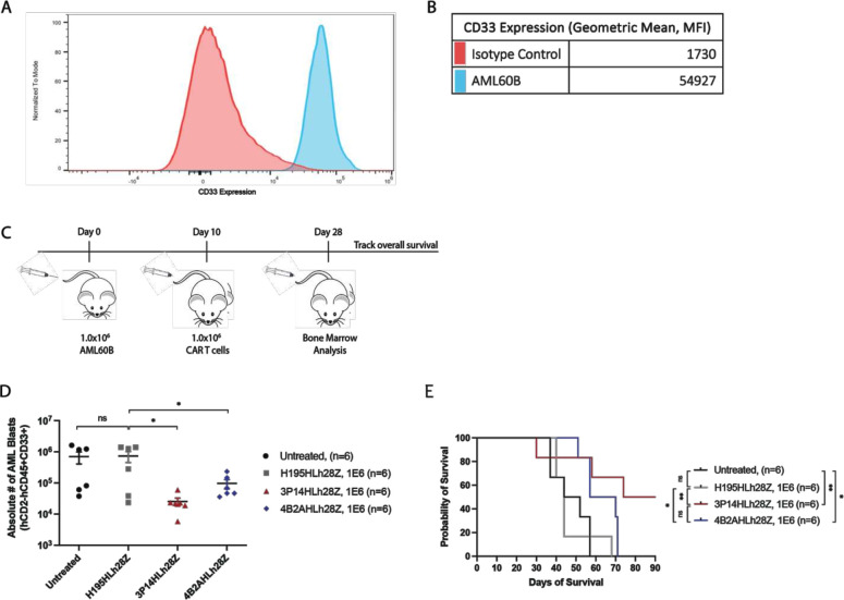
Methods: We developed 3P14HLh28Z, a novel CD33-directed CD28/CD3Z-based CAR T cell derived from a high-affinity binder obtained through membrane-proximal fragment immunization in humanized mice.

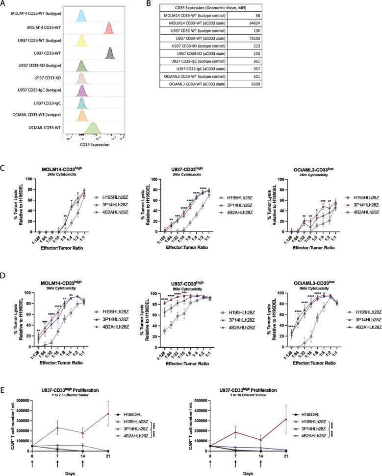
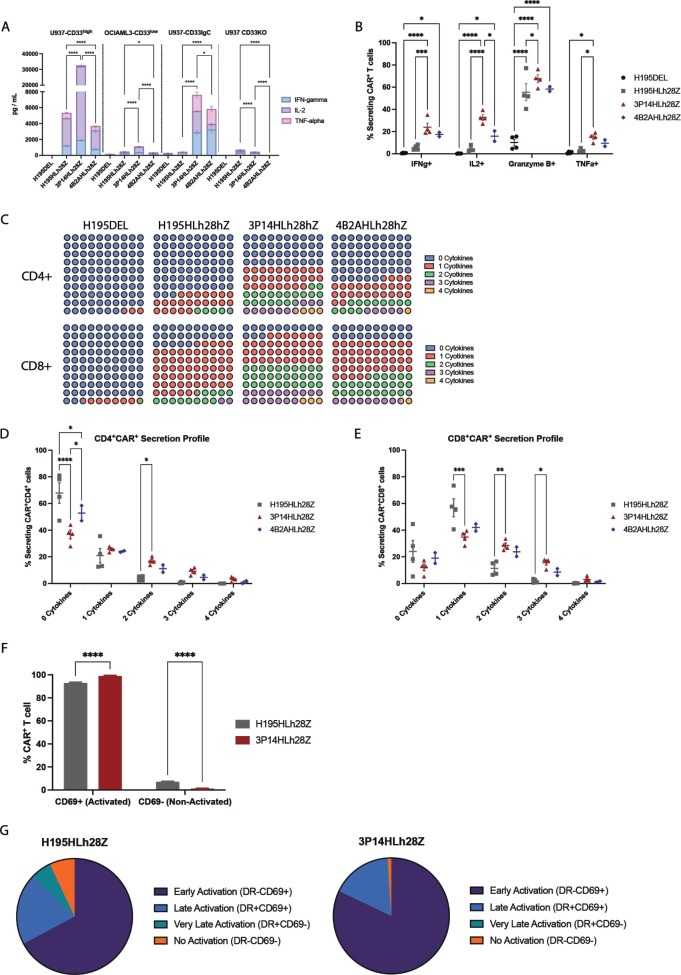
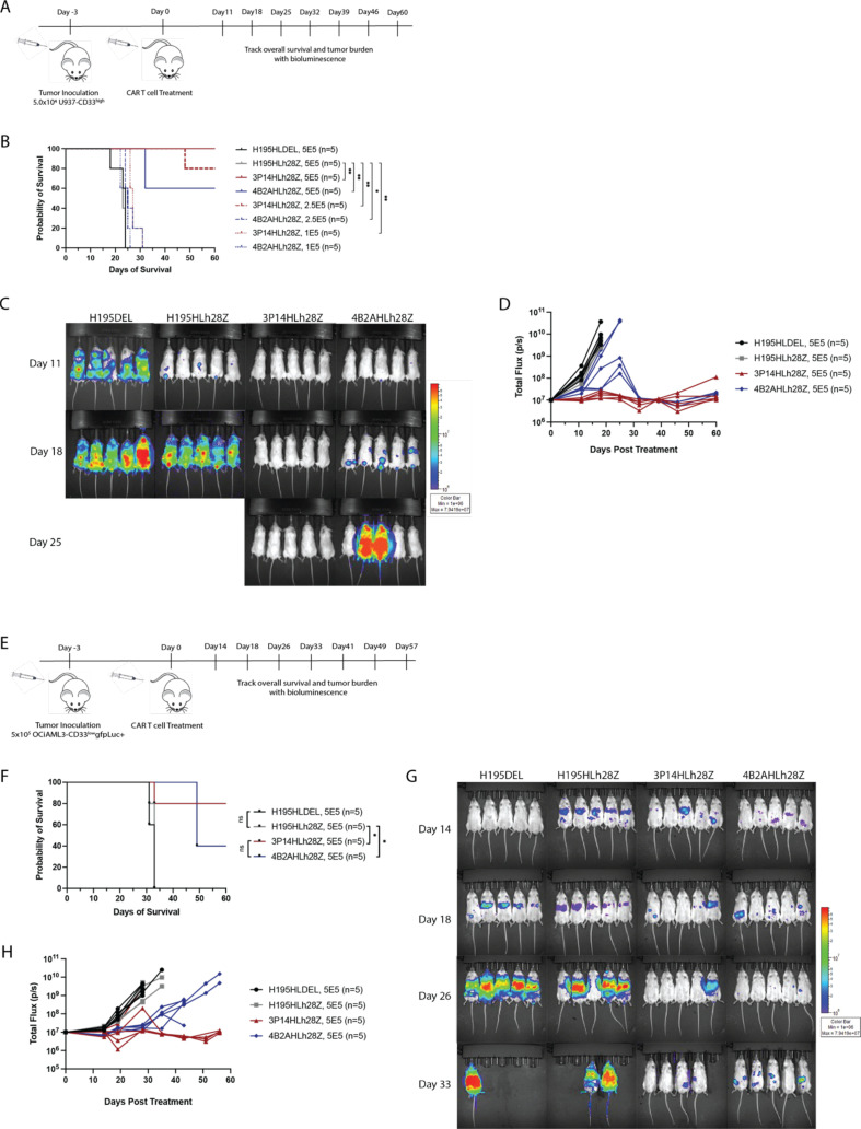
Results: We found that immunization exclusively with the membrane-proximal domain of CD33 is necessary for identification of membrane-proximal binders in humanized mice. Compared with clinically validated lintuzumab-based CAR T cells targeting distal CD33 epitopes, 3P14HLh28Z showed enhanced in vitro functionality as well as superior tumor control and increased overall survival in both low antigen density and clinically relevant patient-derived xenograft models. Increased activation and enhanced polyfunctionality led to enhanced efficacy.

Conclusions: Showing for the first time that a membrane-proximal CAR is superior to a membrane-distal one in the setting of CD33 targeting, our results demonstrate the rationale for targeting membrane-proximal epitopes with high-affinity binders. We also demonstrate the importance of optimizing CAR T cells for functionality in settings of both low antigen density and clinically relevant patient-derived models.

Keywords: Adoptive cell therapy - ACT; Chimeric antigen receptor - CAR; Immunotherapy; Leukemia; T cell.


Conflict of interest statement

Competing interests: AGK, ICL, RJB, and A Daniyan have filed provisional patent applications covering applications of membrane-proximal CD33 binders for cellular therapy. A Dunbar, YP, and RL are supported by National Cancer Institute P01 CA108671 11 (RL). DM and KKH are supported by an MSK Geoffrey Beene Cancer Research Award. RL is on the supervisory board of Qiagen and is a scientific advisor to Imago, Mission Bio, Zentalis, Ajax, Auron, Prelude, C4 Therapeutics and Isoplexis. He receives research support from and consulted for Celgene and Roche and has consulted for Incyte, Janssen, Astellas, Morphosys and Novartis, and has received honoraria from AstraZeneca, Roche, Lilly and Amgen for invited lectures and from Gilead for grant reviews. JJB has consulted for Avrobio, Sobi, BlueRock, Sanofi, Omeros, Advanced Clinical, MERCK, Bluebird Bio and is on the Data Safety Monitoring Board/Advisory Board for Advanced Clinical. RJB has licensed intellectual property to and collects royalties from BMS, Caribou, and Sanofi. RJB received research funding from BMS. RJB is a consultant to BMS, Atara Biotherapeutics Inc, Cargo Tx, Triumvira and was a consultant for CoImmune but ended in the past 3 months and Gracell Biotechnologies Inc but ended employment in the past 24 months. RJB is a member of the scientific advisory board for Triumvira, Cargo Tx and was a member of the scientific advisory board for CoImmune, but that ended in the past 3 months.

Figures

Figure 1
Figure 1
Identification of a membrane-proximal CD33 antibody. (A) Heatmap showing relative binding of selected antibodies to human full-length CD33, CD33-IgC, and U937 CD33-expressing tumor lines. (B) Schematic representation of retroviral vector encoding 5′ and 3′ long terminal repeats sequence (LTR), truncated EGFR, linked with a T2A element to a fully human CD33-targeted single-chain variable fragment, human CD28 hinge, transmembrane, and intracellular domains (CD28), and human zeta chain signaling domain (z chain). A Myc-tag is included for detection of the chimeric antigen receptor (CAR). (C) Summary data of retroviral transduction efficiency of CAR constructs as determined by flow cytometry using cetuximab. P values determined by repeated measures one-way analysis of variance. Data are a mean±SEM of three biological replicates conducted in three independent experiments; ns, non-significant. (D) Representative flow cytometry plot demonstrating CAR expression following human T cell transduction, detected with fluorescently labeled Cetuximab and Myc-Tag-specific antibodies. MFI, mean fluorescence intensity.
Figure 2
Figure 2
Membrane-proximal targeting chimeric antigen receptor (CAR) T cells demonstrate enhanced functionality and proliferative capacity in vitro. (A) Flow cytometry histograms of CD33 expression on acute myeloid leukemia (AML) cells detected with isotype control or fluorescently labeled CD33-specific antibody. (B) Quantitative geometric mean fluorescence intensity (MFI) of CD33 expression on AML cells detected with either isotype control or fluorescently labeled CD33-specific antibody. (C) 24-hour D-luciferin assay demonstrating lysis of CD33-expressing tumor cells (n=4 biological replicates; ****p<0.0001; ***p<0.001; **p<0.01; *p<0.05 by two-way analysis of variance (ANOVA)). Data are a mean±SEM of four biological replicates. (D) 96-hour D-luciferin assay demonstrating lysis of CD33 expressing tumor cells (n=4 biological replicates; ****p<0.0001; ***p<0.001; **p<0.01; *p<0.05 by two-way ANOVA). Data are a mean±SEM of four biological replicates. (E) Quantification of flow cytometric analysis demonstrating enhanced proliferation by membrane-proximal CD33 targeting CAR T cell in the presence of U937-CD33high tumor cells at E:T ratios of 1:2.5 (left) or 1:10 (right) (n=3 biological replicates; ****p<0.0001 by two-way ANOVA at day 21). Arrows indicate when additional target cells were added. Data are a mean±SEM of three biological replicates.
Figure 3
Figure 3
Membrane-proximal targeting chimeric antigen receptor (CAR) T cells are characterized by a unique activation profile in vitro. (A) 24-hour cytokine secretion profile of Tc1/Th1 cytokines when co-cultured with U937-CD33high, OCiAML3-CD33low, U937-CD33IgC, and U937-CD33KO tumor as detected by human 12-plex Luminex panel (n=3; ****p<0.0001; *p<0.05 by Dunnett’s multiple comparison tests against H195DEL control). Data are a mean±SEM of three biological replicates conducted in three independent experiments. (B) Quantification of flow cytometric analysis showing CAR T cell intracellular production of Tc1/Th1 activation cytokines when cultured with U937-CD33high tumor (n=4; ****p<0.0001; **p<0.01; *p<0.05 by two-way analysis of variance (ANOVA); ns, non-significant). Data are a mean±SEM of four biological replicates conducted in three independent experiments. (C) Qualitative representation of CD4+CAR+ and CD8+CAR+ Tc1/Th1 activation cytokines secretion. Cytokines evaluated were IFN-γ, IL-2, granzyme B, and TNF-α. (D) Quantification of flow cytometric analysis showing CD4+ CAR T cell intracellular production of Tc1/Th1 activation cytokines when cultured with U937-CD33high tumor (n=4; ****p<0.0001, *p<0.05 by two-way ANOVA). Data are a mean±SEM of four biological replicates conducted in three independent experiments. Cytokines evaluated were IFN-γ, IL-2, granzyme B, and TNF-α. (E) Quantification of flow cytometric analysis showing CD8+ CAR T cell intracellular production of Tc1/Th1 activation cytokines when cultured with U937-CD33high tumor (n=4; ***p<0.001; **p<0.01; *p<0.05 by two-way ANOVA). Data are a mean±SEM of four biological replicates conducted in three independent experiments. Cytokines evaluated were IFN-γ, IL-2, granzyme B, and TNF-α. (F) Quantification of flow cytometric analysis demonstrating CAR T cell activation status 7 days post-antigen stimulation with U937-CD33high tumor (n=4; ****p<0.0001 by two-way ANOVA). Data are a mean±SEM of four biological replicates conducted in three independent experiments. (G) Qualitative representation of flow cytometric analysis comparing CAR T cell activation profiles. Data are pooled from four biological replicates conducted in three independent experiments.
Figure 4
Figure 4
Membrane-proximal CD33-targeting chimeric antigen receptor (CAR) T cells provide enhanced survival in xenograft mouse model. (A) Schematic diagram of in vivo experimental setup. NCG mice were inoculated with U937-CD33high tumor and subsequently treated with CAR T cells. (B) Survival of NCG mice bearing U937-CD33high tumors and treated with titrated doses of CAR T cells (n=5; **p<0.01, *p<0.05). P values for survival determined by log-rank Mantel-Cox test, with 95% CI. (C) Tumor regression of NCG mice inoculated with U937-CD33high tumor and subsequently treated with 5.0×10⁵ of CAR T cells. (D) Bioluminescence over time of U937-CD33high in tumor-bearing NCG mice treated with 5.0×10⁵ CAR T cells. (E) Schematic diagram of in vivo experimental setup. NCG mice were inoculated with OCiAML3-CD33low tumor and subsequently treated with CAR T cells. (F) Survival of NCG mice inoculated with OCiAML3-CD33low tumors and treated with 5.0×10⁵ CAR T cells (n=5; *p<0.05). P values for survival determined by log-rank Mantel-Cox test, with 95% CI. (G) Tumor regression of NCG mice bearing OCiAML3-CD33low tumors and treated with 5.0×10⁵ CAR T cells. (H) Bioluminescence over time of OCiAML3-CD33low in tumor-bearing NCG mice treated with 5.0×10⁵ CAR T cells.
Figure 5
Figure 5
Membrane-proximal CD33-targeting chimeric antigen receptor (CAR) T cells decrease tumor burden in patient-derived acute myeloid leukemia (AML) xenograft model. (A) Flow cytometry histograms of CD33 expression on AML60B patient sample detected with isotype control or fluorescently labeled CD33-spcific antibody. (B) Quantitative geometric mean fluorescence intensity (MFI) of CD33 expression on AML60B patient sample detected with either isotype control or fluorescently labeled CD33-specific antibody. (C) Schematic diagram of experimental setup of a patient-derived xenograft model. NCG mice were inoculated with patient-derived AML blasts and treated with allogeneic CAR T cells. Bone marrow aspirates were analyzed 28 days post tumor inoculation. (D) Quantification of flow cytometric analysis demonstrating decreased tumor burden in mice treated with membrane-proximal CD33-targeting CAR T cells (n=6; *p<0.05 by unpaired t-test). (E) Survival of NCG mice-bearing AML60B patient-derived tumor and treated with membrane-distal or membrane-proximal CAR T cells (n=6; **p<0.01; *p<0.05). P values for survival determined by log-rank Mantel-Cox test, with 95% CI.

References

    1. Ganzel C, Sun Z, Cripe LD, et al. . Very poor long-term survival in past and more recent studies for relapsed AML patients: the ECOG-ACRIN experience. Am J Hematol 2018;93:1074–81. 10.1002/ajh.25162 - DOI - PMC - PubMed
    1. Vitale C, Strati P. CAR T-cell therapy for B-cell non-Hodgkin lymphoma and chronic lymphocytic leukemia: clinical trials and real-world experiences. Front Oncol 2020;10:849. 10.3389/fonc.2020.00849 - DOI - PMC - PubMed
    1. Martino M, Alati C, Canale FA, et al. . A review of clinical outcomes of CAR T-cell therapies for B-acute lymphoblastic leukemia. Int J Mol Sci 2021;22:2150. 10.3390/ijms22042150 - DOI - PMC - PubMed
    1. Majzner RG, Rietberg SP, Sotillo E, et al. . Tuning the antigen density requirement for CAR T-cell activity. Cancer Discov 2020;10:702–23. 10.1158/2159-8290.CD-19-0945 - DOI - PMC - PubMed
    1. Spiegel JY, Patel S, Muffly L, et al. . CAR T cells with dual targeting of CD19 and CD22 in adult patients with recurrent or refractory B cell malignancies: a phase 1 trial. Nat Med 2021;27:1419–31. 10.1038/s41591-021-01436-0 - DOI - PMC - PubMed

MeSH terms

Substances

LinkOut - more resources