. 2024 May 21;15(1):4341.
doi: 10.1038/s41467-024-48629-y.

Integrative multi-region molecular profiling of primary prostate cancer in men with synchronous lymph node metastasis

Affiliations

Integrative multi-region molecular profiling of primary prostate cancer in men with synchronous lymph node metastasis

Udit Singhal et al. Nat Commun. .

Abstract

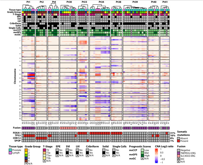
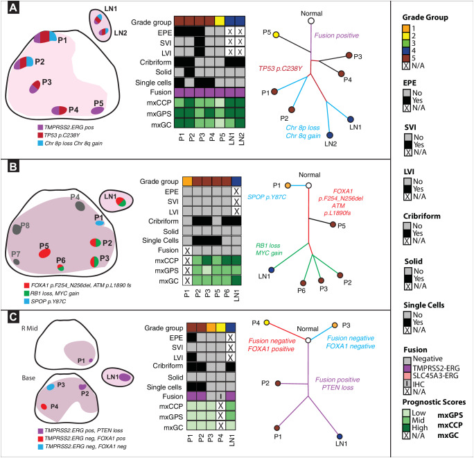
Localized prostate cancer is frequently composed of multiple spatially distinct tumors with significant inter- and intra-tumoral molecular heterogeneity. This genomic diversity gives rise to many competing clones that may drive the biological trajectory of the disease. Previous large-scale sequencing efforts have focused on the evolutionary process in metastatic prostate cancer, revealing a potential clonal progression to castration resistance. However, the clonal origin of synchronous lymph node (LN) metastases in primary disease is still unknown. Here, we perform multi-region, targeted next generation sequencing and construct phylogenetic trees in men with prostate cancer with synchronous LN metastasis to better define the pathologic and molecular features of primary disease most likely to spread to the LNs. Collectively, we demonstrate that a combination of histopathologic and molecular factors, including tumor grade, presence of extra-prostatic extension, cellular morphology, and oncogenic genomic alterations are associated with synchronous LN metastasis.


Conflict of interest statement

The content is solely the responsibility of the authors and does not necessarily represent the official views of U-M Precision Health. JJT is a co-founder with minor equity interest in LynxDx, which has licensed urinary biomarkers unrelated to this project. S.A.T has received travel support from and had a sponsored research agreement with Compendia Bioscience/Life Technologies/ThermoFisher. The University of Michigan and Brigham and Women’s Hospital have been issued a patent on ETS gene fusions (US8969527B2 “Recurrent gene fusions in prostate cancer”) in prostate cancer on which S.A.T. and A.M.C. are co-inventors. The diagnostic field of use was licensed to Hologic/Gen-Probe, Inc., which has sublicensed rights to Roche/Ventana Medical Systems. S.A.T. has served as a consultant for and received honoraria from Janssen, AbbVie, Sanofi, Almac Diagnostics and Astellas/Medivation. S.A.T. has sponsored research agreements with Astellas and GenomeDX. S.A.T. is a co-founder and Chief Medical Officer for Strata Oncology. T.M.M. has received research funding from MDxHealth, Myriad Genetics, and GenomeDx. T.M.M. has served as a consultant for Myriad Genetics. U.S. has received research funding from Merck & Co., Inc. S.S.S. is on a study advisory committee for Bayer Pharma and has a non-sponsored research agreement with GenomeDx. The other authors have declared that no conflict of interest exists.

Figures

Fig. 1
Fig. 1. Integrative comprehensive multi region genomic profiling of primary prostate cancer with synchronous lymph node (LN) metastasis.
We used two targeted DNAseq panels to identify key somatic mutations and copy number alterations (CNA) across 10 patients who passed our custom quality control filtering criteria. For CNA analysis, only the top 445 genes (no. of amplicons per gene > 4 & log10 false discovery rate <0.01 & absolute log2CNvalue > 0.3) with losses and gains are displayed in the heatmap. Unsupervised hierarchical clustering of all tumor regions within each patient was performed to interrogate primary tumor regions that cluster with their respective synchronous LN metastasis regions using log2 normalized data. Genes were ordered by the chromosome number along with their start and end positions within each chromosome. CNA for the known prostate cancer-relevant genes are annotated. ETS gene fusion status was derived from targeted RNAseq data using an in-house fusion quantification pipeline. Relevant clinicopathologic variables such as grade, stage, extraprostatic extension (EPE), seminal vesicle invasion (SVI), lymphovascular invasion (LVI), cribriform pattern, solid pattern, single cells and derived commercially available prognostic scores (mxCCP (derived Cell Cycle Progression score or ProlarisTM), mxGPS (derived Genomic Prostate Score or OncotypeTM), and mxGC (derived Genomic Classifier or DecipherTM) for each sample are annotated on the heatmap. Prognostic scores were categorized into low, mid, and high groups based on their Q1 and Q3 values for comparison among samples with the same patient as well as comparison across different scores within each sample. We observed intra- and inter-patient heterogeneity in histologic grade, genomic alterations, and derived prognostic gene signatures. Source data are provided as a Source Data file.
Fig. 2
Fig. 2. Phylogenetic evolutionary analysis to determine the biologically dominant nodule.
Source data are provided as a Source Data file. A. Spatial spread linked to extra-prostatic extension (EPE). Left panel: In patient #1, 5 primary and 2 LN regions encompassing Grade Groups (GG) 2-5 were analyzed. We identified three distinct molecular subtypes. Middle panel: All tumor regions, including the two LN metastases regions, were TMPRSS2:ERG fusion-positive and with heterogeneous prognostic gene signatures. Right panel: TP53 somatic mutation was detected on all tumor regions, except for P5. Key chromosomal alterations, such as 8p loss and 8q gain, were only detected in regions P1, P2, LN1, LN2. Here, the data suggest that primary tumor regions P1, P2 closely resemble LN1, LN2. Additionally, P5 represents a distinct subclone unrelated to other primary regions or LNs. B. Driver alterations in metastasis to LN. Left panel: In patient #33, 8 primary tumor (GG1-5) and 1 LN metastasis region were analyzed. Gray regions (P4,7,8) designate those with low tumor purity that were excluded. We identified three molecular subtypes. Middle panel: All regions were ETS fusion negative except for P1, P8. P4, P7, and P8 regions were excluded due to low tumor content. Right panel: An SPOP mutation was detected only in P1. Driver alterations: FOXA1, ATM frameshift mutations were detected in regions P2, P3, P5, P6, LN1. RB1 loss and MYC gain were seen in P2, P3, P6, and LN1, but not P5. Here, data suggest that primary regions P2, P3, P6, all GG5 regions with cribriform patterns, closely resemble LN1. C. LN metastasis in multiclonal primary disease. Left panel: In patient #41, 4 primary (GG2-5) and 1 LN metastasis region were analyzed. P1, a region of EPE, was taken from the mid prostate and P2-P4 was taken prostate base. We identified three distinct tumor clones. Middle panel: All samples except P3 and P4 were TMPRSS2:ERG fusion-positive. Right panel: P4 had a FOXA1 somatic mutation which was not seen in other regions. Here, data suggest that primary region P1 closely resembles LN1, as both are TMPRSS2:ERG fusion-positive and harbor PTEN loss.

References

    1. Siegel RL, Miller KD, Fuchs HE, Jemal A. Cancer statistics, 2022. CA. Cancer J. Clin. 2022;72:7–33. doi: 10.3322/caac.21708. - DOI - PubMed
    1. Buyyounouski MK, Pickles T, Kestin LL, Allison R, Williams SG. Validating the interval to biochemical failure for the identification of potentially lethal prostate cancer. J. Clin. Oncol. 2012;30:1857–1863. doi: 10.1200/JCO.2011.35.1924. - DOI - PubMed
    1. D’Amico AV, et al. Cancer-specific mortality after surgery or radiation for patients with clinically localized prostate cancer managed during the prostate-specific antigen era. J. Clin. Oncol. 2003;21:2163–2172. doi: 10.1200/JCO.2003.01.075. - DOI - PubMed
    1. Wei L, et al. Intratumoral and Intertumoral genomic heterogeneity of multifocal localized prostate cancer impacts molecular classifications and genomic prognosticators. Eur. Urol. 2017;71:183–192. doi: 10.1016/j.eururo.2016.07.008. - DOI - PMC - PubMed
    1. Løvf M, et al. Multifocal primary prostate cancer exhibits high degree of genomic heterogeneity. Eur. Urol. 2019;75:498–505. doi: 10.1016/j.eururo.2018.08.009. - DOI - PubMed