

Human CD4-binding site antibody elicited by polyvalent DNA prime-protein boost vaccine neutralizes cross-clade tier-2-HIV strains
- PMID: 38773089
- PMCID: PMC11109196
- DOI: 10.1038/s41467-024-48514-8
Human CD4-binding site antibody elicited by polyvalent DNA prime-protein boost vaccine neutralizes cross-clade tier-2-HIV strains
Abstract
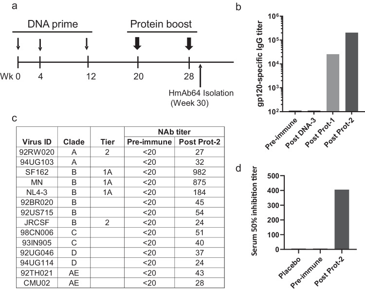
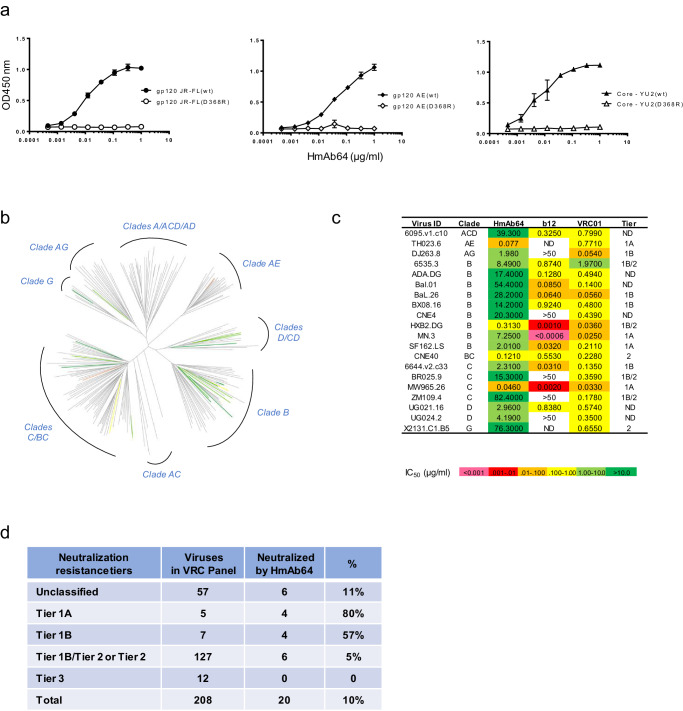
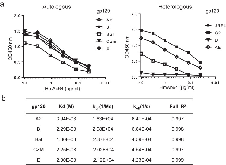
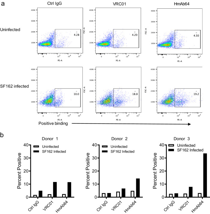
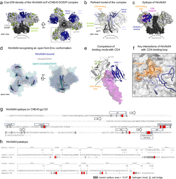
The vaccine elicitation of HIV tier-2-neutralization antibodies has been a challenge. Here, we report the isolation and characterization of a CD4-binding site (CD4bs) specific monoclonal antibody, HmAb64, from a human volunteer immunized with a polyvalent DNA prime-protein boost HIV vaccine. HmAb64 is derived from heavy chain variable germline gene IGHV1-18 and light chain germline gene IGKV1-39. It has a third heavy chain complementarity-determining region (CDR H3) of 15 amino acids. On a cross-clade panel of 208 HIV-1 pseudo-virus strains, HmAb64 neutralized 20 (10%), including tier-2 strains from clades B, BC, C, and G. The cryo-EM structure of the antigen-binding fragment of HmAb64 in complex with a CNE40 SOSIP trimer revealed details of its recognition; HmAb64 uses both heavy and light CDR3s to recognize the CD4-binding loop, a critical component of the CD4bs. This study demonstrates that a gp120-based vaccine can elicit antibodies capable of tier 2-HIV neutralization.
© 2024. The Author(s).
Conflict of interest statement
The authors declare no competing interests.
Figures
Update of
-
Human CD4-Binding Site Antibody Elicited by Polyvalent DNA Prime-Protein Boost Vaccine Neutralizes Cross-Clade Tier-2-HIV Strains.Res Sq [Preprint]. 2023 Oct 20:rs.3.rs-3360161. doi: 10.21203/rs.3.rs-3360161/v1. Res Sq. 2023. Update in: Nat Commun. 2024 May 21;15(1):4301. doi: 10.1038/s41467-024-48514-8. PMID: 37886518 Free PMC article. Updated. Preprint.
Similar articles
-
Human CD4-Binding Site Antibody Elicited by Polyvalent DNA Prime-Protein Boost Vaccine Neutralizes Cross-Clade Tier-2-HIV Strains.Res Sq [Preprint]. 2023 Oct 20:rs.3.rs-3360161. doi: 10.21203/rs.3.rs-3360161/v1. Res Sq. 2023. Update in: Nat Commun. 2024 May 21;15(1):4301. doi: 10.1038/s41467-024-48514-8. PMID: 37886518 Free PMC article. Updated. Preprint.
-
Vaccine-Elicited Tier 2 HIV-1 Neutralizing Antibodies Bind to Quaternary Epitopes Involving Glycan-Deficient Patches Proximal to the CD4 Binding Site.PLoS Pathog. 2015 May 29;11(5):e1004932. doi: 10.1371/journal.ppat.1004932. eCollection 2015 May. PLoS Pathog. 2015. PMID: 26023780 Free PMC article.
-
Boosting of HIV envelope CD4 binding site antibodies with long variable heavy third complementarity determining region in the randomized double blind RV305 HIV-1 vaccine trial.PLoS Pathog. 2017 Feb 24;13(2):e1006182. doi: 10.1371/journal.ppat.1006182. eCollection 2017 Feb. PLoS Pathog. 2017. PMID: 28235027 Free PMC article. Clinical Trial.
-
Structural Features of Broadly Neutralizing Antibodies and Rational Design of Vaccine.Adv Exp Med Biol. 2018;1075:73-95. doi: 10.1007/978-981-13-0484-2_4. Adv Exp Med Biol. 2018. PMID: 30030790 Review.
-
The promise of priming precursors: Advances in inducing CD4 binding site-directed HIV broadly neutralizing antibodies.Sci Immunol. 2024 Aug 30;9(98):eadq8862. doi: 10.1126/sciimmunol.adq8862. Epub 2024 Aug 30. Sci Immunol. 2024. PMID: 39213339 Review.
Cited by
-
Grand Challenges on HIV/AIDS in China - The 5th Symposium, Yunnan 2024.Emerg Microbes Infect. 2025 Dec;14(1):2492208. doi: 10.1080/22221751.2025.2492208. Epub 2025 Apr 22. Emerg Microbes Infect. 2025. PMID: 40202047 Free PMC article.
-
A potent and broad CD4 binding site neutralizing antibody with strong ADCC activity from a Chinese HIV-1 elite neutralizer.Cell Discov. 2025 Jun 10;11(1):55. doi: 10.1038/s41421-025-00808-x. Cell Discov. 2025. PMID: 40490455 Free PMC article.
-
Assessing bnAb potency in the context of HIV-1 envelope conformational plasticity.PLoS Pathog. 2025 Jan 21;21(1):e1012825. doi: 10.1371/journal.ppat.1012825. eCollection 2025 Jan. PLoS Pathog. 2025. PMID: 39836706 Free PMC article.
-
Alterations in BCR heavy chain CDR3 repertoire characteristics in pediatric mycoplasma pneumoniae infection.Front Cell Infect Microbiol. 2025 Jun 12;15:1573511. doi: 10.3389/fcimb.2025.1573511. eCollection 2025. Front Cell Infect Microbiol. 2025. PMID: 40575482 Free PMC article.
-
Enhanced antibody response to the conformational non-RBD region via DNA prime-protein boost elicits broad cross-neutralization against SARS-CoV-2 variants.Emerg Microbes Infect. 2025 Dec;14(1):2447615. doi: 10.1080/22221751.2024.2447615. Epub 2025 Mar 3. Emerg Microbes Infect. 2025. PMID: 39727342 Free PMC article.
References
-
- UNAIDS. UNAIDS Fact Sheet 2022. https://www.unaids.org/sites/default/files/media_asset/UNAIDS_FactSheet_... (accessed September 21, 2022) (2021).
MeSH terms
Grants and funding
- R01AI145655/U.S. Department of Health & Human Services | NIH | National Institute of Allergy and Infectious Diseases (NIAID)
- R33 AI087191/AI/NIAID NIH HHS/United States
- P01 AI100151/AI/NIAID NIH HHS/United States
- U19 AI082676/AI/NIAID NIH HHS/United States
- R01AI65250/U.S. Department of Health & Human Services | NIH | National Institute of Allergy and Infectious Diseases (NIAID)
- R01 AI065250/AI/NIAID NIH HHS/United States
- P01AI100151/U.S. Department of Health & Human Services | NIH | National Institute of Allergy and Infectious Diseases (NIAID)
- Y01 GM000080/GM/NIGMS NIH HHS/United States
- P01 AI082274/AI/NIAID NIH HHS/United States
- R01 AI145655/AI/NIAID NIH HHS/United States
- R21 AI087191/AI/NIAID NIH HHS/United States
- R21/R33 AI087191/U.S. Department of Health & Human Services | NIH | National Institute of Allergy and Infectious Diseases (NIAID)
- N01 AI005394/AI/NIAID NIH HHS/United States
- U19AI082676/U.S. Department of Health & Human Services | NIH | National Institute of Allergy and Infectious Diseases (NIAID)
LinkOut - more resources
Full Text Sources
Research Materials
