. 2024 May 21;15(1):4326.
doi: 10.1038/s41467-024-48138-y.

Post-resolution macrophages shape long-term tissue immunity and integrity in a mouse model of pneumococcal pneumonia

Affiliations

Post-resolution macrophages shape long-term tissue immunity and integrity in a mouse model of pneumococcal pneumonia

Karen T Feehan et al. Nat Commun. .

Abstract

Resolving inflammation is thought to return the affected tissue back to homoeostasis but recent evidence supports a non-linear model of resolution involving a phase of prolonged immune activity. Here we show that within days following resolution of Streptococcus pneumoniae-triggered lung inflammation, there is an influx of antigen specific lymphocytes with a memory and tissue-resident phenotype as well as macrophages bearing alveolar or interstitial phenotype. The transcriptome of these macrophages shows enrichment of genes associated with prostaglandin biosynthesis and genes that drive T cell chemotaxis and differentiation. Therapeutic depletion of post-resolution macrophages, inhibition of prostaglandin E2 (PGE2) synthesis or treatment with an EP4 antagonist, MF498, reduce numbers of lung CD4+/CD44+/CD62L+ and CD4+/CD44+/CD62L-/CD27+ T cells as well as their expression of the α-integrin, CD103. The T cells fail to reappear and reactivate upon secondary challenge for up to six weeks following primary infection. Concomitantly, EP4 antagonism through MF498 causes accumulation of lung macrophages and marked tissue fibrosis. Our study thus shows that PGE2 signalling, predominantly via EP4, plays an important role during the second wave of immune activity following resolution of inflammation. This secondary immune activation drives local tissue-resident T cell development while limiting tissue injury.


Conflict of interest statement

The authors declare no competing interests.

Figures
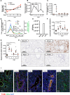
Fig. 1
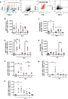
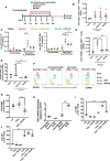
Fig. 1. Intranasal S. pneumoniae causes a self-resolving inflammation.
WT C57BL6/J mice were administered intranasal S. pneumoniae with animals (A) experiencing a transient weight loss. Lungs were digested and samples analysed by polychromatic flow cytometry for markers of acute inflammation including (B) total leucocytes, (C) neutrophils, (D) pro-inflammatory cytokines and (E) bacterial clearance (Colony Forming Units/mL of bronchoalveolar lavage fluid). Flow cytometry was also used to profile (F) total lymphoid and (G) myeloid cells throughout inflammation, resolution and weeks following resolution. The temporal profile of (H) neutrophils (GR1+) as well as (I) macrophages (F4/80+) and CD3 T cells (CD3+) were confirmed at tissue level, panels H-I. A Alveoli, B bronchiole, BV blood vessel. Sections are representative of n = 3 independent experiments. Data were analysed by one-way analysis of variance (ANOVA) and Tukey’s multiple comparisons test. A p value of <0.05 was taken as the threshold of significance with graphical representation as; p < 0.05 = *, p < 0.01 = ** and p < 0.001 = *** and presented as mean ± SEM. (n = 5–8 mice/group).
Fig. 2
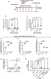
Fig. 2. Resolution of lung inflammation is followed by a second wave of myeloid cell infiltration.
WT C57BL6/J mice were administered intranasal S. pneumoniae with lungs digested for analysis by polychromatic flow cytometry. Using the gating strategy in panel (A) (FMOs are shown in blue to identify positive populations (red)) profiles of (BE) monocytes, interstitial and alveolar macrophage population were identified with panels (FH) providing further analysis in interstitial macrophage sub-types. Data were analysed by one-way analysis of variance (ANOVA) and Tukey’s multiple comparisons test. A p value of <0.05 was taken as the threshold of significance with graphical representation as; p < 0.05 = *, p < 0.01 = ** and p < 0.001 = *** and presented as mean ± SEM (n = 3–8 mice/group).
Fig. 3
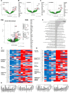
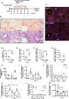
Fig. 3. RNAseq analysis reveals a role for post-resolution macrophage populations in T cell migration/maturation.
WT C57BL6/J mice were administered intranasal S. pneumoniae with lungs digested and macrophage populations including (A) double negative interstitial macrophage, (B) LYVE-1-/MHC-II+ interstitial macrophages, (C) LYVE-1+/MHC-II- interstitial macrophages as well as (D) alveolar macrophages sorted by FACS and subject to analysis by RNAseq followed by bioinformatic analysis using edgeR in RStudio as illustrated (n = 5 mice/group). D qPCR validation (n = 3 mice/group) was used in a separate independent experiment to confirm upregulation of Inhba and Ptgs2 in naïve versus day 14 alveolar macrophages. Students unpaired t-test was used to compare the means of two groups. A p value of <0.05 was taken as the threshold of significance with graphical representation as; p < 0.05 = *, p < 0.01 = ** and p < 0.001 = *** and presented as mean ± SEM. As alveolar macrophages showed the greatest changes post-resolution compared to the naive state these cells were further analysed by using (E) PANTHER to identify GoTerms from upregulated genes at day 14 compared to naïve, (F, G) normalised read counts of five replicates from naïve alveolar macrophages compared to day 14, (H) EdgeR results showing differential gene expression as logFC and p value for genes relating to migration, matrix remodelling, regulation of phenotype and interferon signalling.
Fig. 4
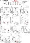
Fig. 4. Therapeutic deletion of blood monocyte reveals the source of post-resolution macrophages and a role for these cells in T cell maturation.
MC-21, which depletes blood monocytes was administered therapeutically (A) six days after intranasal S. pneumoniae and every second day up to day 14. Polychromatic flow cytometry was carried out for profiles of lung (BD) monocytes and macrophages as well as (EG) interstitial macrophage sub-populations. In addition, we determined the impact of depleting post-resolution macrophages on profiles of lung tissue (HL) CD4+ T cell subpopulations. Data were analysed by one-way analysis of variance (ANOVA) and Tukey’s multiple comparisons test. A p value of <0.05 was taken as the threshold of significance with graphical representation as; p < 0.05 = *, p < 0.01 = ** and p < 0.001 = *** and presented as mean ± SEM (n = 5 mice/group).
Fig. 5
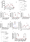
Fig. 5. Post-resolution prostaglandin synthesis regulates monocyte infiltration via EP4/CCL2.
Whole lungs were isolated at the indicated times following intranasal S. pneumoniae for lipid extraction and analysis by LC-MS/MS revealing the profile of (A) PGE2 and (B) a representative reconstructed ion chromatogram. PCR analysis was carried out on (C, D) post-resolution macrophages to identify the source of PGE2 as well as (E, F) the predominant PGE2 receptor (EP) that is expressed on alveolar and interstitial macrophages, respectively, as well as post-resolution (G) T cell populations. A similar biphasic profile for prostacyclin (PGI2), measured as its stable derivative 6-keto PGF1α, is presented in (H, I) along with (J) the post-resolution cells that synthesise it. Data were analysed by one-way analysis of variance (ANOVA) and Tukey’s multiple comparisons test. A p value of <0.05 was taken as the threshold of significance with graphical representation as; p < 0.05 = *, p < 0.01 = ** and p < 0.001 = *** and presented as mean ± SEM(n = 3–5 mice/group).
Fig. 6
Fig. 6. PGE2 via EP4 controls post-resolution T cells populations and phenotype.
A Osmotic pumps loaded with either the pan COX inhibitor naproxen or the EP4 antagonist MF498 were implanted into mice once inflammation resolved (day 4) and their effects of blocking the biological action of post-resolution PGE2 was established 6 weeks later as determined by measuring (B) late effector T cell numbers and (C) their effector function as well as (D) early effector T cells and their expression of (E, F) the α-integrin, CD103. as a marker of T cell residence potential. Linking PGE2 with this post-resolution T cell phenotype, T cells were incubated PGE2 and its direct effect on (G) CD103 expression or with other cytokines expressed during post-resolution that are known to affect lymphocyte function, (H) namely TGFβ, were examined for their collective effects on intracellular (I) IL-17 and (J) IFNγ. Data were analysed by one-way analysis of variance (ANOVA) and Tukey’s multiple comparisons test. A p value of <0.05 was taken as the threshold of significance with graphical representation as; p < 0.05 = *, p < 0.01 = ** and p < 0.001 = *** and presented as mean ± SEM (n = 3–6 mice/group).
Fig. 7
Fig. 7. Post-resolution PGE2 via EP4 determine re-emergence of memory T cells in response to secondary infection.
A Minipumps loaded with either the pan COX inhibitor naproxen or the EP4 antagonist MF498 were implanted into mice post-resolution four days after intranasal S. pneumoniae. 6 weeks after the initial challenge (or 38 days after this intervention), these sensitised mice or their controls were challenged with S. pneumoniae and impact of this intervention on (B) CD4+/CD44+/CD62L+ and (C) CD4+/CD44+/CD62L-/CD27+ T cell numbers was determined 24 h later. Indeed, levels of expression of (D) CD103 and the T cell activation markers (E) CD69 and (F) CD44 was also determined on CD4+/CD44+/CD62L-/CD27+ T cells. Besides lymphocytes we determined how inhibition of PGE2 synthesis or EP4 antagonism also impacted on the ability to recruit (G) neutrophils and ultimately clear the secondary challenge of (H) S. pneumoniae. Data were analysed by one-way analysis of variance (ANOVA) and Tukey’s multiple comparisons test. A p value of <0.05 was taken as the threshold of significance with graphical representation as; p < 0.05 = *, p < 0.01 = ** and p < 0.001 = *** and presented as mean ± SEM (n = 4–5 mice/group).
Fig. 8
Fig. 8. Post-resolution PGE2 via EP4 prevents excessive tissue injury and regulates macrophage trafficking.
The dosing regime in (A) revealed the impact of therapeutically antagonising post-resolution EP4 on (B) fibrosis along with examples of vascular occlusion (arrows) and (C) macrophage infiltration by confocal microscopy as well as (DG) flow cytometry at day 14. These infiltrated macrophages were examined for their (H) phagocytic ability. The infiltration of post-resolution macrophages was associated with a second wave of (I, J) CCL2 expression with its receptor (K, L) CCR2 being negatively controlled by (M) EP4. A Alveoli, B bronchiole, BV blood vessel. Students unpaired t-test was used to compare the means of two groups. Differences between multiple groups were analysed using one-way analysis of variance (ANOVA) and Tukey’s multiple comparisons test. A p value of <0.05 was taken as the threshold of significance with graphical representation as; p < 0.05 = *, p < 0.01 = ** and p < 0.001 = *** and presented as mean ± SEM (n = 3–5 mice/group).

Similar articles

Cited by

References

    1. Fullerton JN, Gilroy DW. Resolution of inflammation: a new therapeutic frontier. Nat. Rev. Drug Discov. 2016;15:551–567. doi: 10.1038/nrd.2016.39. - DOI - PubMed
    1. Serhan CN, Savill J. Resolution of inflammation: the beginning programs the end. Nat. Immunol. 2005;6:1191–1197. doi: 10.1038/ni1276. - DOI - PubMed
    1. Potter PK, Cortes-Hernandez J, Quartier P, Botto M, Walport MJ. Lupus-prone mice have an abnormal response to thioglycolate and an impaired clearance of apoptotic cells. J. Immunol. 2003;170:3223–3232. doi: 10.4049/jimmunol.170.6.3223. - DOI - PubMed
    1. Motwani MP, et al. Prolonged immune alteration following resolution of acute inflammation in humans. PLoS One. 2017;12:e0186964. doi: 10.1371/journal.pone.0186964. - DOI - PMC - PubMed
    1. Newson J, et al. Resolution of acute inflammation bridges the gap between innate and adaptive immunity. Blood. 2014;124:1748–1764. doi: 10.1182/blood-2014-03-562710. - DOI - PMC - PubMed

MeSH terms

Substances