. 2024 May 21;14(1):11591.
doi: 10.1038/s41598-024-62547-5.

A simple protocol to establish a conditionally immortalized mouse podocyte cell line

Affiliations

A simple protocol to establish a conditionally immortalized mouse podocyte cell line

Yujiao Huang et al. Sci Rep. .

Abstract

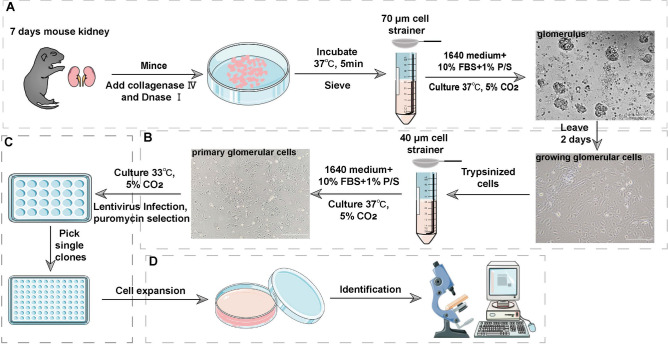
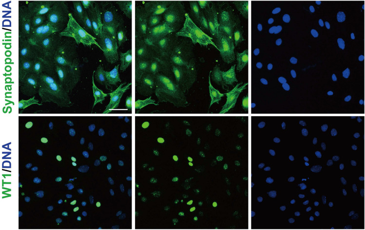
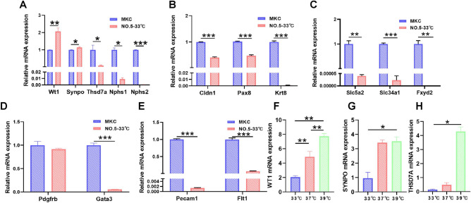
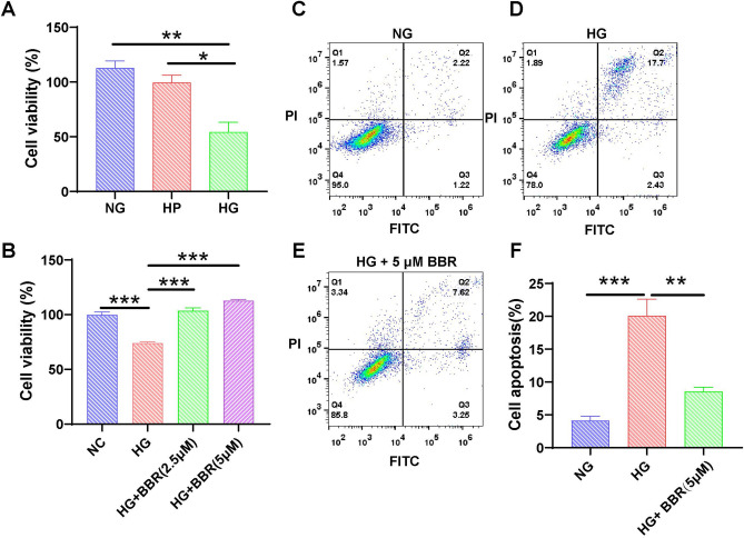
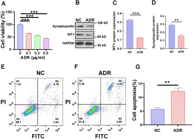
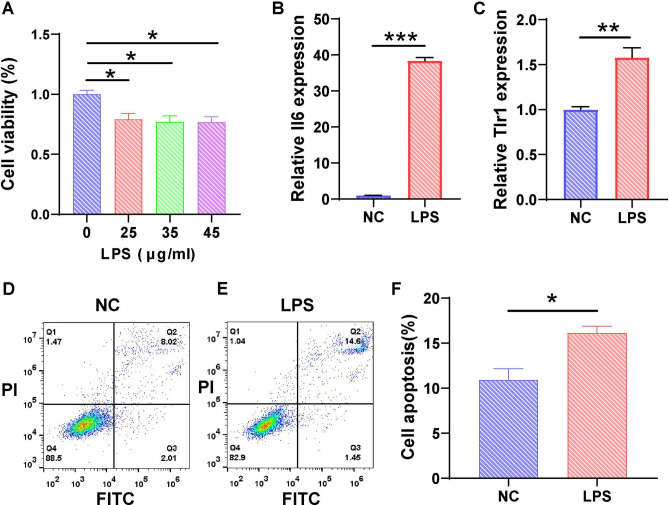
Podocytes are specialized terminally differentiated cells in the glomerulus that are the primary target cells in many glomerular diseases. However, the current podocyte cell lines suffer from prolonged in vitro differentiation and limited survival time, which impede research progress. Therefore, it is necessary to establish a cell line that exhibits superior performance and characteristics. We propose a simple protocol to obtain an immortalized mouse podocyte cell (MPC) line from suckling mouse kidneys. Primary podocytes were cultured in vitro and infected with the SV40 tsA58 gene to obtain immortalized MPCs. The podocytes were characterized using Western blotting and quantitative real-time PCR. Podocyte injury was examined using the Cell Counting Kit-8 assay and flow cytometry. First, we successfully isolated an MPC line and identified 39 °C as the optimal differentiation temperature. Compared to undifferentiated MPCs, the expression of WT1 and synaptopodin was upregulated in differentiated MPCs. Second, the MPCs ceased proliferating at a nonpermissive temperature after day 4, and podocyte-specific proteins were expressed normally after at least 15 passages. Finally, podocyte injury models were induced to simulate podocyte injury in vitro. In summary, we provide a simple and popularized protocol to establish a conditionally immortalized MPC, which is a powerful tool for the study of podocytes.

Keywords: MPC; Podocyte injury; Suckling mouse; Synaptopodin; WT1.


Conflict of interest statement

The authors declare no competing interests.

Figures

Figure 1
Figure 1
The pipeline for establishing conditionally immortalized MPCs. (A) To obtain podocytes for establishing a conditionally immortalized MPC model, glomeruli were harvested from suckling mice. Kidneys were minced to 1-mm3 pieces in a 6-cm cell culture dish. The tissues were digested in collagenase solution. The collagenase-digested tissues were gently pressed through a 70-µm cell strainer, and the glomeruli were cultured at 37 °C. (B) Two days later, outgrowing cells appeared as a monolayer around the individual glomeruli. Trypsin-digested cells were passed through a 40 µm cell strainer to remove the glomerular cores. The primary glomerular cells were cultured at 37 °C until the cell confluence reached 80%. (C) Podocytes were infected with lentivirus carrying SV40 tsA58 and the puromycin resistance gene. We performed limited dilutions in 96-well plates and identified and numbered individual cell wells using microscopy. (D) Thirteen monoclonal cell lines were successfully isolated. The 13 monoclonal cell lines were subjected to preliminary identification using RT-qPCR. A monoclonal podocyte line was used for further identification. Imaging materials were obtained from smart.servier.com. The scale is 100 µm.
Figure 2
Figure 2
Identification of podocytes within glomerular cells cultured in vitro. Indirect immunofluorescent staining revealed strong and weak variations in synaptopodin and WT1 staining in glomerular cells. Synaptopodin showed positive staining in the cytoplasm and at the cell border. WT1 was co-localized in the nucleus with DAPI staining. The scale is 50 µm.
Figure 3
Figure 3
Identification of the monoclonal MPC and its optimum nonpermissive temperature. RT-qPCR was used to measure the expression of markers. (A) The expression of podocyte-specific genes in the No. 5 cell line, including Wt1, Synpo, Thsd7a, Nphs1 and Nphs2. (B) The expression of PEC-specific genes in the No. 5 cell line, including Cldn1, Pax8 and Krt8. (C) The expression of renal tubular epithelial cell-specific genes in the No. 5 cell line, including Slc5a2, Slc34a1and Fxyd2. (D) The expression of mesangial cell-specific genes in the No. 5 cell line, including Padgftb and Gata3. (E) The expression of endothelial cell-specific genes in the No. 5 cell line, including Pecam1 and Flt1. Comparison with permissive temperature, RT-qPCR assays for podocyte-specific gene expression at two nonpermissive temperatures of 37 °C and 39 °C. The expression of Wt1 (F), Synpo (G), and Thsd7a (H) were compared to MKCs. The data are presented as the means ± SEM (n = 3), *P < 0.05, **P < 0.01, ***P < 0.001.
Figure 4
Figure 4
Characteristics of MPCs. (A) Podocyte morphology at 33 °C and 39 °C under a microscope. The scale is 50 µm. (B) WT1 and synaptopodin were detected at 33 °C and 39 °C using Western blot analysis. (C,D) Intensity analyses of WT1 and synaptopodin (B) using ImageJ. (E) Comparison of cell proliferation under permissive and nonpermissive temperatures. (F) The protein expression of SV40 TAg was detected in MPCs cultured at permissive temperature (33 °C) and nonpermissive temperature (39 °C). (G) The mRNA expression of the SV40 tsA58 gene did not differ between differentiated and undifferentiated MPCs according to RT‒qPCR. (H) Changes in WT1 and synaptopodin expression at P10, P15, and P20 in differentiated MPCs. (I,J) Intensity analyses of WT1 and synaptopodin using (H) ImageJ. (K) The growth status of the MPCs (above) and the existing mouse podocyte cell line (below) on days 5, 10, 20, and 30 was evaluated via optical microscopy at 39 °C or 37 °C. The scale is 50 µm. The data are presented as the means ± SEM (n = 3–6), *P < 0.05, **P < 0.01, ***P < 0.001.
Figure 5
Figure 5
In vitro model of podocyte injury induced by high glucose. (A) Viability analysis using the CCK-8 assay showed a reduction in cell viability after treatment with HG. NG, normal glucose; HP, high permeability; HG, high glucose. (B) CCK-8 viability analysis showed alleviation of HG-induced MPCs after BBR treatment. Apoptosis of MPCs was analyzed using flow cytometry: (C) NG, (D) HG, and (E) HG + 5 mM BBR. (F) The percentage of apoptotic MPCs was determined as the average of three independent experiments. The data are presented as the means ± SEM (n = 3–6), *P < 0.05, **P < 0.01, ***P < 0.001.
Figure 6
Figure 6
Induction of podocyte injury model using ADR in vitro. (A) CCK-8 viability analysis showed a reduction in cell viability after treatment with ADR. (B) Changes in WT1 and synaptopodin expression in the NC and ADR groups. (C,D) Intensity analyses of WT1 and synaptopodin in (B) using ImageJ. The apoptosis of MPCs was analyzed using flow cytometry (E) NC and (F) ADR. (G) The percentage of apoptotic MPCs was determined as the average across three independent experiments. The data are presented as the means ± SEM (n = 3–6), **P < 0.01, ***P < 0.001.
Figure 7
Figure 7
Induction of the podocyte injury model using LPS in vitro. (A) Viability analysis using the CCK-8 assay showed a reduction in MPC viability after treatment with LPS. (n = 6) (B,C) RT‒qPCR analysis of Il6 and Tlr1 expression in the NC and LPS groups. Apoptosis of MPCs was analyzed using flow cytometry following treatment with (D) NC or (E) LPS. (F) The percentage of apoptotic MPCs was determined as the average across three independent experiments. The data are presented as the means ± SEM (n = 3–6), *P < 0.05, **P < 0.01, ***P < 0.001.

References

    1. Mundel P, Kriz W. Structure and function of podocytes: An update. Anat Embryol (Berl). 1995;192(5):385–397. doi: 10.1007/BF00240371. - DOI - PubMed
    1. Haraldsson B, Nyström J, Deen WM. Properties of the glomerular barrier and mechanisms of proteinuria. Physiol. Rev. 2008;88(2):451–487. doi: 10.1152/physrev.00055.2006. - DOI - PubMed
    1. Nagata M. Podocyte injury and its consequences. Kidney Int. 2016;89(6):1221–1230. doi: 10.1016/j.kint.2016.01.012. - DOI - PubMed
    1. Tagawa A, Yasuda M, Kume S, et al. Impaired podocyte autophagy exacerbates proteinuria in diabetic nephropathy. Article. Diabetes. 2016;65(3):755–767. doi: 10.2337/db15-0473. - DOI - PubMed
    1. Lenoir O, Jasiek M, Hénique C, et al. Endothelial cell and podocyte autophagy synergistically protect from diabetes-induced glomerulosclerosis. Autophagy. 2015;11(7):1130–1145. doi: 10.1080/15548627.2015.1049799. - DOI - PMC - PubMed