. 2024 May 21;56(1):39.
doi: 10.1186/s12711-024-00909-3.

A recurrent de novo missense mutation in COL1A1 causes osteogenesis imperfecta type II and preterm delivery in Normande cattle

Affiliations

A recurrent de novo missense mutation in COL1A1 causes osteogenesis imperfecta type II and preterm delivery in Normande cattle

Julien Corbeau et al. Genet Sel Evol. .

Abstract

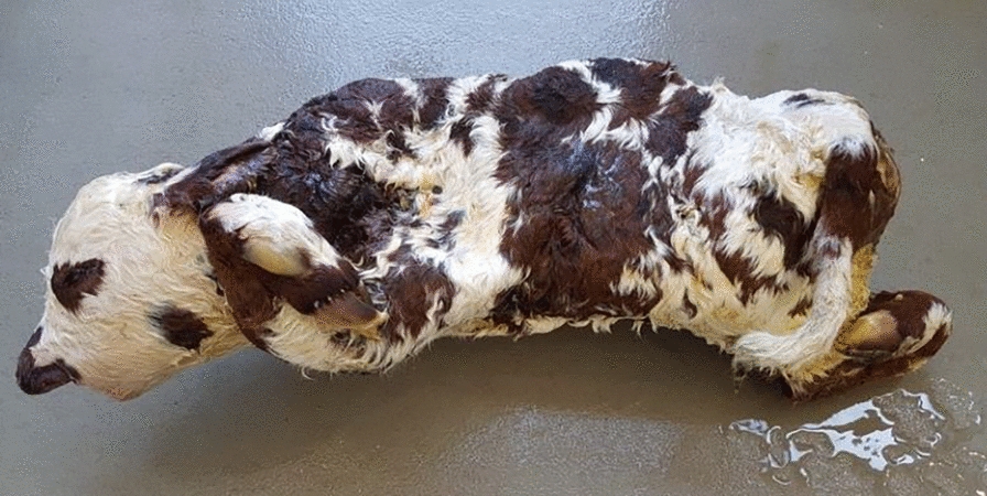
Background: Nine male and eight female calves born to a Normande artificial insemination bull named "Ly" were referred to the French National Observatory of Bovine Abnormalities for multiple fractures, shortened gestation, and stillbirth or perinatal mortality.

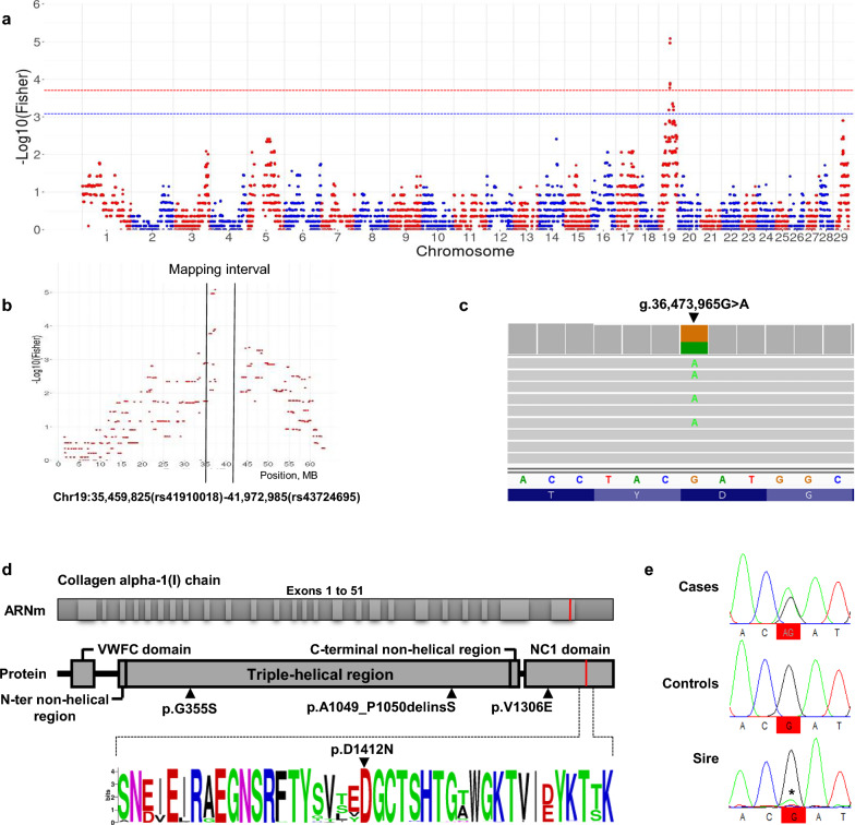
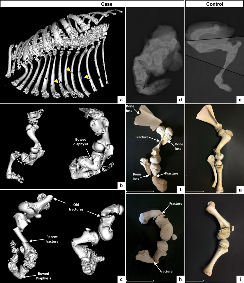
Results: Using Illumina BovineSNP50 array genotypes from affected calves and 84 half-sib controls, the associated locus was mapped to a 6.5-Mb interval on chromosome 19, assuming autosomal inheritance with germline mosaicism. Subsequent comparison of the whole-genome sequences of one case and 5116 control genomes, followed by genotyping in the affected pedigree, identified a de novo missense substitution within the NC1 domain of the COL1A1 gene (Chr19 g.36,473,965G > A; p.D1412N) as unique candidate variant. Interestingly, the affected residue was completely conserved among 243 vertebrate orthologs, and the same substitution in humans has been reported to cause type II osteogenesis imperfecta (OI), a connective tissue disorder that is characterized primarily by bone deformity and fragility. Moreover, three COL1A1 mutations have been described to cause the same syndrome in cattle. Necropsy, computed tomography, radiology, and histology confirmed the diagnosis of type II OI, further supporting the causality of this variant. In addition, a detailed analysis of gestation length and perinatal mortality in 1387 offspring of Ly and more than 160,000 progeny of 63 control bulls allowed us to statistically confirm in a large pedigree the association between type II OI and preterm delivery, which is probably due to premature rupture of fetal membranes and has been reported in several isolated cases of type II OI in humans and cattle. Finally, analysis of perinatal mortality rates and segregation distortion supported a low level of germ cell mosaicism in Ly, with an estimate of 4.5% to 7.7% of mutant sperm and thus 63 to 107 affected calves born. These numbers contrast with the 17 cases reported and raise concerns about the underreporting of congenital defects to heredo-surveillance platforms, even for textbook genetic syndromes.

Conclusions: In conclusion, we describe a large animal model for a recurrent substitution in COL1A1 that is responsible for type II OI in humans. More generally, this study highlights the utility of such datasets and large half-sib families available in livestock species to characterize sporadic genetic defects.


Conflict of interest statement

The authors declare that they have no competing interests.

Figures

Fig. 1
Fig. 1
Picture of the female “corkscrew” calf necropsied in this study. Note the limb deformities that were systematically mentioned in the reports of this defect to the ONAB (see Additional file 1: Table S1), as well as the undershot jaw (brachygnathia inferior) observed in this particular individual
Fig. 2
Fig. 2
Mapping and identification of a de novo missense mutation in the COL1A1 gene. a, b Manhattan plot of the results from transmission disequilibrium mapping, with a zoom on Bos taurus chromosome (BTA)19 (b). The dashed blue and red lines correspond to the 0.05 and 0.001 thresholds, respectively, after adjustment for multiple testing. c Integrative Genomic Viewer screenshot showing heterozygosity for a substitution at position g.36,473,965 of BTA19 in the whole-genome sequence of an affected calf. (d, top) Exon and domain information for the COL1A1 gene and corresponding protein. The position of the p.D1412N substitution caused by the g.36,473,965G > A variant identified in this article and three previously described deleterious mutations in COL1A1 in cattle are indicated on the protein (p.A1049_P1050delinsS in Bourneuf et al. [9]), p.G355S in Petersen et al. [10]), and p.V1306E in Jacinto et al.,[11]. (d, bottom) Logo representation of a multi-species alignment of 243 orthologs showing perfect conservation of the aspartic acid at position 1412 in cattle. Information on the protein ID in Ensembl and the amino acid sequences are provided in Additional file 3: Table S3. e Electropherograms of an affected calf, a control half-sib carrying the at-risk haplotype in its ancestral version (i.e., without the mutation), and their sire Ly for a segment of BTA19 encompassing the candidate variant. *Note the low proportion of allele A versus G in Ly supporting germline mosaicism. Electrophoregrams for a larger window are provided in Additional file 4: Figure S1
Fig. 3
Fig. 3
Computed tomography, radiography and partial skeletal reconstruction of a “corkscrew” calf and a matched control. ac 3D reconstruction of CT images of the affected calf. Detail of the thoracic cavity (a), left thoracic limb in lateral view (b, left), right thoracic limb in medial view (b, right), left pelvic limb in lateral view (c, left) and right pelvic limb in medial view (c, right). Note the presence of multiple chronic and recent fractures and abnormal curvature of the diaphysis of several long bones. Yellow arrows highlight three of the multiple rib fractures seen in panel (a). d, e Radiographs of the posterior limbs of the affected (d) and control (f) calves. Fracture lines are highlighted. Note also the generalized decrease in bone opacity and cortical thinning, i.e., osteopenia, in the case compared to the control. fi Partially reconstructed skeletons of the thoracic (upper panels) and pelvic (lower panels) limbs of the case (f, h) and control (g, i) calves, showing loss of bone substance, fractures, and bone remodeling in the affected calf. Scale bars correspond to 10 cm and their variation in size highlights the shortening of the limbs in the case compared to the control
Fig. 4
Fig. 4
Macroscopic and microscopic structures of the bones of a “corkscrew” calf and a matched control. Macroscopic section of the proximal region of the femur (left panel) and distal region of the tibia (right panel) from the case (a) and control (b) calves. Note the absence of cortical bone and irregular spongiosa (i.e., cancellous bone). Histological sections of the femoral lamellar bone from affected (c) and control (d) calves stained with hematoxylin and eosin (HE). Scale bars in c and d correspond to 200 µm
Fig. 5
Fig. 5
Analysis of the gestation length and perinatal mortality. Distribution of gestation lengths for the 17 reported cases of type II OI and for the calves that died or survived within 48 h of birth in the progeny of Ly or 63 control bulls

References

    1. Sillence DO, Senn A, Danks DM. Genetic heterogeneity in osteogenesis imperfecta. J Med Genet. 1979;16:101–116. doi: 10.1136/jmg.16.2.101. - DOI - PMC - PubMed
    1. Rauch F, Glorieux FH. Osteogenesis imperfecta. Lancet. 2004;363:1377–1385. doi: 10.1016/S0140-6736(04)16051-0. - DOI - PubMed
    1. Marini JC, Forlino A, Cabral WA, Barnes AM, San Antonio JD, Milgrom S, et al. Consortium for osteogenesis imperfecta mutations in the helical domain of type I collagen: regions rich in lethal mutations align with collagen binding sites for integrins and proteoglycans. Hum Mutat. 2007;28:209–221. doi: 10.1002/humu.20429. - DOI - PMC - PubMed
    1. Forlino A, Cabral WA, Barnes AM, Marini JC. New perspectives on osteogenesis imperfecta. Nat Rev Endocrinol. 2011;7:540–557. doi: 10.1038/nrendo.2011.81. - DOI - PMC - PubMed
    1. Valadares ER, Carneiro TB, Santos PM, Oliveira AC, Zabel B. What is new in genetics and osteogenesis imperfecta classification? J Pediatr (Rio J) 2014;90:536–541. doi: 10.1016/j.jped.2014.05.003. - DOI - PubMed