

Brain perfusion changes in beta-thalassemia
- PMID: 38773534
- PMCID: PMC11110312
- DOI: 10.1186/s13023-024-03194-x
Brain perfusion changes in beta-thalassemia
Abstract
Background: Brain injury in hereditary hemoglobinopathies is commonly attributed to anemia-related relative hypoperfusion in terms of impaired oxygen blood supply. Supratentorial and infratentorial vascular watershed regions seem to be especially vulnerable, but data are very scarce.
Aims: We investigated a large beta-thalassemia sample with arterial spin labeling in order to characterize regional perfusion changes and their correlation with phenotype and anemia severity.
Methods: We performed a multicenter single-scanner cross-sectional 3T-MRI study analyzing non-invasively the brain perfusion in 54 transfusion-dependent thalassemia (TDT), 23 non-transfusion-dependent thalassemia (NTDT) patients and 56 Healthy Controls (HC). Age, hemoglobin levels, and cognitive functioning were recorded.
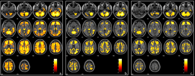
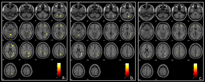
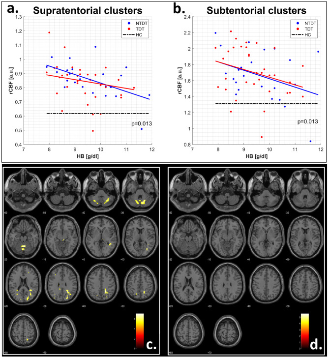
Results: Both TDT and NTDT patients showed globally increased brain perfusion values compared to healthy controls, while no difference was found between patient subgroups. Using age and sex as covariates and scaling the perfusion maps for the global cerebral blood flow, beta-thalassemia patients showed relative hyperperfusion in supratentorial/infratentorial watershed regions. Perfusion changes correlated with hemoglobin levels (p = 0.013) and were not observed in the less severely anemic patients (hemoglobin level > 9.5 g/dL). In the hyperperfused regions, white matter density was significantly decreased (p = 0.0003) in both patient subgroups vs. HC. In NTDT, white matter density changes correlated inversely with full-scale Intelligence Quotient (p = 0.007) while in TDT no correlation was found.
Conclusion: Relative hyperperfusion of watershed territories represents a hemodynamic hallmark of beta-thalassemia anemia challenging previous hypotheses of brain injury in hereditary anemias. A careful management of anemia severity might be crucial for preventing structural white matter changes and subsequent long-term cognitive impairment.
Keywords: Brain; Hemoglobin; Perfusion; Thalassemia; Transfusions.
© 2024. The Author(s).
Conflict of interest statement
The authors declare no conflict of interest.
Figures
References
-
- Tartaglione I, Manara R, Caiazza M, Carafa PA, Caserta V, Ferrantino T, et al. Brain functional impairment in beta-thalassaemia: the cognitive profile in Italian neurologically asymptomatic adult patients in comparison to the reported literature. Br J Haematol. 2019;186:592–607. doi: 10.1111/bjh.15959. - DOI - PubMed
-
- Cappellini MD, Farmakis D, Porter J, Taher A et al. Guidelines for the management of Transfusion Dependent Thalassemia (4th edition - version 2.0). Thalassemia International Federation. 2021.
-
- Tartaglione I, Russo C, Elefante A, Caiazza M, Casale M, Di Concilio R, et al. No evidence of increased cerebrovascular involvement in adult neurologically-asymptomatic β-Thalassaemia. A multicentre multimodal magnetic resonance study. Br J Haematol. 2019;185:733–42. doi: 10.1111/bjh.15834. - DOI - PubMed
MeSH terms
Grants and funding
LinkOut - more resources
Full Text Sources
Medical
