. 2024 May 21;22(1):482.
doi: 10.1186/s12967-024-05254-z.

Unraveling resistance mechanisms in anti-CD19 chimeric antigen receptor-T therapy for B-ALL: a novel in vitro model and insights into target antigen dynamics

Affiliations

Unraveling resistance mechanisms in anti-CD19 chimeric antigen receptor-T therapy for B-ALL: a novel in vitro model and insights into target antigen dynamics

Hongzhe Li et al. J Transl Med. .

Abstract

Background: Cellular immunotherapy, represented by the chimeric antigen receptor T cell (CAR-T), has exhibited high response rates, durable remission, and safety in vitro and in clinical trials. Unfortunately, anti-CD19 CAR-T (CART-19) treatment alone is prone to relapse and has a particularly poor prognosis in relapsed/refractory (r/r) B-ALL patients. To date, addressing or reducing relapse remains one of the research priorities to achieve broad clinical application.

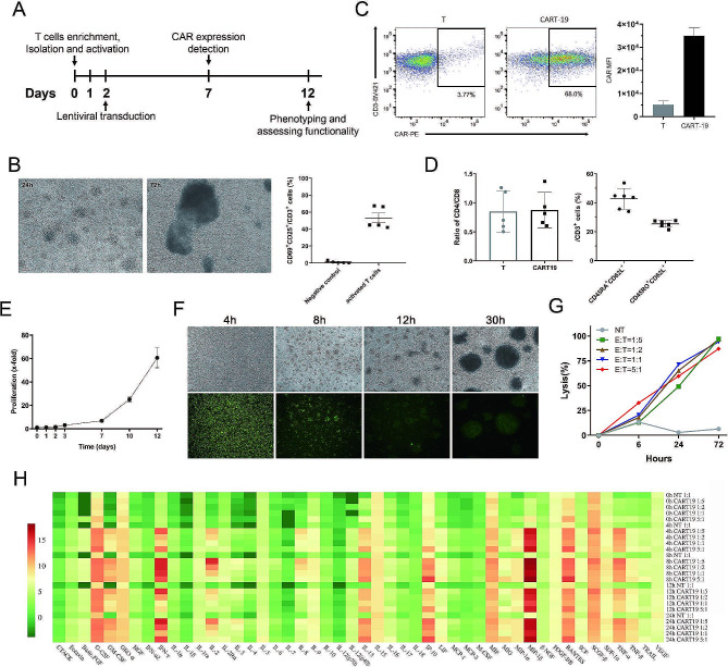
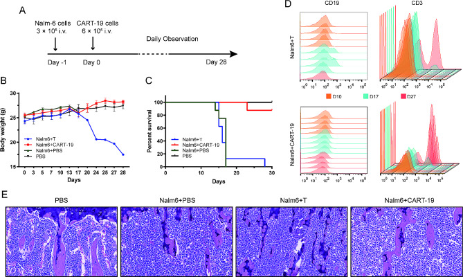
Methods: We manufactured second generation CART-19 cells and validated their efficacy and safety in vitro and in vivo. Through co-culture of Nalm-6 cells with short-term cultured CART-19 cells, CD19-negative Nalm-6 cells were detected by flow cytometry, and further investigation of the relapsed cells and their resistance mechanisms was evaluated in vitro.

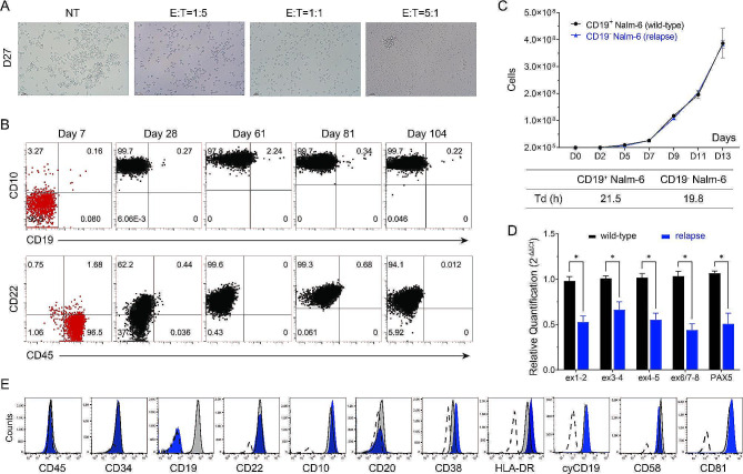
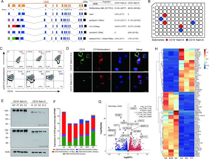
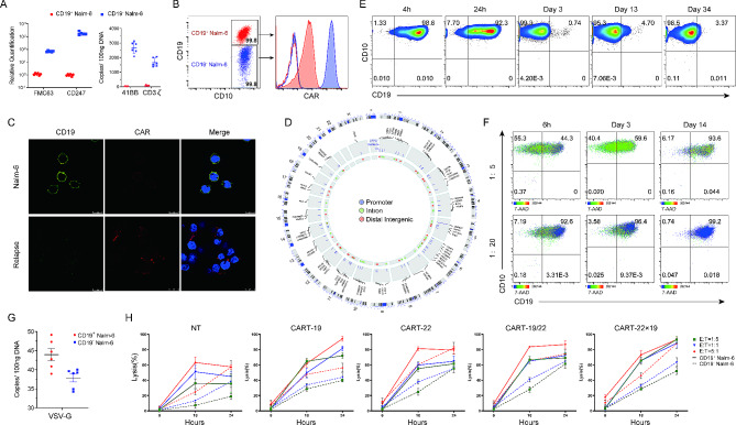
Results: In this study, we demonstrated that CART-19 cells had enhanced and specific antileukemic activities, and the survival of B-ALL mouse models after CART-19 treatment was significantly prolonged. We then shortened the culture time and applied the serum-free culture to expand CAR-T cells, followed by co-culturing CART-19 cells with Nalm-6 cells. Surprisingly, we observed the proliferation of CD19-negative Nalm-6 cells around 28 days. Identification of potential resistance mechanisms showed that the relapsed cells express truncated CD19 proteins with decreased levels and, more importantly, CAR expression was detected on the relapsed cell surface, which may ultimately keep them antigen-negative. Furthermore, it was validated that CART-22 and tandem CART-22/19 cells could effectively kill the relapsed cells, but neither could completely eradicate them.

Conclusions: We successfully generated CART-19 cells and obtained a CD19-negative refractory relapsed B-ALL cell line, providing new insights into the underlying mechanisms of resistance and a new in vitro model for the treatment of r/r B-ALL patients with low antigen density.

Keywords: Antigen negative relapse; B-ALL; CAR-T cell therapy; CD19; Resistant mechanisms.


Conflict of interest statement

The authors declare that they have no conflict of interest.

Figures

Fig. 1
Fig. 1
Proliferation and specific cytotoxic effects of CART-19 cells. A The design of the CAR-T cell construction experiments. B Morphological images of activated T cells clustered after 24 h and 72 h of incubation with TransAct CD3/28 beads. C Flow cytometric analysis of CAR expression on the surface of mock T, and CART-19 cells with biotin-conjugated anti-Fab antibody followed by PE-conjugated streptavidin. Gating was based on the same cells stained with isotype-matched antibody. The median fluorescence intensity (MFI) was calculated for CAR-T population in the PE fluorescence channel (right column). This result is the representative of three separate experiments using cells from healthy volunteer donors. D The phenotypic characterization of CART-19 cells by flow cytometry. The ratio of CD4+ / CD8+ T cells (left) and the proportion of TN/CM (right) are shown. E Growth curves of CAR-T cells. Data represent the mean ± s.d. of three separate experiments. F Cytolytic activities of CART-19 cells in cell assays. Nalm-6 cells were labeled with CFSE labeling reagent (Sigma-Aldrich, USA) and co-cultured with CART-19 cells at the E: T ratio of 1:1 for 30 h. The presence of CFSE-labeled cells was observed by microscopy. Bar, 100 μm. G Cytotoxic activity of mock NT and CART cells against Nalm-6 cells. The effector cells were co-cultured with target cells at E: T ratios of 1:5, 1:2, 1:1 and 5:1 with a total cell number of 1 × 106. H Dynamic changes of cytokine secretion profile of CART-19 cells during 24 h after co-culture with Nalm-6 cells at E: T ratios of 1:5 to 5:1. Data were visualized by heatmap. Concentrations (pg/ml) of cytokines and chemokines in the supernatant were detected by multiplex immunoassay and the values were log2 transformed
Fig. 2
Fig. 2
In vivo functional evaluation of CART-19 cells. A Model system and experimental regimen. NSG mice were injected with PBS (PBS group, n = 4) or Nalm-6 cells on day − 1. One day after tumor implantation, mice were randomized to receive PBS (Nalm6 + PBS group, n = 8), untreated mock T cells (Nalm6 + T group, n = 8), or CART-19 cells (Nalm6 + CART-19 group, n = 8). Animals were monitored daily after tail vein injection. B, C Body weight (B) and survival curves (C) of mice from (A). Values represent mean ± s.e.m. D Persistence of tumor and CAR-T cells from blood of treated mice in the Nalm6 + T and Nalm6 + CART-19 groups by flow cytometry analysis. Tumor cells were identified as CD19+ cells and T cells were identified as CD3+ cells. Representative flow cytometric data from four mice of each group are shown. E Representative morphological images of bone marrow from mice at the end of study. The final magnification of all images was 40×
Fig. 3
Fig. 3
Expansion and phenotypic identification of antigen-negative relapsed cells following CART-19 treatment. A Morphology of CART-19 cells and Nalm-6 cells co-cultured in vitro for 27 days. The CART-19 cells were expanded in the serum-free TexMACS Medium-TransAct-IL-2 culture system. B Long-term immunophenotyping of co-cultured cells of CART-19 and Nalm-6 cells (E: T = 1:5) by flow cytometry. The co-culture cells were marked in red on day 7, while other days of the experiment were marked in black. In vitro proliferation (line) and doubling time analysis (table) of relapsed and wild-type Nalm-6 cells. D qRT-PCR analysis of the expression of different regions of CD19 mRNA and PAX5 mRNA in relapsed CD19 Nalm-6 samples. CD19+ wild-type Nalm-6 cells were applied as the control group while using ACTB as an internal reference, and graphs show mean ± s.e.m. of relative expression levels. *p < 0.05 indicates a significant difference. Data are representative of three independent experiments. E Flow cytometric profiles of immunophenotypes of relapsed CD19 Nalm-6 cells (blue) compared to CD19+ wildtype Nalm-6 cells (grey) after 317 days of continuous culture in vitro. The results of the isotype control are shown (dashed line)
Fig. 4
Fig. 4
Transcription and expression of CD19 gene in the relapsed Nalm-6 cells. A Detection of CD19 transcripts in relapsed CD19 Nalm-6 and wildtype Nalm-6 cells by TA cloning and sequencing. Sequences were compared to the reference sequence gene (NM_001770.6) and visualized by Exon-Intron Graphic Maker from WormWeb.org. Six alternatively spliced isoforms of CD19 mRNA were obtained and the percentages of them were shown separately, including the full-length (FL), skipping exon 2 (Δex2), partial deletion (partΔex2 and partΔex3), retention of intron 2 (in2) and combination of partΔex2 and in2. Of these, partΔex2, in2 and partΔex2 + in2 shift the reading frame, predicting that the expressed CD19 protein will be divided into three shortened proteins (purple, red and green) and a truncated CD19 protein (blue). B Schematic demonstration of single-cell purification of wild-type Nalm-6 cells (red) and relapsed CD19 Nalm-6 cells (blue) by the limiting dilution method. C Flow cytometric profiles of membrane CD19 and cytosolic CD19 in subclones of Nalm-6 (red) and relapsed CD19 Nalm-6 cells (blue). D Fluorescence microscopy of CD19 expression on the surface and cytoplasm using two anti-CD19 antibodies (HIB19, extracellular, green; D4V4B, intracellular, red) and DAPI (blue). E Immunoblotting for CD19 in protein lysates from wild-type Nalm-6 cells and relapsed CD19 Nalm-6 cells using antibodies recognizing the extracellular domain (clone OTI5F3 from Origene, N-terminal) or the cytosolic domain (clone D4V4B from Cell Signaling, intracellular domain). F Expression of CD19 gene transcripts in RNA sequencing data from wild-type Nalm-6 and relapsed Nalm-6 subclones. G, H Volcano (G) and heatmap (H) plots demonstrating changes in differential gene expression of subclones measured by RNA sequencing. Differentially expressed gene (DEG) analysis identified 3453 DEGs with |log2Fc| ≥ 1 and p value < 0.05, and the heatmap for top 75 DEGs is shown
Fig. 5
Fig. 5
Observation of CD19-BBζ-CAR expression in relapsed Nalm-6 cells and salvage treatment. A Detection of FMC63 and CD247 transcripts and 4-1BB gene of CAR in CD19+ Nalm-6 (red) and relapsed CD19 Nalm-6 cells (blue) by qRT-PCR. Data of left bar graph represent the relative quantification using ACTB as the internal reference. Error bars represent s.d. The data are the representative of three independent experiments. B Expression of CD19 and CAR on CD19+ Nalm-6 cells and relapsed CD19 Nalm-6 cells analyzed by flow cytometry (representative of 3 experiments). Merge Graphs, the blue dots represent CD19 Nalm-6 cells and the red dots represent Nalm-6 cells. C Confocal imaging of Nalm-6 cells and relapsed CD19 Nalm-6 cells using Alexa Flour 488-conjugated anti-CD19 antibody (green), Alexa Flour 647-conjugated anti-CAR19 antibody (red), and DAPI (blue). D Lentiviral integration sites of CAR transduced Nalm-6 cells were analyzed by linear-amplification mediated PCR (LAM-PCR) and visualized with Circos plots. The integration sites across the genome and genomic features were shown from outer to inner circle: (1) cytogenetic bands; (2) genes that harbor these integration sites along with a bar chart showing the reads of integration sites; (3) the distribution of integration sites, with colored circles representing different gene functional regions of the host sequence: purple for promoter region, green for intron region, and red for distal intergenic region. E Phenotype changes of Nalm-6 cells transduced with small amount of CD19 CAR lentiviruses detected by flow cytometry over time. Gating was based on the same cells stained with isotype-matched antibody. F Dynamics of CD19 B phenotype in relapsed cells after co-culture with different ratios (5×, 20×) of Nalm-6 cells. Gating was based on the same cells stained with isotype-matched antibody. G Relapsed CD19 Nalm-6 cells were tested by qPCR specific for VSV-G sequence. H Comparison of in vitro efficacy of CD19-, CD22-, CD19/CD22- and CD22×CD19- CAR T cells. Cocultures with the relapsed cells were performed at 1:5, 1:1, and 5:1 E: T ratios, and lysis efficacies were detected by the LDH release assay Declarations

Similar articles

References

    1. Arber DA, Orazi A, Hasserjian R, Thiele J, Borowitz MJ, Le Beau MM, Bloomfield CD, Cazzola M, Vardiman JW. The 2016 revision to the World Health Organization classification of myeloid neoplasms and acute leukemia. Blood. 2016;127(20):2391–405. doi: 10.1182/blood-2016-03-643544. - DOI - PubMed
    1. Bhojwani D, Yang JJ, Pui CH. Biology of childhood acute lymphoblastic leukemia. Pediatr Clin North Am. 2015;62(1):47–60. doi: 10.1016/j.pcl.2014.09.004. - DOI - PMC - PubMed
    1. Inaba H, Mullighan CG. Pediatric acute lymphoblastic leukemia. Haematologica. 2020;105(11):2524–39. doi: 10.3324/haematol.2020.247031. - DOI - PMC - PubMed
    1. Hunger SP, Mullighan CG. Acute lymphoblastic leukemia in children. N Engl J Med. 2015;373(16):1541–52. doi: 10.1056/NEJMra1400972. - DOI - PubMed
    1. Inaba H, Pui CH. Immunotherapy in pediatric acute lymphoblastic leukemia. Cancer Metastasis Rev. 2019;38(4):595–610. doi: 10.1007/s10555-019-09834-0. - DOI - PMC - PubMed

Publication types

MeSH terms

LinkOut - more resources