. 2024 Sep;57(9):e13656.
doi: 10.1111/cpr.13656. Epub 2024 May 21.

Melatonin promotes hair regeneration by modulating the Wnt/β-catenin signalling pathway

Affiliations

Melatonin promotes hair regeneration by modulating the Wnt/β-catenin signalling pathway

Yi-Lin Niu et al. Cell Prolif. 2024 Sep.

Abstract

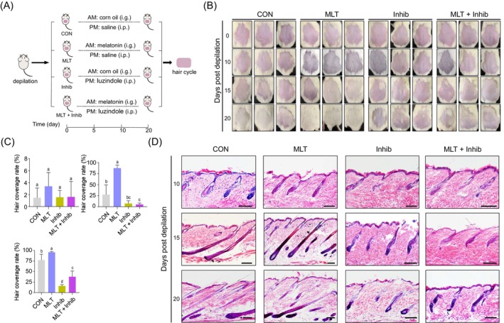
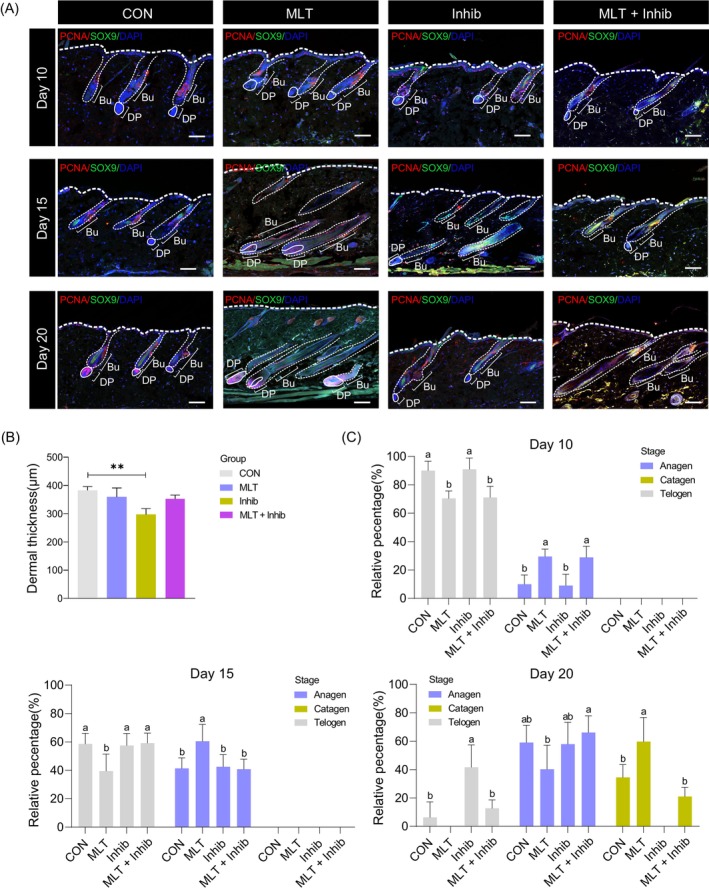
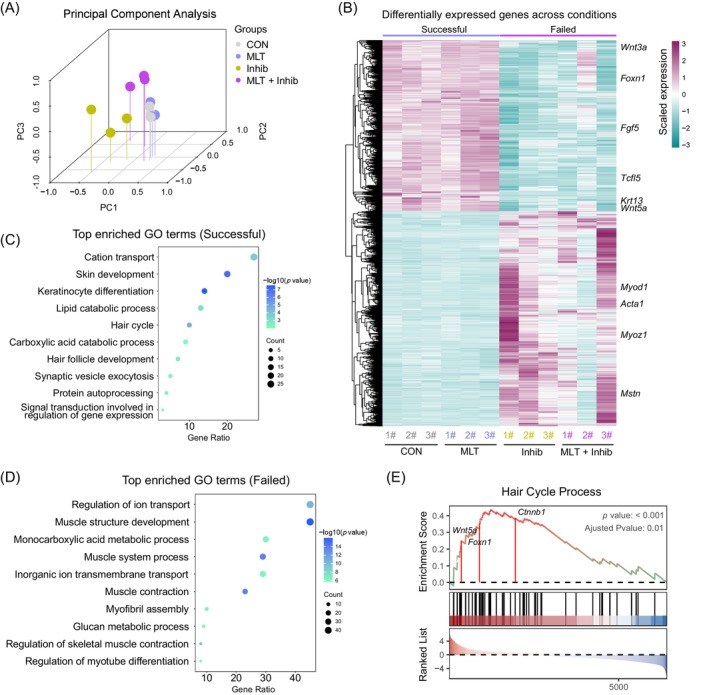
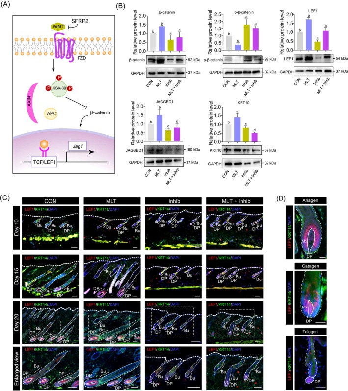
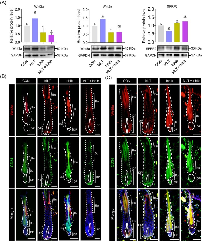
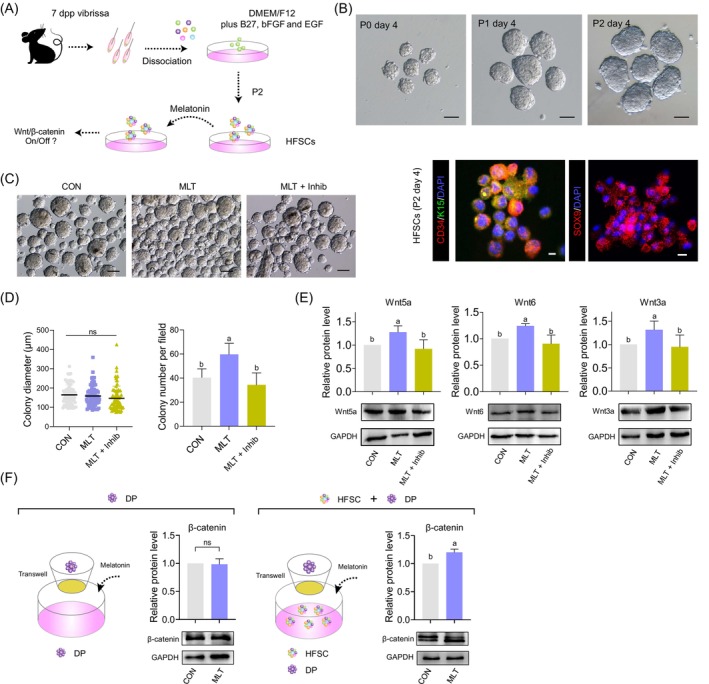
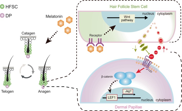
Melatonin (MLT) is a circadian hormone that reportedly influences the development and cyclic growth of secondary hair follicles; however, the mechanism of regulation remains unknown. Here, we systematically investigated the role of MLT in hair regeneration using a hair depilation mouse model. We found that MLT supplementation significantly promoted hair regeneration in the hair depilation mouse model, whereas supplementation of MLT receptor antagonist luzindole significantly suppressed hair regeneration. By analysing gene expression dynamics between the MLT group and luzindole-treated groups, we revealed that MLT supplementation significantly up-regulated Wnt/β-catenin signalling pathway-related genes. In-depth analysis of the expression of key molecules in the Wnt/β-catenin signalling pathway revealed that MLT up-regulated the Wnt/β-catenin signalling pathway in dermal papillae (DP), whereas these effects were facilitated through mediating Wnt ligand expression levels in the hair follicle stem cells (HFSCs). Using a DP-HFSCs co-culture system, we verified that MLT activated Wnt/β-catenin signalling in DPs when co-cultured with HFSCs, whereas supplementation of DP cells with MLT alone failed to activate Wnt/β-catenin signalling. In summary, our work identified a critical role for MLT in promoting hair regeneration and will have potential implications for future hair loss treatment in humans.


Conflict of interest statement

The authors declare no conflicts of interest.

Figures

FIGURE 1
FIGURE 1
Preparation and handling of mouse models. (A) The mice were randomly divided into four groups after depilation, and the treatments were divided into morning and afternoon. The groups were handled as shown, continuously for 20 days. (B) Hair coverage of mice during treatment. Mice were imaged on days 0, 10, 15 and 20. (C) Quantification of hair coverage on days 10, 15 and 20. Hair growth on the backs of all individuals was recorded. For each group, at least five mice were included for statistical analysis; statistical significance was considered at p < 0.05. (D) Haematoxylin and eosin staining of different groups of skin at 10, 15 and 20 days after depilation. Scale bars, 100 μm. i.g., intragastrical administration; i.p., intraperitioneal injection.
FIGURE 2
FIGURE 2
Immunofluorescence of the skin and statistics of the hair follicle stage. (A) Immunofluorescence co‐staining of PCNA and SOX9 on skin samples of different groups at 10, 15 and 20 days after depilation. Scale bars, 100 μm. (B) Statistics of dermal layer thickness, **p < 0.01. (C) Statistical analysis of hair follicle developmental stage in different groups at 10, 15 and 20 days after depilation. For each group, at least four mice were included for statistical analysis. Bu, bulge; DP, dermal papillae.
FIGURE 3
FIGURE 3
Transcriptome analysis of skin tissues from different groups. (A) The principal component analysis (PCA) plot of different groups at 20 days after depilation. (B) Heatmap showing the top 200 differentially expressed genes on day 20 skin between the Successful group and the Failed group. Control group (CON) and melatonin (MLT) groups were placed into the Successful group, while the MLT receptor inhibitor (Inhib) and negative control (MLT + Inhib) groups were assigned to the Failed group, based on hair growth status. (C) Gene Ontology (GO) enrichment results of up‐regulated genes in the Successful group. (D) GO enrichment results of up‐regulated genes in the Failed group. (E) The gene set enrichment analysis (GSEA) plot illustrating the enrichment of hair cycle process‐related genes between the Successful group and the Failed group.
FIGURE 4
FIGURE 4
Wnt/β‐catenin signalling pathway regulates hair regeneration. (A) Schematic diagram illustrating how Wnt ligands activate the Wnt/β‐catenin signalling pathway. (B) Western blot showing β‐catenin, p‐β‐catenin, LEF1, JAGGED1 and keratin‐10 (KRT10) expression levels in skin tissues of different groups at 20 days after depilation. (C) Immunofluorescence co‐staining of LEF1 and cytokeratin‐14 (KRT14) in skin samples from different groups. The magnified view at the bottom corresponds to the area indicated by the white dashed rectangle on the 20th day after depilation. Scale bars, 100 μm. (D) Localization of LEF1 at different stages of the hair cycle; the solid lines indicate the dermal papillae (DP) region. Scale bars, 50 μm. FZD, frizzled receptor; APC, adenomatous polyposis coli; GSK‐3β, glycogen synthase kinase; Bu, bulge; Mx, matrix.
FIGURE 5
FIGURE 5
Expression levels and location of Wnt ligands. (A) The protein levels of Wnt3a, Wnt5a and SFRP2 in different groups at 20 days after depilation. (B) Immunofluorescence co‐staining of Wnt3a and CD34 of skin samples at 20 days after depilation. Scale bars, 50 μm. (C) Immunofluorescence co‐staining of Wnt5a and CD34 of skin samples at 20 days after depilation. Scale bars, 50 μm. Bu, bulge; DP, dermal papillae.
FIGURE 6
FIGURE 6
The impact of melatonin (MLT) on the interaction between hair follicle stem cells (HFSCs) and dermal papillae (DP). (A) Schematic diagram illustrating the major steps in hair follicle stem cells isolation and culture in vitro. (B) Top panel: The morphology of colonies formed by HFSCs cultured for 4 days at passages P0, P1 and P2. Scale bars, 100 μm. Bottom panel: Immunofluorescence staining of CD34, keratin‐15 (K15) and SOX9 of HFSCs cultured for 4 days at P2. Scale bars, 10 μm. (C) The morphology of colonies formed by HFSCs cultured for 4 days at passage P2 after MLT and luzindole exposure. Scale bars, 100 μm. (D) Statistical analysis of colony diameter and colony number per field after MLT and luzindole exposure. (E) Western blotting and quantification of Wnt5a, Wnt6 and Wnt3a levels in the HFSCs in different groups. (F) Western blot illustrating the effects of MLT on the expression level of β‐catenin in dermal papillae (DP) cells based on a DP‐HFSCs Transwell co‐culture system. dpp, days post partum.
FIGURE 7
FIGURE 7
A schematic diagram illustrating how melatonin (MLT) activates the Wnt/β‐catenin signalling pathway in dermal papillae (DP) cells by promoting the release of Wnt ligands from hair follicle stem cells (HFSCs), ultimately leading to enhanced hair regeneration.

Similar articles

Cited by

References

    1. Schmidt‐Ullrich R, Paus R. Molecular principles of hair follicle induction and morphogenesis. Bioessays. 2005;27(3):247‐261. - PubMed
    1. Rishikaysh P, Dev K, Diaz D, Qureshi WM, Filip S, Mokry J. Signaling involved in hair follicle morphogenesis and development. Int J Mol Sci. 2014;15(1):1647‐1670. - PMC - PubMed
    1. Stenn KS, Paus R. Controls of hair follicle cycling. Physiol Rev. 2001;81(1):449‐494. - PubMed
    1. Li S, Chen J, Chen F, et al. Liposomal honokiol promotes hair growth via activating Wnt3a/beta‐catenin signaling pathway and down regulating TGF‐beta1 in C57BL/6N mice. Biomed Pharmacother. 2021;141:111793. - PubMed
    1. Geyfman M, Plikus MV, Treffeisen E, Andersen B, Paus R. Resting no more: re‐defining telogen, the maintenance stage of the hair growth cycle. Biol Rev Camb Philos Soc. 2015;90(4):1179‐1196. - PMC - PubMed

LinkOut - more resources