. 2024 May 8;10(10):e30743.
doi: 10.1016/j.heliyon.2024.e30743. eCollection 2024 May 30.

Formulation optimization of lyophilized aptamer-gold nanoparticles: Maintained colloidal stability and cellular uptake

Affiliations

Formulation optimization of lyophilized aptamer-gold nanoparticles: Maintained colloidal stability and cellular uptake

Dalya Saidi et al. Heliyon. .

Abstract

Anti-nucleolin (NCL) aptamer AS1411 is the first anticancer aptamer tested in clinical trials. Gold nanoparticles (AuNP) have been widely exploited for various biomedical applications due to their unique functional properties. In this study, we evaluated the colloidal stability and targeting capacity of AS1411-funtionalized AuNP (AuNP/NCL-Apt) against MCF-7 breast cancer cell line before and after lyophilization. Trehalose, mannitol, and sucrose at various concentrations were evaluated to determine their cryoprotection effects. Our results indicate that sucrose at 10 % (w/v) exhibits the best cryoprotection effect and minimal AuNP/NCL-Apt aggregation as confirmed by UV-Vis spectroscopy and dynamic light scattering (DLS) measurements. Moreover, the lyophilized AuNP/NCL-Apt at optimized formulation maintained its targeting and cytotoxic functionality against MCF-7 cells as proven by the cellular uptake assays utilizing flow cytometry and confocal laser scanning microscopy (CLSM). Quantitative PCR (qPCR) analysis of nucleolin-target gene expression also confirmed the effectiveness of AuNP/NCL-Apt. This study highlights the importance of selecting the proper type and concentration of cryoprotectant in the typical nanoparticle lyophilization process and contributes to our understanding of the physical and biological properties of functionalized nanoparticles upon lyophilization.

Keywords: Aggregation; Aptamer; Cryoprotectants; Gold nanoparticles; Lyophilization.


Conflict of interest statement

The authors declare that they have no known competing financial interests or personal relationships that could have appeared to influence the work reported in this paper.

Figures

Fig. 1
Fig. 1
Characteristics of AuNP and AuNP/NCL-APT preperations. (A) Representative TEM images of Citrate-capped AuNP (top). (Inset: Higher magnification with scale bar = 100 nm) and AuNP/NCL-APT (bottom) (B) A schematic view of AS1411 aptamer conjugation to a gold nanoparticle. (C) A representative photograph of AuNP suspension (left vial) and AuNP/NCL-APT suspension (right vial).(D) Absorption spectra of AuNP and AuNP/NCL-APT suspensions as labeled. (E) A ssDNA standard graph of the recommended standards (400, 200, 100, 50, 25, 12.5, 6.25 ng/well) and an AuNPs/NCL-APT sample. The values represent mean ± SD of 3 independent experiments. (For interpretation of the references to color in this figure legend, the reader is referred to the Web version of this article.)
Fig. 2
Fig. 2
Assesment of the colloidal stability and cellular uptake of AuNP/NCL-APT conjugates. (A) Absorption spectra and absorption peak and (B) The visual observation of AuNPs conjugated to different concentrations of NCL-APT, and (C) Mean Fluorescent Intensity (MFI) for Alexa-Flour 647 obtained from flow cytometry analysis after treatment of MCF-7 cells with AuNP (30 nM) conjugated to Alexa-NCL-APT at different concentrations (10, 25, 50, 100 nM). The values represent mean ± SD of 3 independent experiments.
Fig. 3
Fig. 3
(A) Photographs of AuNP and AuNP/NCL-APT before lyophilization (upper panel), after lyophilization (middle panel), and after reconstitution with water (bottom panel). Lyophilization was carried out in the absence or presence of different cryoprotectants as labeled. (B) Absorption spectra and absorption peak of AuNP and AuNP/NCL-APT before lyophilization in the absence and presence of different cryoprotectants, and (C) after Lyophilization and reconstitution with water.
Fig. 4
Fig. 4
(A) Photographs of AuNP and AuNP/NCL-APT before lyophilization (upper panel), after lyophilization (middle panel), and after reconstitution with water (bottom panel). Lyophilization was carried out in the absence or presence of different concentrations of Sucrose. (B and C) Absorption spectra and absorption peak of AuNP and AuNP/NCL-APT in the absence and presence of different precents of Sucrose before lyophilization and fter lyophilization and reconstitution with water, respectively.
Fig. 5
Fig. 5
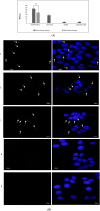
Cellular and functional analysis of AuNP/NCL-APT upon lyophilization. (A) Mean Fluorescent Intensity (MFI) of Alexa-Flour 647 for AuNP/NCL-APT before and after lyophilization with 10 % sucrose obtained from flow cytometry analysis of treated MCF-7 cells. Values represent mean ± SD of 3 independent experiments (P > 0.05). (B) Confocal images of MCF-7 cells. Cell nuclei are depicted in blue and AS1411 is depicted in red. 1) Cells treated with non-lyophilized AuNP/NCL -APT, 2) Cells treated with lyophilized AuNP/NLC -APT, 3) Cells treated with free NCL-APT.4) Cells treated with free AuNP, and 5) Untreated cells. (C) Q-RT-PCR analysis of Bcl-2 expression in MCF-7 cells treated with non-lyophilized or lyophilized AuNP/NCL-APT, free NCL-APT, and free AuNP, as compared to untreated cells. Data represent Mean ± SD of 3 independent experiments, P < 0.05. (For interpretation of the references to color in this figure legend, the reader is referred to the Web version of this article.)
Fig. 5
Fig. 5
Cellular and functional analysis of AuNP/NCL-APT upon lyophilization. (A) Mean Fluorescent Intensity (MFI) of Alexa-Flour 647 for AuNP/NCL-APT before and after lyophilization with 10 % sucrose obtained from flow cytometry analysis of treated MCF-7 cells. Values represent mean ± SD of 3 independent experiments (P > 0.05). (B) Confocal images of MCF-7 cells. Cell nuclei are depicted in blue and AS1411 is depicted in red. 1) Cells treated with non-lyophilized AuNP/NCL -APT, 2) Cells treated with lyophilized AuNP/NLC -APT, 3) Cells treated with free NCL-APT.4) Cells treated with free AuNP, and 5) Untreated cells. (C) Q-RT-PCR analysis of Bcl-2 expression in MCF-7 cells treated with non-lyophilized or lyophilized AuNP/NCL-APT, free NCL-APT, and free AuNP, as compared to untreated cells. Data represent Mean ± SD of 3 independent experiments, P < 0.05. (For interpretation of the references to color in this figure legend, the reader is referred to the Web version of this article.)

References

    1. Blair J. Making magic bullets. Nat Microbiol. 2017;2 - PubMed
    1. Farokhzad O.C., Langer R. Impact of nanotechnology on drug delivery. ACS Nano. 2009;3(1):16–20. - PubMed
    1. Alshaer W., Hillaireau H., Fattal E. Aptamer-guided nanomedicines for anticancer drug delivery. Adv. Drug Deliv. Rev. 2018;134:122–137. - PubMed
    1. Mitchell M.J., et al. Engineering precision nanoparticles for drug delivery. Nat. Rev. Drug Discov. 2021;20(2):101–124. - PMC - PubMed
    1. Alkilany A.M., et al. Ligand density on nanoparticles: a parameter with critical impact on nanomedicine. Adv. Drug Deliv. Rev. 2019;143:22–36. - PubMed

LinkOut - more resources