. 2024 May 7:14:1369301.
doi: 10.3389/fcimb.2024.1369301. eCollection 2024.

Pleiotropic roles of LAMMER kinase, Lkh1 in stress responses and virulence of Cryptococcus neoformans

Affiliations

Pleiotropic roles of LAMMER kinase, Lkh1 in stress responses and virulence of Cryptococcus neoformans

Sunhak Kwon et al. Front Cell Infect Microbiol. .

Abstract

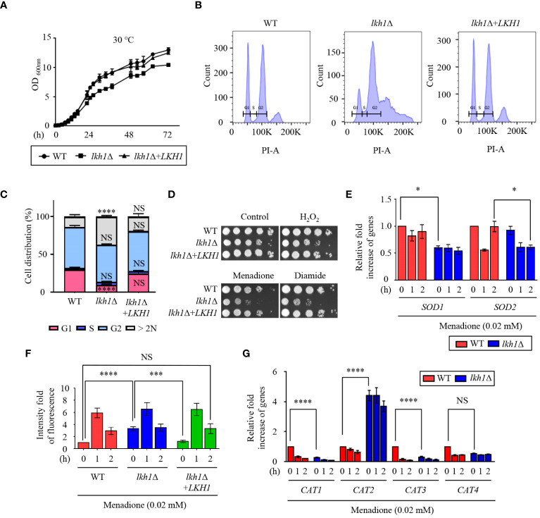
Dual-specificity LAMMER kinases are highly evolutionarily conserved in eukaryotes and play pivotal roles in diverse physiological processes, such as growth, differentiation, and stress responses. Although the functions of LAMMER kinase in fungal pathogens in pathogenicity and stress responses have been characterized, its role in Cryptococcus neoformans, a human fungal pathogen and a model yeast of basidiomycetes, remains elusive. In this study, we identified a LKH1 homologous gene and constructed a strain with a deleted LKH1 and a complemented strain. Similar to other fungi, the lkh1Δ mutant showed intrinsic growth defects. We observed that C. neoformans Lkh1 was involved in diverse stress responses, including oxidative stress and cell wall stress. Particularly, Lkh1 regulates DNA damage responses in Rad53-dependent and -independent manners. Furthermore, the absence of LKH1 reduced basidiospore formation. Our observations indicate that Lkh1 becomes hyperphosphorylated upon treatment with rapamycin, a TOR protein inhibitor. Notably, LKH1 deletion led to defects in melanin synthesis and capsule formation. Furthermore, we found that the deletion of LKH1 led to the avirulence of C. neoformans in a systemic cryptococcosis murine model. Taken together, Lkh1 is required for the stress response, sexual differentiation, and virulence of C. neoformans.

Keywords: Cryptococcus; LAMMER kinase; antifungal drug resistance; stress response; virulence.


Conflict of interest statement

The authors declare that the research was conducted in the absence of any commercial or financial relationships that could be construed as a potential conflict of interest. The author(s) declared that they were an editorial board member of Frontiers, at the time of submission. This had no impact on the peer review process and the final decision.

Figures

Figure 1
Figure 1
Lkh1 regulated growth and oxidative stress response. (A) WT (H99), lkh1Δ (KW1451), and lkh1Δ+LKH1 (KW1633) strains were grown in liquid YPD medium for 16 h at 30°C and subcultured in fresh liquid YPD medium at 30°C. Cell density was measured at an optical density of 600 nm every 2 (h) (B, C) Histogram of cell count in WT and lkh1Δ mutant by propidium iodide using flow cytometry. The provided images represent data from three biological replicates (B). Population proportion of cells were quantified. One-way ANOVA was used to dertermine statistical differences among strains (NS: not significant; * P < 0.05; *** P < 0.001; **** P < 0.0001) (D) WT (H99), lkh1Δ (KW1451), and lkh1Δ+LKH1 (KW1633) strains were cultured in liquid YPD medium overnight at 30°C, 10-fold serially diluted and spotted on YPD medium containing 0.02 mM menadione, 2 mM diamide, or 2 mM H2O2. (E, G) Expression levels of SOD genes and CAT genes were verified by qRT-PCR analysis using cDNA of WT (H99) and lkh1Δ strains with or without treatment of menadione (0.02 mM). Three independent biological experiments with duplicate technical replicates were performed. Error bars indicate the standard error of the mean (S.E.M). The statistical significance of the differences was determined by one-way ANOVA with Bonferroni’s multiple-comparison test. (NS: not significant; * P < 0.05; *** P < 0.001; **** P < 0.0001) (F) Intracellular ROS levels of WT, lkh1Δ, and lkh1Δ+LKH1 strains treated with or without MD (0.02 mM) were measured using H2DCFDA.
Figure 2
Figure 2
Lkh1 regulated DNA damage response. (A) WT (H99), lkh1Δ (KW1451), and lkh1Δ+LKH1 (KW1633) strains were cultured in a liquid YPD medium overnight at 30°C, 10-fold serially diluted and spotted on YPD medium containing the indicated concentration of DNA damage insults. For UV-C and γ-radiation tests, the serially diluted cells were spotted onto YPD medium and exposed to the indicated dose of UV-C (200 J/m2) or γ-radiation (2.5 kGy). Next, cells were further incubated at 30 °C and photographed daily for 2-4 days. (B) Expression levels of the BDR1 gene and Rad53-Bdr1 pathway downstream genes were verified by qRT-PCR analysis using cDNA of WT and lkh1Δ mutant with or without treating MMS (0.02%). Three independent biological experiments with duplicate technical replicates were performed. Error bars indicate S.E.M. Statistical significances of the differences were determined by one-way ANOVA with Bonferroni’s multiple-comparison test. (* P < 0.05; ** P < 0.01; *** P < 0.001; **** P < 0.0001) (C) Phosphorylation of Rad53 was monitored by analysis of the reduced electrophoretic migration using western blotting with anti-FLAG antibody. The Rad53-4×FLAG strain (YSB3806) and lkh1Δ Rad53-4×FLAG (KW1806) were grown to the mid-logarithmic phase and the cell extract was incubated at 30°C for 1 h with or without λ-phosphatase (PPase). (D) WT (H99), lkh1Δ (KW1451), rad53Δ (YSB3785), and rad53Δ lkh1Δ (KW1610 and KW1612) strains were cultured in a liquid YPD medium overnight at 30°C, 10-fold serially diluted and spotted on YPD medium containing the indicated concentration of DNA damage insults. For UV-C and γ-radiation tests, the serially diluted cells were spotted onto a YPD medium and exposed to the indicated dose of UV-C or γ-radiation. Next, cells were further incubated at 30°C and photographed daily for 2-4 days.
Figure 3
Figure 3
Lkh1 was involved in the cell wall damage stress. (A) WT (H99), lkh1Δ (KW1451), and lkh1Δ+LKH1 (KW1633) strains were cultured in a liquid YPD medium overnight at 30°C, 10-fold serially diluted and spotted on a YPD medium containing 60 mg/mL CFW, 1 mg/mL caffeine, or 1% congo-red. (B) Mpk1 phosphorylation was monitored using western blotting with an anti-phospho-Erk1 antibody. The WT (H99) and lkh1Δ strains were grown to the mid-logarithmic phase and then treated with caffeine (1 mg/mL) for 3 h. Next, total protein was extracted from each strain for immunoblot analysis. (C) Expression levels of chitin synthesis-related genes and glucan synthesis-related genes were verified by qRT-PCR analysis using cDNA of WT (H99) and lkh1Δ strains. Three independent biological experiments with duplicate technical replicates were performed. Error bars indicate S.E.M. Statistical significances of the differences were determined by one-way ANOVA with Bonferroni’s multiple-comparison test. (* P < 0.05; ** P < 0.01; **** P < 0.0001; NS: not significant) (D) WT (H99), lkh1Δ (KW1451), mpk1Δ (YSB3814), and mpk1Δ lkh1Δ (KW1858 and KW1860) strains were cultured in liquid YPD medium overnight at 30°C, 10-fold serially diluted and spotted on a YPD medium containing 1 mg/mL CFW, 0.3 mg/mL caffeine, or 0.04% congo-red. (E) Chitin staining with CFW. The quantitative fluorescence intensity of 50 individual cells of each strain were measured suing ImageJ/Fiji software. The error bar indicates the standard deviations (SD). The statistical significance of the differences was determined by one-way ANOVA using Bonferroni’s multiple-comparison test. (*** P < 0.001; **** P < 0.0001; NS: not significant).
Figure 4
Figure 4
Lkh1 was involved in antifungal drug resistance. (A, B) WT (H99), lkh1Δ (KW1451), and lkh1Δ+LKH1 (KW1633) strains were cultured in a liquid YPD medium overnight at 30°C, 10-fold serially diluted and spotted on YPD medium containing 0.02% SDS, 1.5 μg/mL amphotericin B, 14 μg/mL fluconazole. (C, E) Expression levels of ergosterol synthesis-related genes and ABC transporter genes were verified by qRT-PCR analysis using cDNA of WT (H99) and lkh1Δ strains with or without treatment of fluconazole (16 μg/mL). Three independent biological experiments with duplicate technical replicates were performed. Error bars indicate S.E.M. Statistical significances of the differences were determined by one-way ANOVA with Bonferroni’s multiple-comparison test. (* P < 0.05; ** P < 0.01; *** P < 0.001; NS: not significant) (D) The relative ergosterol contents of WT, lkh1Δ, and lkh1Δ+LKH1 strains were assessed with or without fluconazole treatment.
Figure 5
Figure 5
Lkh1 was required for basidiospores formation in C neoformans. (A) Serotype A MATα and MATa strains were co-cultured on a V8 medium (pH 5.0) for 5 weeks at room temperature in the dark: WT α × WT a (H99 and KN99), WT α × a lkh1Δ (H99 and KW1529), α lkh1Δ × a (KW1451 and KN99), α lkh1Δ × a lkh1Δ (KW1451 and KW1529), α lkh1Δ+LKH1 × a (KW1633 and KN99), and α lkh1Δ+LKH1 × a lkh1Δ (KW1633 and KW1529). The images were photographed after 18 days and 36 days. The black arrows and red arrows indicate basidium and basidiospores, respectively. (B) WT, lkh1Δ, and lkh1Δ+LKH1 strains were cultured in a liquid YPD medium, and each strain was normalized to an OD600 of 1.5. The OD600 was measured to determine flocculation at the indicated time points. The error bars indicate the standard error of the means (S.E.M). (C) The expression levels of adhesion-related genes in lkh1Δ mutant. Three independent biological experiments with duplicate technical replicates were performed. Error bars indicate the S.E.M. The statistical significance of the differences was determined by one-way ANOVA with Bonferroni’s multiple-comparison test. (NS: not significant; * P < 0.05; ** P < 0.01) (D) The same concentration of each strain was spotted on filament agar. After 5 days of incubation at room temperature, cells were washed with flowing water, followed by a subsequent incubation at 30°C for 2 days.
Figure 6
Figure 6
Phosphorylation of Lkh1 in a TOR1-dependent manner. (A) WT (H99), lkh1Δ (KW1451), and lkh1Δ+LKH1 (KW1633) strains were cultured in a liquid YPD medium overnight at 30°C, 10-fold serially diluted and spotted on a YPD medium containing rapamycin (9 μg/mL). (B–D) Lkh1 phosphorylation was monitored using western blotting with an anti-FLAG antibody. The Lkh1-4×FLAG strain (KW1755), sit4Δ Lkh1-4×FLAG strain (KW2058), and sch9Δ Lkh1-4×FLAG strain (KW2091) were grown to the mid-logarithmic phase and then treated with rapamycin (3 ng/mL) for 2 h. The cell extract was incubated at 30°C for 1 h with or without λ-phosphatase (PPase) and PPase inhibitor.
Figure 7
Figure 7
Kinase activity of Lkh1 was required for Lkh1 function. (A) Sequence alignment of the catalytic region for the kinase activity of S. cerevisiae Kns1 and C neoformans Lkh1. (B) The LKH1 expression level in the LKH1KD strain. The qRT-PCR analysis was performed with cDNA synthesized from WT (H99), lkh1Δ (KW1451), and LKH1KD (KW1958) strains grown to the mid-log phase. Error bars indicate standard deviation. The statistical significance of difference was determined by one-way analysis of variance with Bonferroni’s multiple comparison tests. (* P < 0.05 and NS: non-significant) (C) WT (H99), lkh1Δ (KW1451), lkh1Δ+LKH1 (KW1633), and LKH1KD (KW1958) strains were cultured in liquid YPD medium overnight at 30°C, 10-fold serially diluted and spotted on YPD medium containing stress-inducing agents. For the UV-C resistance test, the serially-diluted cells were spotting onto the YPD medium, and the indicated dose of UV-C was exposed. The plates were further incubated at 30°C and daily photographed for 2-4 days.
Figure 8
Figure 8
Transcriptome analysis of lkh1Δ mutant in response to rapamycin. (A) Significant differences in KEGG pathway analysis were observed between basal and rapamycin-treatment conditions (3 ng/mL) in lkh1Δ mutants. (B, C) Expression levels of genes encoding genes related to ribosome synthesis, small nuclear RNA, and amino acid transporter were verified by qRT-PCR analysis using cDNA of WT and lkh1Δ strains with or without rapamycin treatment (3 ng/mL). Three independent biological experiments with duplicate technical replicates were performed. Error bars indicate S.E.M. Statistical significances of the differences were determined by one-way ANOVA with Bonferroni’s multiple-comparison test (NS: not significant; ** P < 0.01; **** P < 0.0001). (D) Growth yield was calculated by measuring the growth curve of each strain. Each strain was grown in a minimal medium containing certain nitrogen sources (2 mM proline, 2 mM isoleucine, 2 mM lysine, and all together) Error bars indicate S.E.M. Statistical significances of the differences were determined by one-way ANOVA with Bonferroni’s multiple-comparison test. (* P < 0.05; ** P < 0.01; *** P < 0.001).
Figure 9
Figure 9
Lkh1 was involved in capsule and melanin production of C. neoformans. (A) WT (H99), lkh1Δ (KW1451), and lkh1Δ+LKH1 (KW1633) strains were spotted on L-DOPA medium (0.1%, 0.2% glucose, and without glucose) at 30°C for 3 days. (B, D) Expression levels of laccase genes (LAC1 and LAC2) and capsule-related genes (CAP10, CAP59, CAP60, and CAP64) were verified by qRT-PCR analysis using cDNA of WT and lkh1Δ mutant. Three independent biological experiments with duplicate technical replicates were performed. Error bars indicate S.E.M. Statistical significances of the differences were determined by one-way ANOVA with Bonferroni’s multiple-comparison test. (* P < 0.05; *** P < 0.001; **** P < 0.0001; NS: not significant) (C) The WT, lkh1Δ, and lkh1Δ+LKH1 strains were cultured on a Littman medium for capsule production at 30°C for 3 days. The scale bar indicates 10 μm.
Figure 10
Figure 10
Lkh1 was essential for the virulence of C neoformans. (A) C neoformans WT (H99), lkh1Δ (KW1451), and lkh1Δ+LKH1 (KW1633) strains were nasally inoculated into the 7-week-old female Balb/c mice and monitored. Survival curve of infected mice (n=7). (B) Mice were sacrificed for histochemistry at 16 dpi (n=5). (B) Comparison of lung tissue weight measurements. (C) Grocott’s methenamine silver (GMS) and Periodic Acid-Schiff (PAS) staining of lung tissues of mice sacrificed 16 days after infection. Red arrows indicate the walls of small bronchioles. Scale bar means 0.04 mm. (D) BBB translocation assays using hCMEC/D3-coated transwell. Plates with an inoculum of 105 cells of WT, lkh1Δ, lkh1 Δ+LKH1, and sit4Δ strains were incubated at 37°C in a CO2 incubator for 24 h, and the number of yeast cells passing through the hCMEC/D3-coated transwell was measured by CFU. The sit4Δ mutant was used as a negative control. The left and right Y axes indicate the migrated CFU and the trans-endothelial electrical resistance (TEER) value, respectively. Three independent biological experiments with duplicate technical replicates were performed. Error bars indicate S.E.M. Statistical significances of the differences were determined by one-way ANOVA with Bonferroni’s multiple-comparison test. (* P < 0.05; *** P < 0.001).
Figure 11
Figure 11
The proposed model of Lkh1-dependent pathway in C. neoformans. In response to extraneous stress stimuli, Lkh1 orchestrates a myriad of stress responses including oxidative stress response, preservation of cell wall integrity, DNA damage response, and modulation of amino acid metabolism via the regulation of downstream target gene expression. Additionally, Lkh1 contributes to antifungal drug resistance, mating, flocculation, adhesion, and intrinsic growth. Notably, Lkh1 plays a pivotal role in virulence by modulating the translocalization of the blood-brain barrier (BBB).

Similar articles

References

    1. Arthington-Skaggs B. A., Jradi H., Desai T., Morrison C. J. (1999). Quantitation of ergosterol content: novel method for determination of fluconazole susceptibility of Candida albicans . J. Clin. Microbiol. 37, 3332–3337. doi: 10.1128/JCM.37.10.3332-3337.1999 - DOI - PMC - PubMed
    1. Bahmed K., Bonaly R., Coulon J. (2003). Relation between cell wall chitin content and susceptibility to amphotericin B in Kluyveromyces, Candida and Schizosaccharomyces species. Res. Microbiol. 154, 215–222. doi: 10.1016/S0923-2508(03)00049-4 - DOI - PubMed
    1. Banks I. R., Specht C. A., Donlin M. J., Gerik K. J., Levitz S. M., Lodge J. K. (2005). A chitin synthase and its regulator protein are critical for chitosan production and growth of the fungal pathogen Cryptococcus neoformans . Eukaryot. Cell 4, 1902–1912. doi: 10.1128/EC.4.11.1902-1912.2005 - DOI - PMC - PubMed
    1. Bender J., Fink G. R. (1994). Afc1, A LAMMER kinase from Arabidopsis thaliana, activates ste12-dependent processes in yeast. Proc. Natl. Acad. Sci. U. S. A., 12105–12109, 91. doi: 10.1073/pnas.91.25.12105 - DOI - PMC - PubMed
    1. Chang M., Sionov E., Khanal Lamichhane A., Kwon-Chung K. J., Chang Y. C. (2018). Roles of three Cryptococcus neoformans and Cryptococcus gattii efflux pump-coding genes in response to drug treatment. Antimicrob. Agents Chemother. 62, 10.1128/Aac. 01751–17. doi: 10.1128/AAC.01751-17 - DOI - PMC - PubMed

MeSH terms