. 2024 May;29(5):050501.
doi: 10.1117/1.JBO.29.5.050501. Epub 2024 May 21.

Use of topical methylene blue to image nuclear morphometry with a low-cost scanning darkfield microendoscope

Affiliations

Use of topical methylene blue to image nuclear morphometry with a low-cost scanning darkfield microendoscope

Huayu Hou et al. J Biomed Opt. 2024 May.

Abstract

Significance: Fiber-optic microendoscopy is a promising approach to noninvasively visualize epithelial nuclear morphometry for early cancer and precancer detection. However, the broader clinical application of this approach is limited by a lack of topical contrast agents available for in vivo use.

Aim: The aim of this study was to evaluate the ability to image nuclear morphometry in vivo with a novel fiber-optic microendoscope used together with topical application of methylene blue (MB), a dye with FDA approval for use in chromoendoscopy in the gastrointestinal tract.

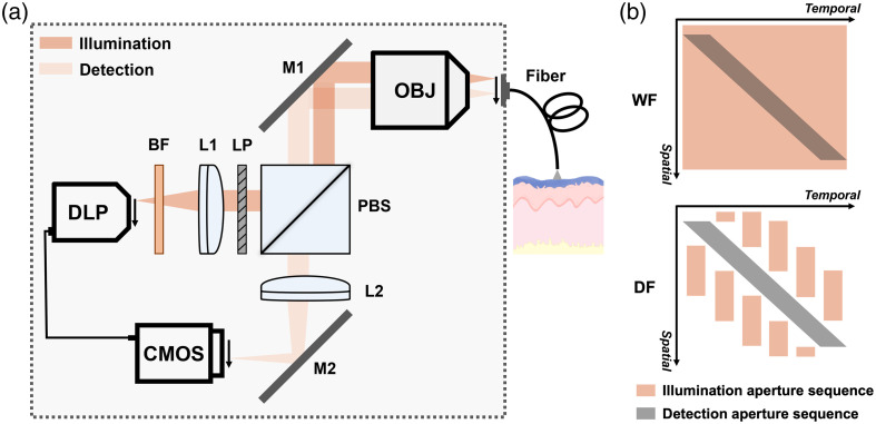
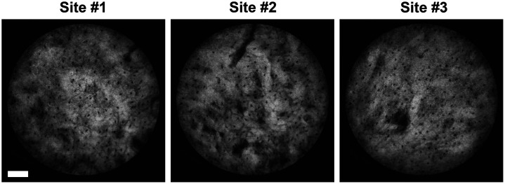
Approach: The low-cost, high-resolution microendoscope implements scanning darkfield imaging without complex optomechanical components by leveraging programmable illumination and the rolling shutter of the image sensor. We validate the integration of our system and MB staining for visualizing epithelial cell nuclei by performing ex vivo imaging on fresh animal specimens and in vivo imaging on healthy volunteers.

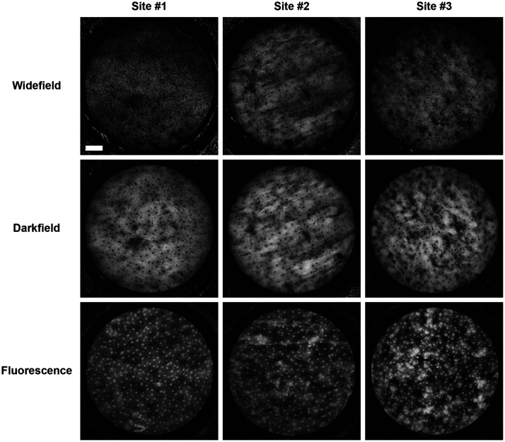
Results: The results indicate that scanning darkfield imaging significantly reduces specular reflection and resolves epithelial nuclei with enhanced image contrast and spatial resolution compared to non-scanning widefield imaging. The image quality of darkfield images with MB staining is comparable to that of fluorescence images with proflavine staining.

Conclusions: Our approach enables real-time microscopic evaluation of nuclear patterns and has the potential to be a powerful noninvasive tool for early cancer detection.

Keywords: cancer detection; fiber-optic microendoscopy; methylene blue; scanning darkfield.


Figures

Fig. 1
Fig. 1
DF-HRME implements scanning darkfield imaging to reduce internal reflection and enhance image contrast. (a) Optical schematic of DF-HRME. To enable scanning darkfield imaging, a spatiotemporal offset is introduced between illumination and detection by programming and synchronizing the DLP illumination and CMOS camera rolling shutter. Arrows show the scanning direction. A thin, flexible fiber bundle is placed in contact with tissue epithelium stained by MB for real-time imaging of cell nuclei. The dashed square indicates the optical system enclosure. (b) Spatiotemporal design of illumination and detection aperture sequences. Overlapped illumination and detection aperture in non-scanning widefield imaging captures the strong specular reflection generated at fiber surfaces, degrading image quality. In scanning darkfield imaging, a sequence of illumination line pair patterns is designed to avoid overlap with the detection aperture and suppress specular reflection background. DF-HRME, scanning darkfield high-resolution microendoscope; DLP, digital light projector; CMOS, complementary metal-oxide semiconductor camera; BF, bandpass filter; LP, linear polarizer; L1, collimating lens; L2, tube lens; PBS, polarizing beamsplitter; M1, M2, mirrors; OBJ, microscope objective; WF, non-scanning widefield imaging; DF, scanning darkfield imaging.
Fig. 2
Fig. 2
Comparison of representative images acquired from porcine tongue specimens using non-scanning widefield imaging, scanning darkfield imaging, and fluorescence imaging. The image contrast of MB stained cell nuclei is significantly improved by darkfield imaging compared to widefield imaging. Cell nuclei are clearly resolved with comparable image quality in darkfield images and corresponding fluorescence images. (Scale bar: 100  μm)
Fig. 3
Fig. 3
Representative DF-HRME images acquired in the oral cavity of healthy volunteers. The morphology of cell nuclei stained by MB is clearly resolved by in vivo DF-HRME imaging. (Scale bar: 100  μm)

References

    1. Kumar V., Abbas A. K., Aster J. C., Robbins & Cotran Pathologic Basis of Disease, 9th ed., Elsevier; (2014).
    1. Yang E. C., et al. , “Development of an integrated multimodal optical imaging system with real-time image analysis for the evaluation of oral premalignant lesions,” J. Biomed. Opt. 24(02), 025003 (2019).JBOPFO10.1117/1.JBO.24.2.025003 - DOI - PMC - PubMed
    1. Villard A., et al. , “Confocal laser endomicroscopy and confocal microscopy for head and neck cancer imaging: recent updates and future perspectives,” Oral Oncol. 127, 105826 (2022).EJCCER10.1016/j.oraloncology.2022.105826 - DOI - PubMed
    1. Wellikoff A. S., et al. , “Comparison of in vivo probe-based confocal laser endomicroscopy with histopathology in lung cancer: a move toward optical biopsy,” Respirology 20(6), 967–974 (2015).10.1111/resp.12578 - DOI - PubMed
    1. Napoléon B., et al. , “A novel approach to the diagnosis of pancreatic serous cystadenoma: needle-based confocal laser endomicroscopy,” Endoscopy 47(01), 26–32 (2014).ENDCAM10.1055/s-0034-1390693 - DOI - PubMed

Publication types