. 2024 Apr 15;21(6):1037-1048.
doi: 10.7150/ijms.94177. eCollection 2024.

METTL3 Deficiency Aggravates Hepatic Ischemia/Reperfusion Injury in Mice by Activating the MAPK Signaling Pathway

Affiliations

METTL3 Deficiency Aggravates Hepatic Ischemia/Reperfusion Injury in Mice by Activating the MAPK Signaling Pathway

Yang Gao et al. Int J Med Sci. .

Abstract

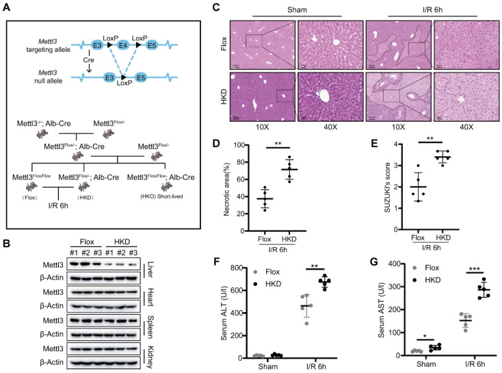
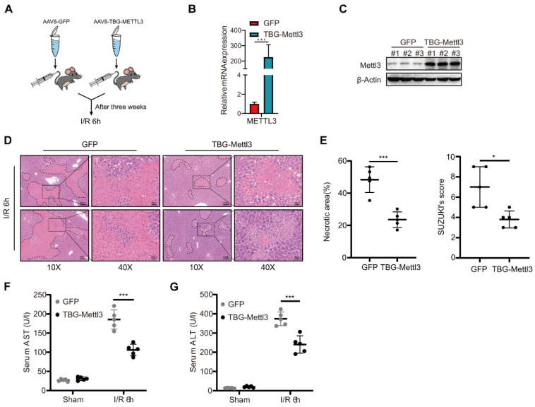
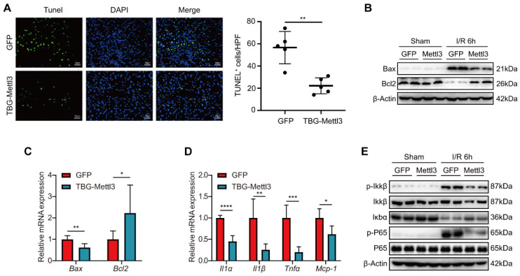
Background: Inflammatory responses, apoptosis, and oxidative stress, are key factors that contribute to hepatic ischemia/reperfusion (I/R) injury, which may lead to the failure of liver surgeries, such as hepatectomy and liver transplantation. The N6-methyladenosine (m6A) modification has been implicated in multiple biological processes, and its specific role and mechanism in hepatic I/R injury require further investigation. Methods: Dot blotting analysis was used to profile m6A levels in liver tissues at different reperfusion time points in hepatic I/R mouse models. Hepatocyte-specific METTL3 knockdown (HKD) mice were used to determine the function of METTL3 during hepatic I/R. RNA sequencing and western blotting were performed to assess the potential signaling pathways involved with the deficiency of METTL3. Finally, AAV8-TBG-METTL3 was injected through the tail vein to further elucidate the role of METTL3 in hepatic I/R injury. Results: The m6A modification levels and the expression of METTL3 were upregulated in mouse livers during hepatic I/R injury. METTL3 deficiency led to an exacerbated inflammatory response and increased cell death during hepatic I/R, whereas overexpression of METTL3 reduced the extent of liver injury. Bioinformatic analysis revealed that the MAPK pathway was significantly enriched in the livers of METTL3-deficient mice. METTL3 protected the liver from I/R injury, possibly by inhibiting the phosphorylation of JNK and ERK, but not P38. Conclusions: METTL3 deficiency aggravates hepatic I/R injury in mice by activating the MAPK signaling pathway. METTL3 may be a potential therapeutic target in hepatic I/R injury.

Keywords: Hepatic ischemia/reperfusion injury; MAPK.; METTL3; apoptosis; inflammatory response; m6A.


Conflict of interest statement

Competing Interests: The authors have declared that no competing interest exists.

Figures

Figure 1
Figure 1
m6A levels and METTL3 expression were upregulated during liver I/R. (A-H) Wild-type C57BL/6 mice were subjected to sham or 1 h of ischemia and subsequent reperfusion for 3 h, 6 h, 12 h, and 24 h (n = 5 each time point). Relevant indicators were detected. (A-B) Serum ALT/AST levels. (C) Representative HE staining images of liver sections. (D) Necrotic area proportion and (E) The Suzuki's scores of liver sections. (F) The m6A methylation of total RNA was detected by Dot Blot analysis. (G-H) The mRNA level (G) and protein level (H) of METTL3 in the liver tissue harvested from sham and I/R group. (I) Western blot analysis of METTL3 expression in primary hepatocytes or L02 cells after 6 h of hypoxia followed by 6 h of reoxygenation.
Figure 2
Figure 2
METTL3 deficiency exacerbated liver damage after I/R injury. (A) Schematic representation of the generation of METTL3-HKD and METTL3-Flox mice. (B) The expression of METTL3 in major organs of HKD mice. (C-E) Representative HE staining images (C), necrotic area pro-portion (D) and Suzuki' score (E) of liver sections, and serum AST/ALT levels (F-G) of METTL3-HKD mice and METTL3-Flox mice in the sham group and I/R 6 h group (n = 5 per group).
Figure 3
Figure 3
METTL3 deficiency aggravated inflammatory response and cell death during hepatic I/R injury. (A-E) Mice were subjected to sham or I/R 6 h injury (n = 5 per group). (A) Representative TUNEL staining images and number of TUNEL-positive cells from liver sections of METTL3-HKD and METTL3-Flox mice at 6 h after suffering I/R injury. (B-C) The mRNA levels (B) and protein levels (C) of apoptosis-related factors in the liver. (D-E) The mRNA levels of inflammatory factors (D) and protein expression of NF-κB signaling pathway (E) in the liver. (F) L02 cells were transfected with three different shMettl3 plasmids. (G-J) Cell death and inflammation-related factors were detected in L02 cells subjected to 6 h of hypoxia and 6 h of reoxygenation after being transfected with shMettl3 or control plasmids.
Figure 4
Figure 4
AAV-mediated hepatocyte-specific METTL3 overexpression attenuated hepatic I/R injury in mice. (A) Wild type C57BL/6 mice were injected with AAV8-GFP or AAV-TBG-METTL3 through tail vein and then subjected to I/R 6 h injury (n = 5 per group). (B-C) Western blot (C) and qPCR (B) analysis of METTL3 in the liver of mice injected with AAV8-GFP or AAV-TBG-METTL3. (D-G) Representative HE staining images (D), necrotic area proportion and Suzuki' score (E) of liver sections, and serum AST/ALT levels (F-G) of mice injected with AAV8-GFP or AAV-TBG-METTL3 and further subjected to I/R 6 h injury.
Figure 5
Figure 5
Overexpression of METTL3 inhibited inflammation and apoptosis during hepatic I/R injury. (A) Representative TUNEL staining images and numbers of TUNEL-positive cells from liver sections, which were obtained from mice injected with AAV8-GFP or AAV-TBG-METTL3 and further subjected to I/R 6 h injury (n = 5 per group). (B-C) The protein levels (B) and mRNA levels (C) of apoptosis-related factors in the liver of mice injected with AAV8-GFP or AAV-TBG-METTL3 and further subjected to I/R 6 h injury. (D-E) The mRNA levels of inflammatory factors (D) and protein expressions of NF-κB signaling pathway (E) in the liver of mice injected with AAV8-GFP or AAV-TBG-METTL3 and further subjected to I/R 6 h injury.
Figure 6
Figure 6
METTL3 participated in hepatic I/R injury by inhibiting the MAPK pathway. (A-B) GSEA analysis of the biological processes related to apoptosis (A) and inflammatory response (B). (C) Volcano plots showing the significantly differential genes between the METTL3-HKD and METTL3-Flox mice subjected to I/R 6 h injury. MAPK signaling pathway-related genes are high-lighted in green. (D) Major biological pathways determined by KEGG enrichment analysis of RNA-seq of liver tissues from METTL3-HKD and METTL3-Flox mice subjected to I/R 6 h injury. (E-F) Heatmap generated based on the expression of apoptosis-related genes (E) and inflammation-related genes (F) in the livers from METTL3-HKD and METTL3-Flox mice detected by RNA-seq analyses. (G-I) Western blot analyses of the total and phosphorylated protein levels of ERK, JNK, and P38 in liver and L02 cells after I/R or H/R insult.

Similar articles

References

    1. Wisse E, Braet F. et al. Structure and function of sinusoidal lining cells in the liver. Toxicol Pathol. 1996;24:100–11. - PubMed
    1. Shao JL, Wang LJ, Xiao J, Yang JF. Non-coding RNAs: The potential biomarker or therapeutic target in hepatic ischemia-reperfusion injury. World J Gastroenterol. 2023;29:4927–41. - PMC - PubMed
    1. Datta G, Fuller BJ, Davidson BR. Molecular mechanisms of liver ischemia reperfusion injury: insights from transgenic knockout models. World J Gastroenterol. 2013;19:1683–98. - PMC - PubMed
    1. Yu B, Zhang Y, Wang T, Guo J, Kong C, Chen Z. et al. MAPK Signaling Pathways in Hepatic Ischemia/Reperfusion Injury. J Inflamm Res. 2023;16:1405–18. - PMC - PubMed
    1. Pretzsch E, Niess H, Khaled NB, Bosch F, Guba M, Werner J, Molecular Mechanisms of Ischaemia-Reperfusion Injury and Regeneration in the Liver-Shock and Surgery-Associated Changes. Int J Mol Sci. 2022. 23. - PMC - PubMed

MeSH terms