Biomaterial strategies for regulating the neuroinflammatory response
- PMID: 38774837
- PMCID: PMC11103561
- DOI: 10.1039/d3ma00736g
Biomaterial strategies for regulating the neuroinflammatory response
Abstract
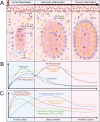
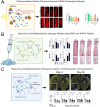
Injury and disease in the central nervous system (CNS) can result in a dysregulated inflammatory environment that inhibits the repair of functional tissue. Biomaterials present a promising approach to tackle this complex inhibitory environment and modulate the mechanisms involved in neuroinflammation to halt the progression of secondary injury and promote the repair of functional tissue. In this review, we will cover recent advances in biomaterial strategies, including nanoparticles, hydrogels, implantable scaffolds, and neural probe coatings, that have been used to modulate the innate immune response to injury and disease within the CNS. The stages of inflammation following CNS injury and the main inflammatory contributors involved in common neurodegenerative diseases will be discussed, as understanding the inflammatory response to injury and disease is critical for identifying therapeutic targets and designing effective biomaterial-based treatment strategies. Biomaterials and novel composites will then be discussed with an emphasis on strategies that deliver immunomodulatory agents or utilize cell-material interactions to modulate inflammation and promote functional tissue repair. We will explore the application of these biomaterial-based strategies in the context of nanoparticle- and hydrogel-mediated delivery of small molecule drugs and therapeutic proteins to inflamed nervous tissue, implantation of hydrogels and scaffolds to modulate immune cell behavior and guide axon elongation, and neural probe coatings to mitigate glial scarring and enhance signaling at the tissue-device interface. Finally, we will present a future outlook on the growing role of biomaterial-based strategies for immunomodulation in regenerative medicine and neuroengineering applications in the CNS.
This journal is © The Royal Society of Chemistry.
Conflict of interest statement
There are no conflicts to declare.
Figures


Similar articles
-
Biomaterials and glia: Progress on designs to modulate neuroinflammation.Acta Biomater. 2019 Jan 1;83:13-28. doi: 10.1016/j.actbio.2018.11.008. Epub 2018 Nov 7. Acta Biomater. 2019. PMID: 30414483 Review.
-
Biomaterial-supported MSC transplantation enhances cell-cell communication for spinal cord injury.Stem Cell Res Ther. 2021 Jan 7;12(1):36. doi: 10.1186/s13287-020-02090-y. Stem Cell Res Ther. 2021. PMID: 33413653 Free PMC article. Review.
-
Recent advances in immunomodulatory hydrogels biomaterials for bone tissue regeneration.Mol Immunol. 2023 Nov;163:48-62. doi: 10.1016/j.molimm.2023.09.010. Epub 2023 Sep 22. Mol Immunol. 2023. PMID: 37742359 Review.
-
Biomaterial-Driven Immunomodulation: Cell Biology-Based Strategies to Mitigate Severe Inflammation and Sepsis.Front Immunol. 2020 Aug 4;11:1726. doi: 10.3389/fimmu.2020.01726. eCollection 2020. Front Immunol. 2020. PMID: 32849612 Free PMC article. Review.
-
Mesenchymal stem cell-derived exosomes regulate microglia phenotypes: a promising treatment for acute central nervous system injury.Neural Regen Res. 2023 Aug;18(8):1657-1665. doi: 10.4103/1673-5374.363819. Neural Regen Res. 2023. PMID: 36751776 Free PMC article. Review.
Cited by
-
Immunosuppressive Formulations for Immunological Defense against Traumatic Brain Injury.Adv Healthc Mater. 2025 Jul;14(19):e2501417. doi: 10.1002/adhm.202501417. Epub 2025 May 29. Adv Healthc Mater. 2025. PMID: 40437894 Free PMC article.
-
Poly(curcumin-co-poly(ethylene glycol)) films provide neuroprotection following reactive oxygen species insultin vitro.J Neural Eng. 2025 Jan 27;22(1):10.1088/1741-2552/ada8df. doi: 10.1088/1741-2552/ada8df. J Neural Eng. 2025. PMID: 39793199
-
Nano-scaffold containing functional motif of stromal cell-derived factor 1 enhances neural stem cell behavior and synaptogenesis in traumatic brain injury.Sci Rep. 2025 Feb 17;15(1):5811. doi: 10.1038/s41598-025-85698-5. Sci Rep. 2025. PMID: 39962142 Free PMC article.
-
Emerging Insights into Brain Inflammation: Stem-Cell-Based Approaches for Regenerative Medicine.Int J Mol Sci. 2025 Apr 1;26(7):3275. doi: 10.3390/ijms26073275. Int J Mol Sci. 2025. PMID: 40244116 Free PMC article. Review.
References
-
- Report to Congress: Traumatic Brain Injury in the United States | Concussion|Traumatic Brain Injury | CDC Injury Center [Internet]. 2019 [cited 2023 Sep 10]. Available from: https://www.cdc.gov/traumaticbraininjury/pubs/tbi_report_to_congress.html
-
- TBI Data | Concussion | Traumatic Brain Injury | CDC Injury Center [Internet]. 2023 [cited 2023 Sep 10]. Available from: https://www.cdc.gov/traumaticbraininjury/data/index.html
-
- Thorpe K. E., Levey A. I. and Thomas J. U. S. BURDEN OF Neurodegenerative Disease. 2021. Available from: https://www.fightchronicdisease.org/sites/default/files/May%202021%20Neu...
Publication types
Grants and funding
LinkOut - more resources
Full Text Sources
