. 2024 May 7:15:1380628.
doi: 10.3389/fimmu.2024.1380628. eCollection 2024.

Differential regulation of lung homeostasis and silicosis by the TAM receptors MerTk and Axl

Affiliations

Differential regulation of lung homeostasis and silicosis by the TAM receptors MerTk and Axl

Kamila Guimarães-Pinto et al. Front Immunol. .

Abstract

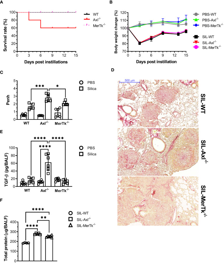
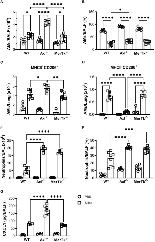
Introduction: TAM receptor-mediated efferocytosis plays an important function in immune regulation and may contribute to antigen tolerance in the lungs, a site with continuous cellular turnover and generation of apoptotic cells. Some studies have identified failures in efferocytosis as a common driver of inflammation and tissue destruction in lung diseases. Our study is the first to characterize the in vivo function of the TAM receptors, Axl and MerTk, in the innate immune cell compartment, cytokine and chemokine production, as well as the alveolar macrophage (AM) phenotype in different settings in the airways and lung parenchyma.

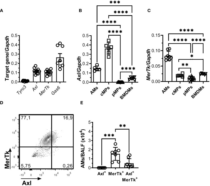
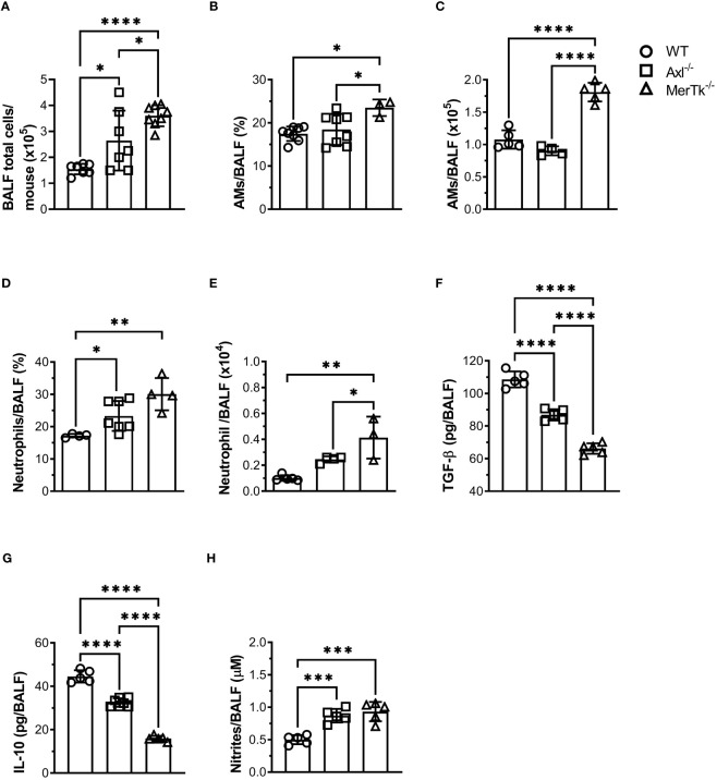
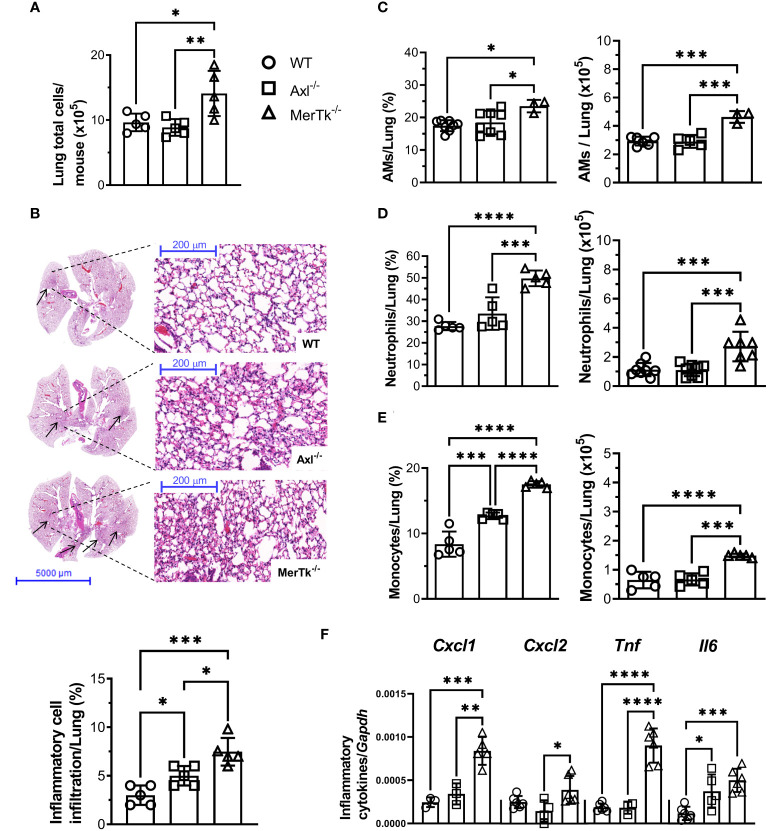
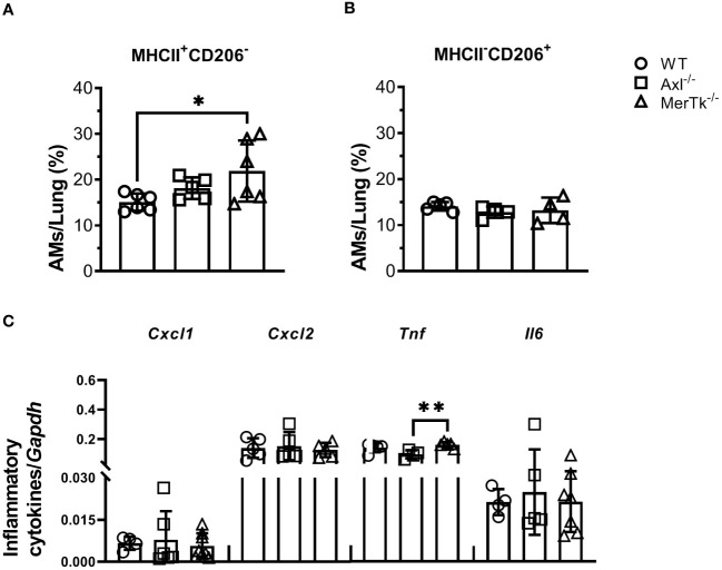
Methods: We employed MerTk and Axl defective mice to induce acute silicosis by a single exposure to crystalline silica particles (20 mg/50 μL). Although both mRNA levels of Axl and MerTk receptors were constitutively expressed by lung cells and isolated AMs, we found that MerTk was critical for maintaining lung homeostasis, whereas Axl played a role in the regulation of silica-induced inflammation. Our findings imply that MerTk and Axl differently modulated inflammatory tone via AM and neutrophil recruitment, phenotype and function by flow cytometry, and TGF-β and CXCL1 protein levels, respectively. Finally, Axl expression was upregulated in both MerTk-/- and WT AMs, confirming its importance during inflammation.

Conclusion: This study provides strong evidence that MerTk and Axl are specialized to orchestrate apoptotic cell clearance across different circumstances and may have important implications for the understanding of pulmonary inflammatory disorders as well as for the development of new approaches to therapy.

Keywords: airways homeostasis; alveolar macrophage; efferocytosis; immunoregulation; silicosis.


Conflict of interest statement

The authors declare that the research was conducted in the absence of any commercial or financial relationships that could be construed as a potential conflict of interest. The author(s) declared that they were an editorial board member of Frontiers, at the time of submission. This had no impact on the peer review process and the final decision.

Figures

Figure 1
Figure 1
The mucosal microenvironment regulates TAM receptors’ expression pattern during homeostasis. (A) TAM receptors and their ligand, Gas6, mRNA expression in isolated total lung cells from WT mice. Data are shown as means ± SD. (B, C) Axl and MerTk mRNA expression in different macrophage subsets (AMs, cMPs, pMPs, and BMDMs) from WT mice by RT-qPCR. (D) Representative FACS plot of Axl+, MerTk+, and Axl+MerTk+ cells gated on AMs (SiglecF+CD11c+) from WT BALFs. (E) Absolute number of Axl+, MerTk+, and Axl+MerTk+ of AMs. (A-D) Data were obtained from three independent experiments with similar results (n ≥ 7 per group). (E) Data shown were combined from three independent experiments with similar results (n ≥ 6 per group). Data are shown as means ± SD. Statistical differences were analyzed by unpaired t-tests (B, C) or one-way ANOVA with Tukey’s multiple comparison test (E) and depicted as *p<0.05, **p<0.01, ***p<0.001, ****p<0.0001. AMs, Alveolar macrophages; cMPs, colonic macrophages; pMPs, peritoneal macrophages; BMDM: bone marrow-derived macrophages.
Figure 2
Figure 2
MerTk receptor deficiency causes airway homeostasis imbalance. (A) Total BALF cell numbers from WT, Axl-/-, and MerTk-/- mice. Data are shown as means ± SD. (B, C) Frequencies and absolute numbers of AMs (SiglecF+CD11c+) of total live cells gate from WT, Axl-/-, and MerTk-/- mice BALFs determined by flow cytometry. (D, E) Frequencies and absolute numbers of neutrophils (SiglecF-CD11c-Ly6G+) within CD11b+ gate from WT, Axl-/-, and MerTk-/- mice BALFs analyzed by flow cytometry. (F) Levels of active TGF-β, (G) IL-10, and (H) nitrites in BAL fluids from WT, Axl-/-, and MerTk-/- mice, measured by ELISA or Griess Assay, respectively. (A, F-H) Data were obtained from four independent experiments with similar results (n ≥ 5 per group). (B-E) Data were obtained from three independent experiments with similar results (n ≥ 3 per group). Data are shown as means ± SD. Statistical differences were analyzed by one-way ANOVA with Tukey’s multiple comparison test (A-H) and depicted as *p<0.05, **p<0.01, ***p<0.001, ****p<0.0001. AMs, Alveolar macrophages; BALFs, Bronchoalveolar lavage fluid.
Figure 3
Figure 3
MerTk receptor deficiency causes lung parenchyma homeostasis imbalance. (A) Total lung cell numbers from WT, Axl-/-, and MerTk-/- mice. (B) Representative lung histology images and percentage of lung area occupied by inflammatory cell recruitment based on H&E staining in WT, Axl-/-, and MerTk-/- mice. Frequencies and absolute numbers of (C) AMs (SiglecF+CD11c+) of total live cells gate, (D) neutrophils (SiglecF-CD11c-Ly6G+), and (E) monocytes (SiglecF-CD11c-Ly6C+) within CD11b+ gate from total lung tissue of WT, Axl-/-, and MerTk-/- mice acquired by flow cytometry. (F) Total lung cells mRNA expression of CXCL1, CXCL2, TNF-α, and IL-6 from WT, Axl-/-, and MerTk-/- mice analyzed by RT-qPCR. (A, B) Data were obtained from three independent experiments with similar results (n = 5 per group). (C-F) Data were obtained from four independent experiments with similar results (n ≥ 3 per group). Data are shown as means ± SD. Statistical differences were analyzed by one-way ANOVA with Tukey’s multiple comparison test (A-F) and depicted as *p<0.05, **p<0.01, ***p<0.001, ****p<0.0001. AMs, Alveolar macrophages.
Figure 4
Figure 4
Expression of the MerTk receptor regulates AMs functional phenotype during homeostasis. (A, B) MHCII and CD206 expression in BALF-isolated AMs (SiglecF+CD11c+) from WT, Axl-/-, and MerTk-/- mice. (C) CXCL1, CXCL2, TNF, and IL-6 mRNA expression by RT-qPCR of AMs from WT, Axl-/-, and MerTk-/- mice. (A-C) Data were obtained from two independent experiments with similar results (n ≥ 4per group). Data are shown as means ± SD. Statistical differences were analyzed by one-way ANOVA with Tukey’s multiple comparison test (A-C) depicted as *p<0.05, **p<0.01. AMs, Alveolar macrophages.
Figure 5
Figure 5
Acute induced-silica inflammation disrupts pulmonary structure and function in the airways. WT, Axl-/-, and MerTk-/- mice were i.t. injected with 20 mg/50 µL of a sterile silica suspension or 50 µL of PBS. Mice were sacrificed 15 days after the injection. (A) Survival curve and (B) body weight change of mice over 15 days following PBS or silica i.t. instillation. (C) Non-invasive Penh measurements of airway resistance on day 7 following PBS or silica i.t. injection measured by whole-body plethysmography (Buxco system). (D) Representative images of lung lobes stained with Picro Sirius from silica WT, Axl-/-, and MerTk-/- mice for assessment of visible collagen deposition areas. Scale bars = 500 μm. (E) ELISA of active TGF-β amounts and (F) total protein levels in BALFs from WT, Axl-/-, and MerTk-/- mice following PBS or silica i.t. injection. (A-F) Data were obtained from four independent experiments with similar results (n ≥ 4per group). Data are shown as means ± SD. Statistical differences were analyzed by one-way ANOVA with Tukey’s multiple comparison test and depicted as *p<0.05, **p<0.01, ***p<0.001, ****p<0.0001. BALFs, Bronchoalveolar lavage fluid.
Figure 6
Figure 6
Silica-induced inflammation enhanced airway cell recruitment and is associated with AMs phenotype shift. (A) Absolute numbers and (B) frequencies of AMs (SiglecF+CD11c+) of total live cells from WT, Axl-/-, and MerTk-/- mice BALFs determined by flow cytometry 15 days following PBS or silica i.t. injection. (C) Frequencies of MHCII+ and (D) CD206+ AMs analyzed by flow cytometry from WT, Axl-/-, and MerTk-/- mice BALFs on day 15 following i.t. PBS or silica administration. (E) Absolute numbers and (F) frequencies of neutrophils (SiglecF-CD11c-Ly6G+) within CD11b+ gate from WT, Axl-/-, and MerTk-/- mice BALFs determined by flow cytometry 15 days following PBS or silica i.t. injection. (G) ELISA of CXCL1 amounts in BAL fluids from WT, Axl-/-, and MerTk-/- mice following PBS or silica i.t. injection. (A, B, E, F) Data were obtained from three independent experiments with similar results (n ≥ 5 per group). (C, D, G) Data were obtained from four independent experiments with similar results (n ≥ 4 per group). Data are shown as means ± SD. Statistical differences were analyzed by one-way ANOVA with Tukey’s multiple comparison test and depicted as *p<0.05, **p<0.01, ***p<0.001, ****p<0.0001. AMs, Alveolar macrophages; BALFs, Bronchoalveolar lavage fluid.
Figure 7
Figure 7
Axl receptor regulates apoptotic cell removal during acute silica inflammation. (A-C) Absolute numbers of apoptotic (Annexin V+7AAD-), late apoptotic (Annexin V+7AAD+), and necrotic (Annexin V-7AAD+) AMs (SiglecF+CD11c+) and (D-F) neutrophils (Ly6G+CD11b+) from WT, Axl-/-, and MerTk-/- mice by flow cytometry 15 days following PBS or silica i.t. injection. (G) Axl mRNA expression by RT-qPCR of total lung cells from WT, and MerTk-/- mice 15 days following silica i.t. injection. (A-G) Data were obtained from three independent experiments with similar results (n ≥ 4 per group). Data are shown as means ± SD. Statistical differences were analyzed by one-way ANOVA with Tukey’s multiple comparison test (A-F) or unpaired t-tests (G) and depicted as *p<0.05, **p<0.01, ***p<0.001, ****p<0.0001. AMs, Alveolar macrophages; BALFs, Bronchoalveolar lavage fluid.

Similar articles

Cited by

References

    1. Bowden DH. Cell turnover in the lung. Am Rev Respir Dis. (1983) 128. doi: 10.1164/ARRD.1983.128.2P2.S46 - DOI - PubMed
    1. Grabiec AM, Hussell T. The role of airway macrophages in apoptotic cell clearance following acute and chronic lung inflammation. Semin Immunopathol. (2016) 38:409–23. doi: 10.1007/s00281-016-0555-3 - DOI - PMC - PubMed
    1. Guimarães-Pinto K, Maia EP, Ferreira JRM, Filardy AA. Efferocytosis in lung mucosae: implications for health and disease. Immunol Lett. (2022) 248:109–18. doi: 10.1016/J.IMLET.2022.07.005 - DOI - PubMed
    1. Demedts IK, Demoor T, Bracke KR, Joos GF, Brusselle GG. Role of apoptosis in the pathogenesis of COPD and pulmonary emphysema. Respir Res. (2006) 7:53. doi: 10.1186/1465-9921-7-53 - DOI - PMC - PubMed
    1. Li J, Shao R, Xie Q, Qin K, Ming SP, Xie Y, et al. . Ulinastatin promotes macrophage efferocytosis and ameliorates lung inflammation via the ERK5/Mer signaling pathway. FEBS Open Bio. (2022) 12:1498–508. doi: 10.1002/2211-5463.13461 - DOI - PMC - PubMed

MeSH terms