

Comparative analysis of primer sets for the assessment of clonality in feline lymphomas
- PMID: 38774911
- PMCID: PMC11106357
- DOI: 10.3389/fvets.2024.1356330
Comparative analysis of primer sets for the assessment of clonality in feline lymphomas
Abstract
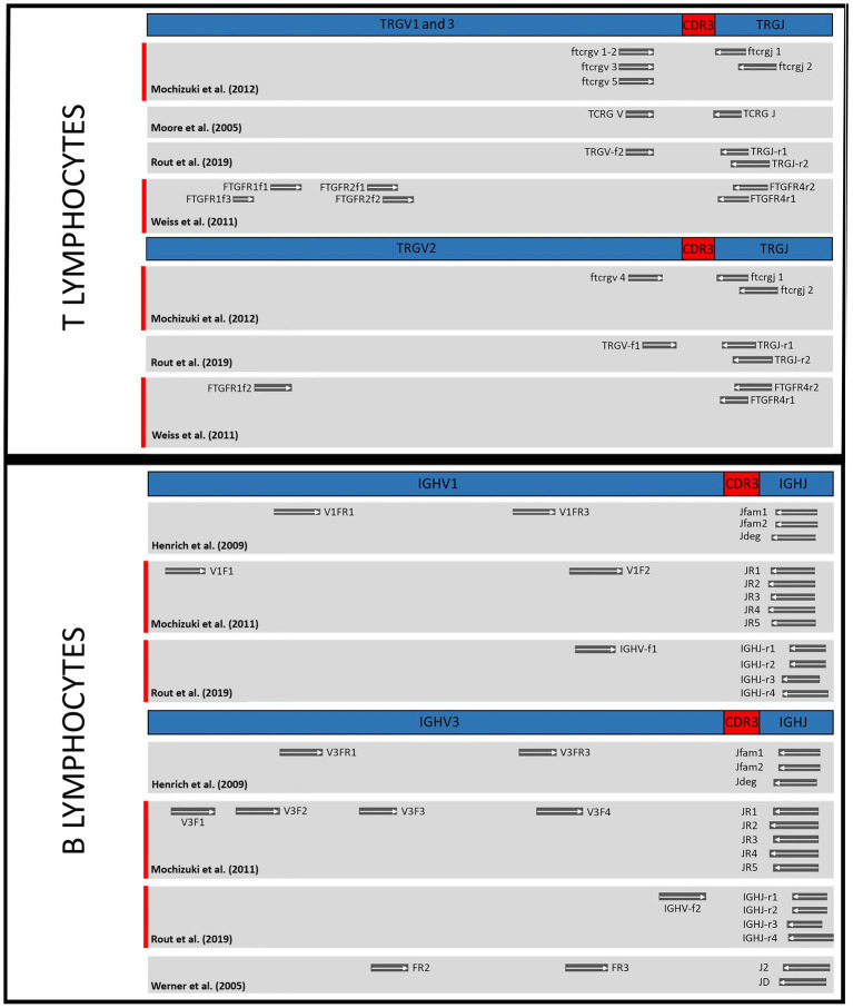
Introduction: Lymphomas are among the most important and common malignant tumors in cats. Differentiating lymphomas from reactive lymphoid proliferations can be challenging, so additional tools such as clonality assessment by PCR are important in diagnosis finding. Several PCR assays have been developed to assess clonality in feline lymphomas. For T-cell lymphomas TRG (T-cell receptor gamma) genes are the preferred target whereas for B-cell lymphomas most primer sets target immunoglobulin heavy chain (IGH) genes. Here we compare commonly used diagnostic primer sets for the assessment of clonality in feline lymphomas under controlled conditions (i.e., identical sample set, PCR setup, amplicon detection system).
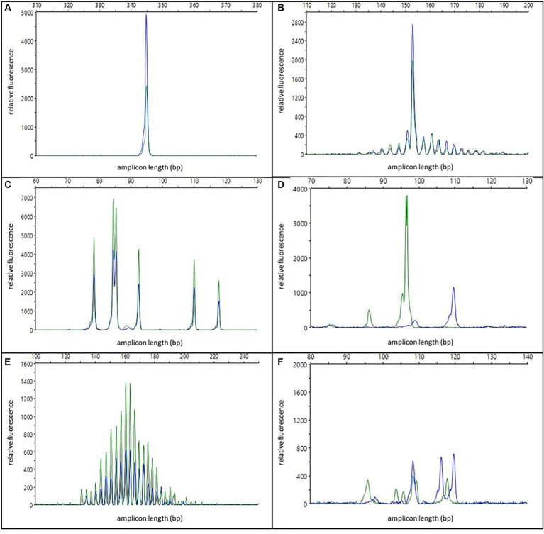
Methods: Formalin-fixed and paraffin-embedded samples from 31 feline T-cell lymphomas, 29 B-cell lymphomas, and 11 non-neoplastic controls were analyzed by PCR combined with capillary electrophoresis.
Results and discussion: We show that the combination of the primer sets published by Weiss et al. and Mochizuki et al. provided the best results for T-cell clonality, i.e., correctly assigns most populations as clonal or polyclonal. For B-cell clonality, the combination of the primer sets by Mochizuki et al. and Rout et al. gave the best results when omitting the Kde gene rearrangement due to its low specificity. This study rigorously evaluated various primer sets under uniform experimental conditions to improve accuracy of lymphoma diagnostic and provides a recommendation for achieving the highest diagnostic precision in lymphoma clonality analysis.
Keywords: Felis catus; PARR; PCR; antigen receptor rearrangements; cat; clonality; lymphoma.
Copyright © 2024 Weyrich, Hecht, Köhler, Herden and Henrich.
Conflict of interest statement
The authors declare that the research was conducted in the absence of any commercial or financial relationships that could be construed as a potential conflict of interest. The author(s) declared that they were an editorial board member of Frontiers, at the time of submission. This had no impact on the peer review process and the final decision.
Figures
Similar articles
-
Assessment of immunoglobulin heavy chain, immunoglobulin light chain, and T-cell receptor clonality testing in the diagnosis of feline lymphoid neoplasia.Vet Clin Pathol. 2019 Oct;48 Suppl 1:45-58. doi: 10.1111/vcp.12767. Epub 2019 Sep 2. Vet Clin Pathol. 2019. PMID: 31478220
-
Enhanced sensitivity with a novel TCRgamma PCR assay for clonality studies in 569 formalin-fixed, paraffin-embedded (FFPE) cases.Mol Diagn. 1999 Jun;4(2):119-33. doi: 10.1016/s1084-8592(99)80036-8. Mol Diagn. 1999. PMID: 10462627
-
A new subgroup of immunoglobulin heavy chain variable region genes for the assessment of clonality in feline B-cell lymphomas.Vet Immunol Immunopathol. 2009 Jul 15;130(1-2):59-69. doi: 10.1016/j.vetimm.2009.01.006. Epub 2009 Feb 24. Vet Immunol Immunopathol. 2009. PMID: 19243841
-
A review of canine B cell clonality assays and primer set optimization using large-scale repertoire data.Vet Immunol Immunopathol. 2019 Mar;209:45-52. doi: 10.1016/j.vetimm.2019.01.002. Epub 2019 Jan 25. Vet Immunol Immunopathol. 2019. PMID: 30885305 Review.
-
[Analyses of the rearrangement of T-cell receptor- and immunoglobulin genes in the diagnosis of lymphoproliferative disorders].Veroff Pathol. 1995;144:1-109. Veroff Pathol. 1995. PMID: 7856305 Review. German.
Cited by
-
Investigating comparative polymerase chain reaction for antigen receptor rearrangement analysis in different types of feline lymphoma samples.Front Vet Sci. 2024 Aug 30;11:1439068. doi: 10.3389/fvets.2024.1439068. eCollection 2024. Front Vet Sci. 2024. PMID: 39280837 Free PMC article.
References
-
- Valli VE. Histological classification of hematopoietic tumors of domestic animals. Washington, DC: Armed Forces Institute of Pathology, in cooperation with the American Registry of Pathology and the World Health Organization Collaborating Center for Worldwide Reference on Comparative Oncology; (2002).
-
- Briscoe KA, Krockenberger M, Beatty JA, Crowley A, Dennis MM, Canfield PJ, et al. . Histopathological and immunohistochemical evaluation of 53 cases of feline lymphoplasmacytic enteritis and low-grade alimentary lymphoma. J Comp Pathol. (2011) 145:187–98. doi: 10.1016/j.jcpa.2010.12.011, PMID: - DOI - PubMed
-
- Mooney SC, Patnaik AK, Hayes AA, MacEwen EG. Generalized lymphadenopathy resembling lymphoma in cats: six cases (1972-1976). J Am Vet Med Assoc. (1987) 190:897–900. PMID: - PubMed
LinkOut - more resources
Full Text Sources
Miscellaneous