. 2024 Aug 15;111(2):376-390.
doi: 10.1093/biolre/ioae081.

Mapping the follicle-specific regulation of extracellular vesicle-mediated microRNA transport in the southern white rhinoceros (Ceratotherium simum simum)†

Affiliations

Mapping the follicle-specific regulation of extracellular vesicle-mediated microRNA transport in the southern white rhinoceros (Ceratotherium simum simum)†

Ahmed Gad et al. Biol Reprod. .

Abstract

Efforts to implement effective assisted reproductive technologies (ARTs) for the conservation of the northern white rhinoceros (NWR; Ceratotherium simum cottoni) to prevent its forthcoming extinction, could be supported by research conducted on the closely related southern white rhinoceros (SWR; Ceratotherium simum simum). Within the follicle, extracellular vesicles (EVs) play a fundamental role in the bidirectional communication facilitating the crucial transport of regulatory molecules such as microRNAs (miRNAs) that control follicular growth and oocyte development. This study aimed to elucidate the dynamics of EV-miRNAs in stage-dependent follicular fluid (FF) during SWR ovarian antral follicle development. Three distinct follicular stages were identified based on diameter: Growing (G; 11-17 mm), Dominant (D; 18-29 mm), and Pre-ovulatory (P; 30-34 mm). Isolated EVs from the aspirated FF of segmented follicle stages were used to identify EV-miRNAs previously known via subsequent annotation to all equine (Equus caballus; eca), bovine (Bos taurus; bta), and human (Homo sapiens; hsa) miRNAs. A total of 417 miRNAs were detected, with 231 being mutually expressed across all three stages, including eca-miR-148a and bta-miR-451 as the top highly expressed miRNAs. Distinct expression dynamics in miRNA abundance were observed across the three follicular stages, including 31 differentially expressed miRNAs that target various pathways related to follicular growth and development, with 13 miRNAs commonly appearing amidst two different comparisons. In conclusion, this pioneering study provides a comprehensive understanding of the stage-specific expression dynamics of FF EV-miRNAs in the SWR. These findings provide insights that may lead to novel approaches in enhancing ARTs to catalyze rhinoceros conservation efforts.

Keywords: extracellular vesicles; follicular fluid; miRNA; ovarian follicle; rhinoceros.


Figures

Graphical Abstract
Graphical Abstract
Figure 1
Figure 1
Schematic experimental illustration of the data analysis pipeline. Experimental pipeline outlining the design and workflow for the analysis of extracellular vesicle-coupled miRNAs from stage-specific rhinoceros FF. Given the rairity of the sample collections, following in vivo FF collections and subsequent EV isolation, the first-factor variable of differences in individual animals were removed. Total RNA including small RNAs were isolated from equal volumes of pooled EV technical replicates of two biologically dissimilar females (three pooled replicates/follicle stage, pool consisting of two animals).
Figure 2
Figure 2
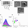
Rhinoceros extracellular vesicle morphological and biochemical validation. (A) Immunoblots indicating traditionally expressed EV-associated marker proteins (TSG101 and HSP70), and the absence of cellular contamination marker (CYCS). (B) Representative transmission electron microscopy images revealing the ultrastructure cup-like morphology of rhinoceros FF-derived EVs, indicated by arrows. Scale bars: 100 nm. Nanoparticle tracking analysis of the concentration (C) and size distribution (D) of rhinoceros FF-derived EVs from diverging stages. (E) Representative graphical overview of the quality and integrity of exosomal RNA samples used for small RNA sequencing.
Figure 3
Figure 3
Small-RNA sequencing data overview. (A) Principal component analysis. (B) Heatmap and hierarchical clustering of expressed miRNAs. (C) Venn diagram for commonly and exclusively expressed miRNAs in the FF EVs at different stages of follicular development. MicroRNAs with a value of CPM >10 in at least two replicates of the three biological replicates was considered detected. Exclusively expressed miRNAs that exhibited a significant difference are presented in the Venn diagram with arrows indicating significantly up or downregulated miRNAs in the corresponding comparison. G: Growing; D: dominant; P: preovulatory.
Figure 4
Figure 4
DE miRNAs. (A) All DE-miRNAs from the three different comparisons. Up- and down regulated miRNAs from each comparison are presented as red and green lines, respectively. Volcano plots of expressed miRNAs. Up- (upper right), and downregulated (upper left) miRNAs in (B) G vs. D, (C) P vs. G, and (D) P vs. D comparisons. The number of up and downregulated miRNAs in each comparison are presented on the up and down arrows, respectively. G: growing; D: dominant; P: preovulatory.
Figure 5
Figure 5
Clustering analysis of EV-coupled miRNAs across the stages of follicular development. (A) Nine different miRNA expression patterns in the FF EVs during follicular development. (B) The counts of total and DE miRNAs in each cluster. G: growing; D: dominant; P: preovulatory.
Figure 6
Figure 6
Cluster 3 consists of 67 miRNAs. (A) A group of 67 miRNAs exhibited an increase in their expression from the G until the D stage and then a reduction at the P stage. (B) Among this cluster, eight miRNAs were differentially or exclusively expressed amidst the different comparisons. (C) Pathways associated with the exclusive and DE-miRNA target genes of this cluster. G: growing; D: dominant; P: preovulatory.
Figure 7
Figure 7
Cluster 5 consists of 53 miRNAs. (A) A group of 53 miRNAs exhibited a sharp reduction in their expression in the D and P stages, however, not in the G stage. (B) Among this cluster, seven miRNAs were differentially or exclusively expressed in different comparisons. (C) Pathways associated with the exclusive and DE-miRNA target genes of this cluster. G: growing; D: dominant; P: preovulatory.
Figure 8
Figure 8
Cluster 6 consists of 47 miRNAs. (A) A group of 47 miRNAs exhibited a slight increase in their expression from the G to the D stage and then a sharp reduction at the P stage. (B) Among this cluster, 10 miRNAs were differentially or exclusively expressed in the different comparisons. (C) Pathways associated with the exclusive and DE-miRNA target genes of this cluster. G: growing; D: dominant; P: preovulatory.

References

    1. Hildebrandt TB, Hermes R, Colleoni S, Diecke S, Holtze S, Renfree MB, Stejskal J, Hayashi K, Drukker M, Loi P, Göritz F, Lazzari G, et al. Embryos and embryonic stem cells from the white rhinoceros. Nat Commun 2018; 9:2589. - PMC - PubMed
    1. Hildebrandt TB, Holtze S, Colleoni S, Hermes R, Stejskal J, Lekolool I, Ndereeh D, Omondi P, Kariuki L, Mijele D, Mutisya S, Ngulu S, et al. In vitro fertilization (IVF) program in white rhinoceros. Reproduction 2023; 166:383–399. - PMC - PubMed
    1. Hildebrandt TB, Hermes R, Goeritz F, Appeltant R, Colleoni S, de Mori B, Diecke S, Drukker M, Galli C, Hayashi K, Lazzari G, Loi P, et al. The ART of bringing extinction to a freeze - history and future of species conservation, exemplified by rhinos. Theriogenology 2021; 169:76–88. - PubMed
    1. Radcliffe RW, Czekala NM, Osofsky SA. Combined serial ultrasonography and fecal progestin analysis for reproductive evaluation of the female white rhinoceros (Ceratotherium simum simum): Preliminary results. Zoo Biol 1997; 16:445–456.
    1. Pryor P, Tibary A. Management of estrus in the performance mare. Clin Tech Equine Pract 2005; 4:197–209.