Meta-Analysis
. 2024 Sep 1;63(9):2387-2398.
doi: 10.1093/rheumatology/keae286.

Disruption of memory B-cell trafficking by belimumab in patients with systemic lupus erythematosus

Affiliations
Meta-Analysis

Disruption of memory B-cell trafficking by belimumab in patients with systemic lupus erythematosus

Eline J Arends et al. Rheumatology (Oxford). .

Abstract

Objectives: Autoreactive memory B cells (MBCs) contribute to chronic and progressive courses in autoimmune diseases like SLE. The efficacy of belimumab (BEL), the first approved biologic treatment for SLE and LN, is generally attributed to depletion of activated naïve B cells and inhibition of B-cell activation. BEL's effect on MBCs is currently unexplained. We performed an in-depth cellular and transcriptomic analysis of BEL's impact on the blood MBC compartment in patients with SLE.

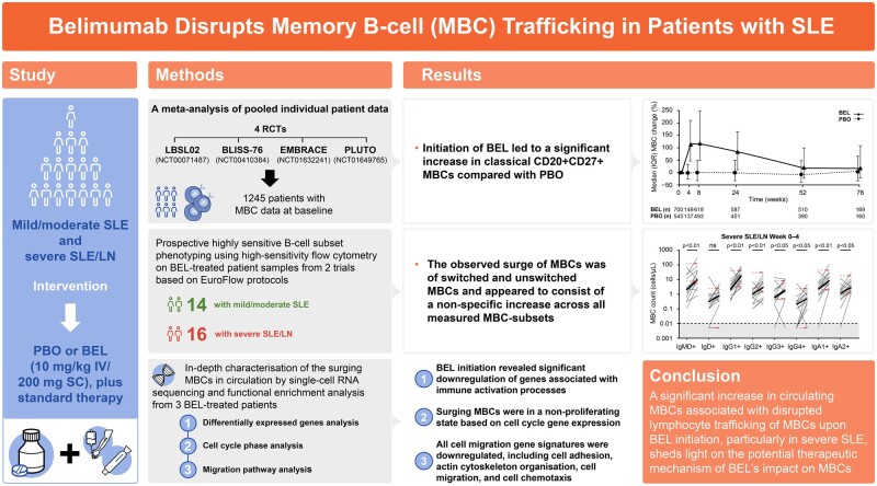
Methods: A retrospective meta-analysis was conducted, pooling flow cytometry data from four randomized trials involving 1245 patients with SLE treated with intravenous BEL or placebo. Then, extensive MBC phenotyping was performed using high-sensitivity flow cytometry in patients with mild/moderate SLE and severe SLE/LN treated with subcutaneous BEL. Finally, transcriptomic characterization of surging MBCs was performed by single-cell RNA sequencing.

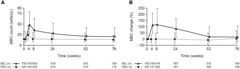
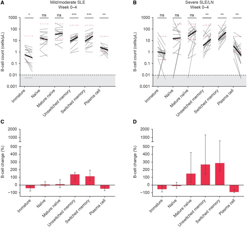
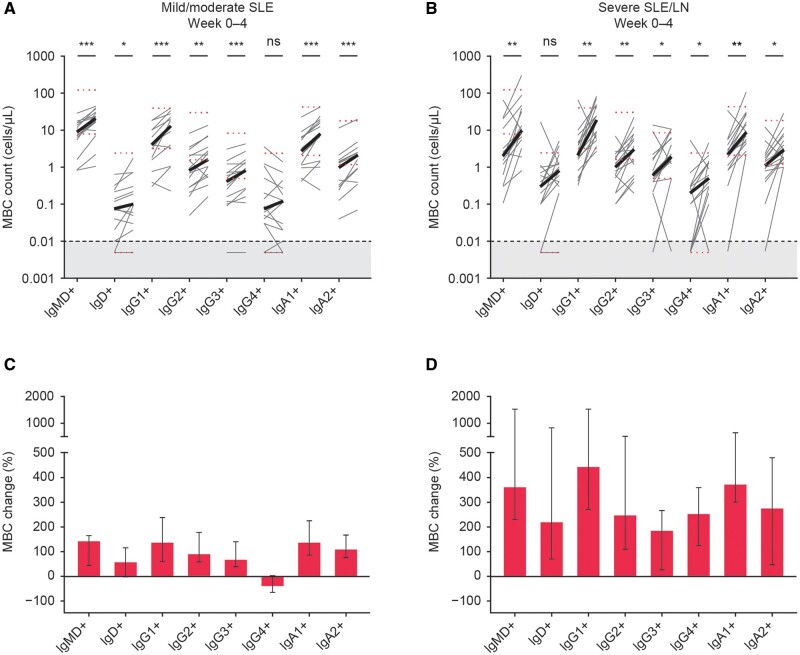
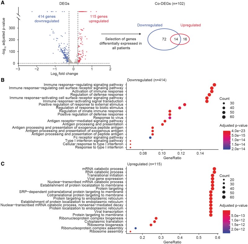
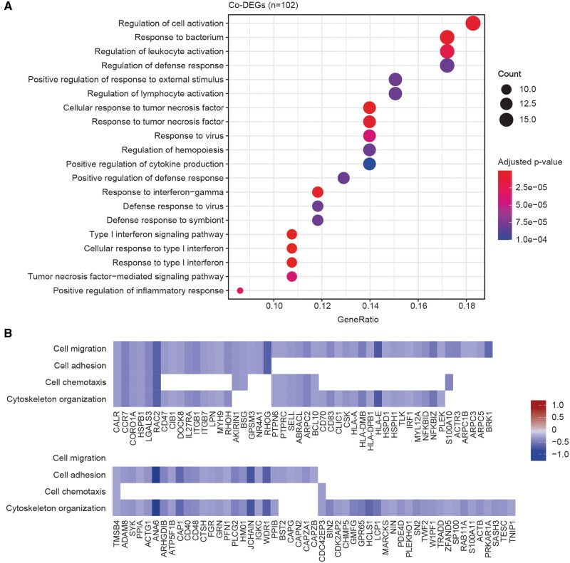
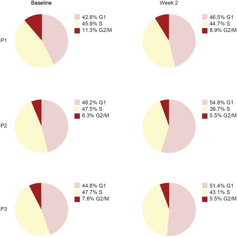
Results: In BEL-treated patients, a significant increase in circulating MBCs, in a broad range of MBC subsets, was established at week 2, gradually returning to baseline by week 52. The increase was most prominent in patients with higher SLE disease activity, serologically active patients and patients aged ≤18 years. MBCs had a non-proliferating phenotype with a prominent decrease in activation status and downregulation of numerous migration genes.

Conclusion: Upon BEL initiation, an increase of MBCs was firmly established. In the small cohort investigated, circulating MBCs were de-activated, non-proliferative and demonstrated characteristics of disrupted lymphocyte trafficking, expanding on our understanding of the therapeutic mechanism of B-cell-activating factor inhibition by BEL.

Trial registration: ClinicalTrials.gov, http://clinicaltrials.gov, NCT00071487, NCT00410384, NCT01632241, NCT01649765, NCT03312907, NCT03747159.

Keywords: B-lymphocyte; LN; SLE; biologicals; gene expression.


Figures

None
Graphical abstract
Figure 1.
Figure 1.
Median (IQR) MBC changes in patients from four pooled SLE clinical trials treated with BEL 10 mg/kg IV or PBO: absolute cell count (post hoc analysis) (A), and percentage change from baseline (B). Week 4 data are from studies LBSL02 [6] and PLUTO [17] only, and Week 76 data are from BLISS-76 [3] only. The apparent increase in absolute cell counts at week 4 in the PBO arm is an artefact attributable to minor variations in baseline cell counts in the included studies. Patients with a baseline MBC count of zero were excluded from percentage change calculations in post-baseline visits (B) because of division with zero. BEL: belimumab; IQR: interquartile range; IV: intravenous; MBC: memory B-cell; PBO: placebo
Figure 2.
Figure 2.
B-cell changes after initiation of BEL measured by high-sensitivity FACS by EuroFlow protocol on samples from 14 patients with mild/moderate SLE (A, C) and 16 patients with severe SLE/LN (B, D). Depicted are changes in B-cell subsets at baseline compared with week 4 in absolute cell counts with the median in bold (A, B) and in median (IQR) percentage change (C, D). The grey area indicates 1–20 analyzed events that were excluded from the analysis. Black dotted line at 0.01 cells/µL indicates the detection limit. Red dotted lines indicate the proposed normal values for adults 18–59 years of age [21]. *P < 0.05; **P < 0.01; ***P < 0.001. BEL: belimumab; FACS: fluorescence-activated cell-sorting; IQR: interquartile range; ns: not significant
Figure 3.
Figure 3.
MBC changes after initiation of BEL measured by HSFC by EuroFlow protocol on samples from two different clinical trials including 14 patients with mild/moderate SLE (A, C) vs 16 patients with severe SLE/LN (B, D). Depicted are changes in MBCs subclassified according to their Ig isotypes at baseline compared with week 4 in absolute cell counts with the median in bold (A, B) and in median (IQR) percentage change (C, D). The grey area indicates 1–20 analyzed events that were excluded from the analysis. Black dotted line at 0.01 cells/µL indicates the detection limit. Red dotted lines indicate the proposed normal values for adults 18–59 years of age [21]. *P < 0.05; **P < 0.01; ***P < 0.001. BEL: belimumab; HSFC: high-sensitivity flow cytometry; MBC: memory B-cell; ns: not significant
Figure 4.
Figure 4.
Identification of DEGs and co-DEGs at 2 weeks after BEL initiation. Depicted are the average log2 fold changes from genes with a minimal percentage expression of 10% and a change above 0.25 in all analyzed patients. Downregulation is shown in blue, upregulation in red (A), with the corresponding top 20 significant biological processes assessed from functional enrichment analysis on 414 downregulated DEGs (B) and 115 upregulated DEGs (C). BEL: belimumab; DEG: differentially expressed gene; mRNA, messenger RNA; RNA: ribonucleic acid; SRP: signal-recognition particle
Figure 5.
Figure 5.
In-depth analysis on DEGs and co-DEGs using FEA on GO terms and migration analysis at 2 weeks after BEL initiation. The top 20 significant biological processes associated with the 102 co-DEGs are shown (A). The average log2 fold changes at 2 weeks after BEL initiation of 99 DEGs associated with migration processes are depicted, subdivided into four categories: cell migration, cell adhesion, cell chemotaxis, and actin cytoskeleton organization (B). BEL: belimumab; DEG: differentially expressed gene; FEA: functional enrichment analysis; GO: gene ontology
Figure 6.
Figure 6.
Identification of the cell cycle phase of each cell analyzed from three patients (P1, P2 and P3) with severe SLE at 2 weeks after BEL initiation, shown as percentage of MBCs per sample that are predicted to be in G1, S or G2/M phase. BEL: belimumab; MBC: memory B-cell

References

    1. Anolik JH. B cell biology: implications for treatment of systemic lupus erythematosus. Lupus 2013;22:342–9. - PubMed
    1. Levy RA, Gonzalez-Rivera T, Khamashta M. et al. 10 Years of belimumab experience: what have we learnt? Lupus 2021;30:1705–21. - PMC - PubMed
    1. Furie R, Petri M, Zamani O. et al. A phase III, randomized, placebo-controlled study of belimumab, a monoclonal antibody that inhibits B lymphocyte stimulator, in patients with systemic lupus erythematosus. Arthritis Rheum 2011;63:3918–30. - PMC - PubMed
    1. Rauch M, Tussiwand R, Bosco N, Rolink AG.. Crucial role for BAFF-BAFF-R signaling in the survival and maintenance of mature B cells. PLoS One 2009;4:e5456. - PMC - PubMed
    1. Chatham WW, Wallace DJ, Stohl W. et al. Effect of belimumab on vaccine antigen antibodies to influenza, pneumococcal, and tetanus vaccines in patients with systemic lupus erythematosus in the BLISS-76 trial. J Rheumatol 2012;39:1632–40. - PubMed

Publication types

MeSH terms

Associated data