Case Reports
. 2024 May 22;18(1):44.
doi: 10.1007/s12105-024-01650-5.

Acinic cell Carcinoma with high-grade Squamoglandular and Chondrosarcomatous Transformation Mimicking 'Carcinosarcoma ex-pleomorphic Adenoma': A Wrinkle in the Proposed Nomenclature Revision for Sarcomatoid Salivary Gland Neoplasms

Affiliations
Case Reports

Acinic cell Carcinoma with high-grade Squamoglandular and Chondrosarcomatous Transformation Mimicking 'Carcinosarcoma ex-pleomorphic Adenoma': A Wrinkle in the Proposed Nomenclature Revision for Sarcomatoid Salivary Gland Neoplasms

Rayan Rammal et al. Head Neck Pathol. .

Abstract

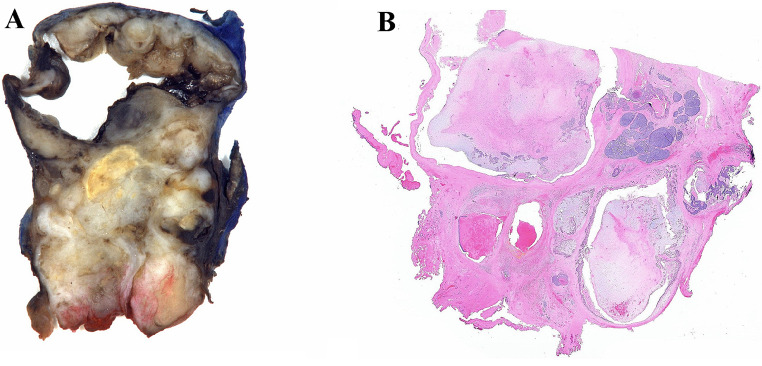
While acinic cell carcinoma (AciCC) can undergo high-grade transformation (HGT) to high-grade adenocarcinoma or poorly differentiated carcinoma, other morphologies such as spindle cell/sarcomatoid carcinoma are rare and not well-characterized. We herein report a novel case of AciCC with squamoglandular and chondrosarcomatous HGT mimicking a so-called 'carcinosarcoma ex-pleomorphic adenoma'. The patient is an 81-year-old male with a two-month history of neck swelling and referred otalgia who presented with a left parapharyngeal space mass extending into retropharyngeal space and pterygoid muscles. On resection, the tumor showed considerable morphologic diversity with high-grade serous and mucous acinar components as well as cribriform to solid apocrine-like components with comedonecrosis and squamous differentiation, all of which were embedded in a chondromyxoid background ranging from paucicellular and bland to a high-grade chondrosarcoma/pleomorphic sarcoma-like appearance. Only a minor conventional AciCC component was noted. Immunostains were negative for AR and only focally positive for GCDFP-15 arguing against a true apocrine phenotype, while PLAG1 and HMGA2 were negative arguing against an antecedent pleomorphic adenoma. On the other hand, SOX-10, DOG-1 and PAS after diastase highlighted serous acinar differentiation, and mucicarmine, and NKX3.1 highlighted mucous acinar differentiation. NR4A3 immunohistochemical staining and NR4A3 fluorescence in situ hybridization were positive in the carcinomatous and sarcomatoid components while sequencing analysis of both components revealed identical alterations involving TP53, PIK3CB, ARID1A, and STK11. This unique case warrants caution in designating all salivary sarcomatoid carcinomas with heterologous elements as part of the 'carcinoma ex-pleomorphic adenoma' family.

Keywords: Acinic cell carcinoma; Chondrosarcoma; Heterologous; High-grade; Molecular; Sarcoma.


Conflict of interest statement

The authors declare that they have no conflict of interest. Dr. Seethala is currently a member of the Editorial Board for Head and Neck Pathology Journal.

Figures

Fig. 1
Fig. 1
(A) Gross examination of the left parapharyngeal mass reveals a firm partially encapsulated and focally disrupted mass with a lobulated heterogeneous cut surface with areas of necrosis and cystic change. (B) Low power histologic examination shows a predominantly solid mass with hemorrhage, necrosis, and cystic degeneration as well as lobulated areas reminiscent of pleomorphic adenoma
Fig. 2
Fig. 2
(A) The carcinoma showed eosinophilic/oncocytoid cells with prominent nucleoli, frequent mitoses, and necrosis, resembling a salivary duct (apocrine) carcinoma, however, it was (B) negative for AR, (C) positive for SOX-10 and (D) showed focal but moderate to strong positivity for GCDFP-15 (400x). (E) Elsewhere, the carcinoma was mucinous/glandular with tubular/cribriform growth (300x); this component showed positivity for (F) intracytoplasmic mucicarmine, (G) SOX-10 and (H) NKX3.1 (400x). (I) This mucinous also showed focally solid growth with comedo-type necrosis but (J) retained mucicarmine staining. K) Foci of squamous differentiation were also noted and L) confirmed with p40 IHC. M) Areas with a squamoglandular appearance consisting of: cells with foamy to clear cytoplasm and a combination of gland formation and cells dense eosinophilic cytoplasm and intercellular bridging (arrows) were embedded in a pleomorphic adenoma-like chondromyxoid background with atypical spindle cells, which led to an initial working diagnosis of ‘carcinosarcoma ex-pleomorphic adenoma’
Fig. 3
Fig. 3
(A) A purely conventional AciCC was present in one 0.2 cm focus, consisting of cells with bland nuclei, amphophilic to lightly basophilic granular and vacuolated cytoplasm, in a solid-microcystic growth pattern (400x). Similar cells were admixed with the (B) the mucinous tubular-cribriform component, located mostly at the periphery of the nests and showed (C) abundant cytoplasmic PAS-D positivity, consistent with zymogen granules, while the tubular/cribriform areas showed more apical accentuation of granular PAS-D staining. These areas showed (D) luminal DOG-1 positivity as well as (E) diffuse NR4A3 positivity (400x). Bland serous acinar cells were also scattered in (F) within the solid apocrine like areas and also showed (G) zymogen granules with PAS-D. Interestingly, this component demonstrated (H) complete membranous staining for DOG-1 and was (I) positive for NR4A3 by IHC (400x). (J) The cells with squamous differentiation retained K) rare PAS-D positivity (inset), L) DOG-1 negativity but M) NR4A3 positivity by IHC. These different carcinoma areas were sent for break-apart FISH studies and all revealed an N)NR4A3 translocation denoted by a split orange green signal (circled cells)
Fig. 4
Fig. 4
(A) The carcinoma component (right upper corner) transitioned to pleomorphic sarcoma (left), with ‘pleomorphic adenoma-like’ chondromyxoid transitional area (100x). (B) A high-power magnification of the transition point shows deceptively bland ovoid to spindled cells in a chondromyxoid background with some lacunae formation (400x). (C) The more cellular pleomorphic sarcomatoid areas showed islands with chondroid matrix surrounding tumor cells compatible with a high-grade chondrosarcomatous element. (D) The chondrosarcoma component was positive for NR4A3 by IHC and FISH confirmed NR4A3 rearrangement (inset)

References

    1. Stevens TM, Agaimy A, Faquin WC, Simpson RHW, Hellquist H, Hsieh M, Kiss K Acinic cell carcinoma. In: WHO Classification of Tumours Editorial Board. Head and neck tumours. Lyon (France): International Agency for Research on Cancer; 2022.(WHO classification of tumours series, 5th ed.; vol. 9). https://publications.iarc.fr
    1. Roy S, Dhingra KK, Gupta P, Khurana N, Gupta B, Meher R Acinic cell carcinoma with extensive neuroendocrine differentiation: a diagnostic challenge. Head Neck Pathol 2009 Jun. 3(2): p. 163–168. 10.1007/s12105-009-0114-5 - PMC - PubMed
    1. Shah AA, Seethala RR Squamoglandular variant of Acinic Cell Carcinoma: a Case Report of a novel variant. Head Neck Pathol 2022 Sep. 16(3): p. 870–875. 10.1007/s12105-021-01399-1 - PMC - PubMed
    1. Romanish MG, Seethala RR (2023) Sep True Oncocytic Acinic Cell Carcinoma: A Case Image. Head Neck Pathol. 17(3): pp. 883–885. 10.1007/s12105-023-01578-2 - PMC - PubMed
    1. Chiosea SI, Griffith C, Assaad A, Seethala RR The profile of acinic cell carcinoma after recognition of mammary analog secretory carcinoma. Am J Surg Pathol 2012 Mar 36(3): p. 343–350. 10.1097/PAS.0b013e318242a5b0 - PubMed

Publication types

MeSH terms