. 2024 Sep 1;81(9):870-881.
doi: 10.1001/jamapsychiatry.2024.1112.

Key Roles of CACNA1C/Cav1.2 and CALB1/Calbindin in Prefrontal Neurons Altered in Cognitive Disorders

Affiliations

Key Roles of CACNA1C/Cav1.2 and CALB1/Calbindin in Prefrontal Neurons Altered in Cognitive Disorders

Dibyadeep Datta et al. JAMA Psychiatry. .

Abstract

Importance: The risk of mental disorders is consistently associated with variants in CACNA1C (L-type calcium channel Cav1.2) but it is not known why these channels are critical to cognition, and whether they affect the layer III pyramidal cells in the dorsolateral prefrontal cortex that are especially vulnerable in cognitive disorders.

Objective: To examine the molecular mechanisms expressed in layer III pyramidal cells in primate dorsolateral prefrontal cortices.

Design, setting, and participants: The design included transcriptomic analyses from human and macaque dorsolateral prefrontal cortex, and connectivity, protein expression, physiology, and cognitive behavior in macaques. The research was performed in academic laboratories at Yale, Harvard, Princeton, and the University of Pittsburgh. As dorsolateral prefrontal cortex only exists in primates, the work evaluated humans and macaques.

Main outcomes and measures: Outcome measures included transcriptomic signatures of human and macaque pyramidal cells, protein expression and interactions in layer III macaque pyramidal cells using light and electron microscopy, changes in neuronal firing during spatial working memory, and working memory performance following pharmacological treatments.

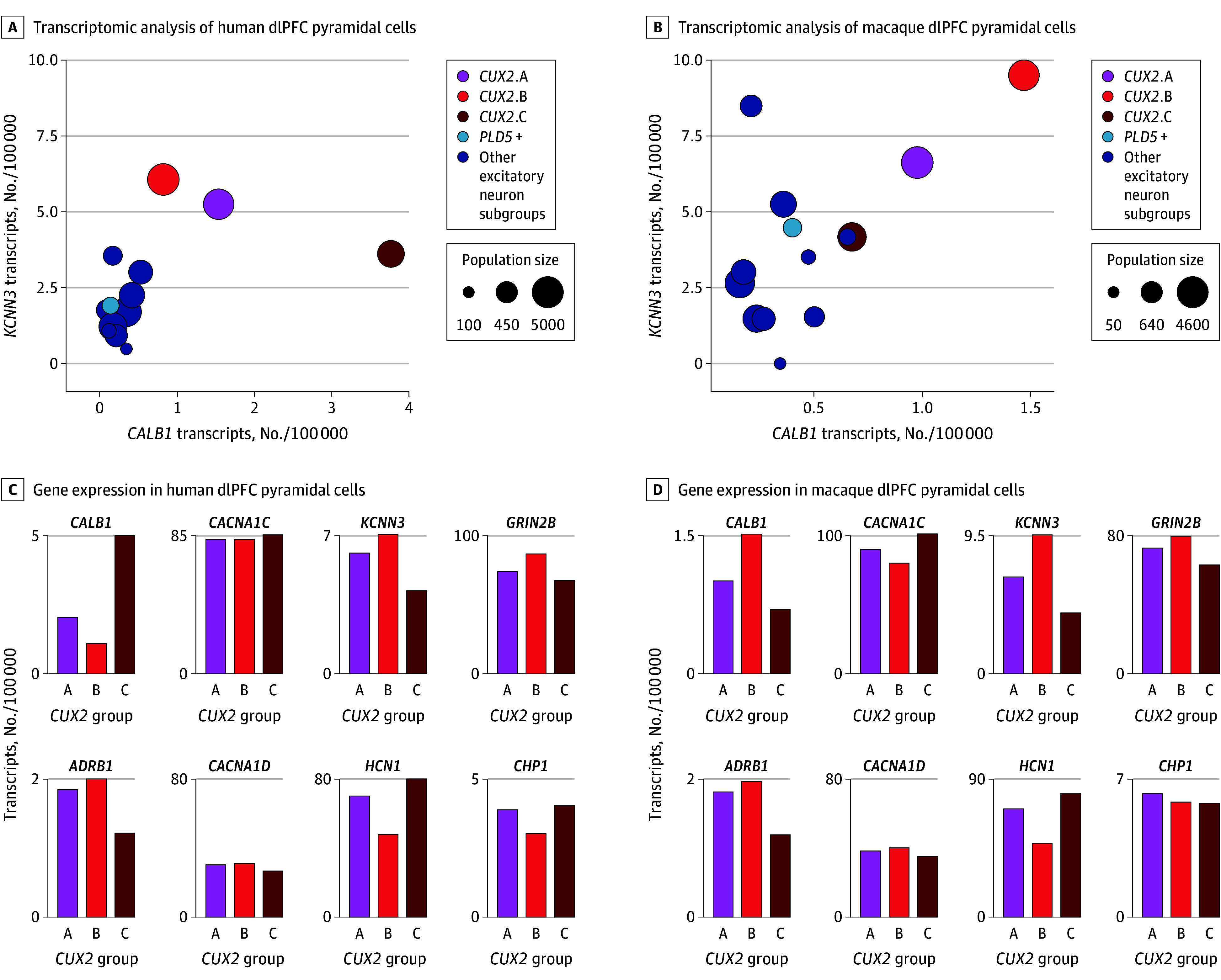
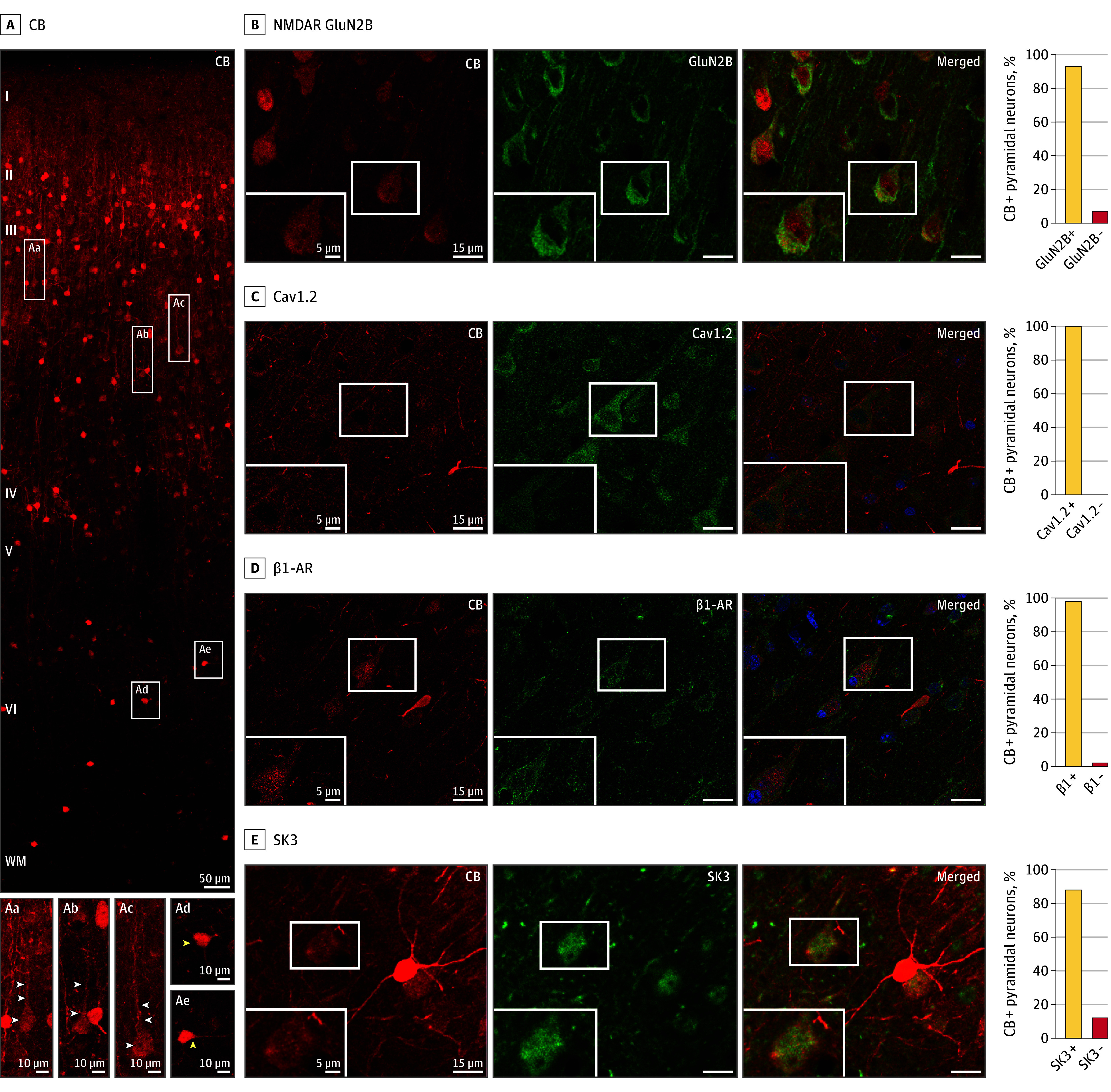
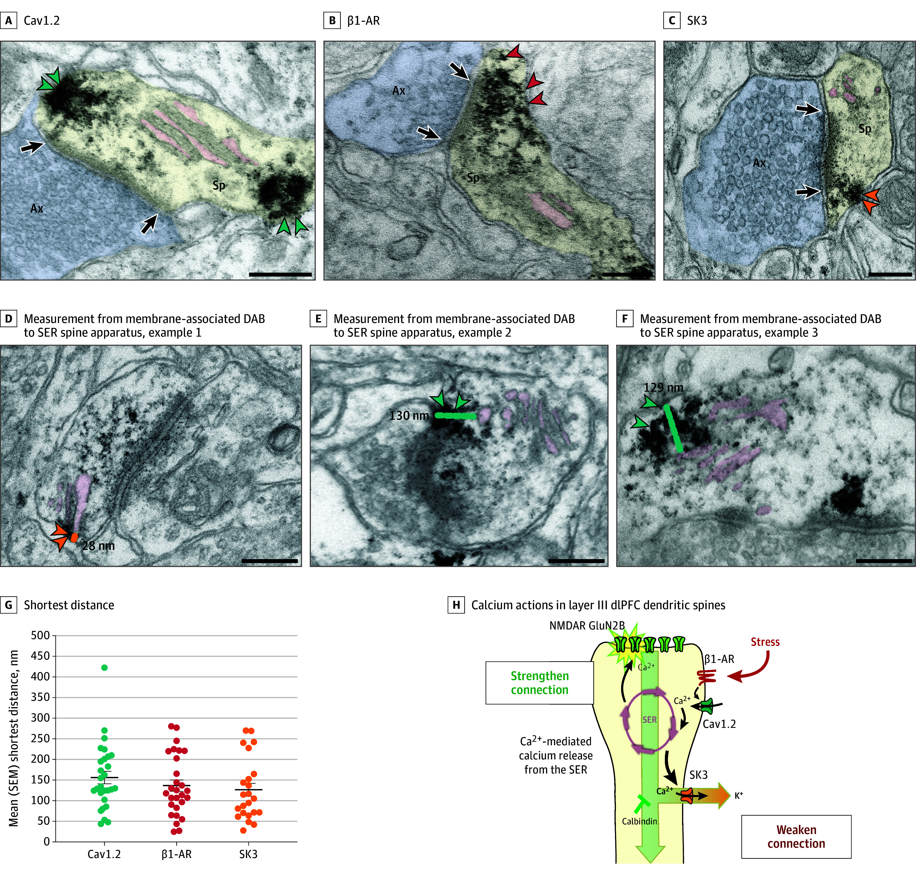
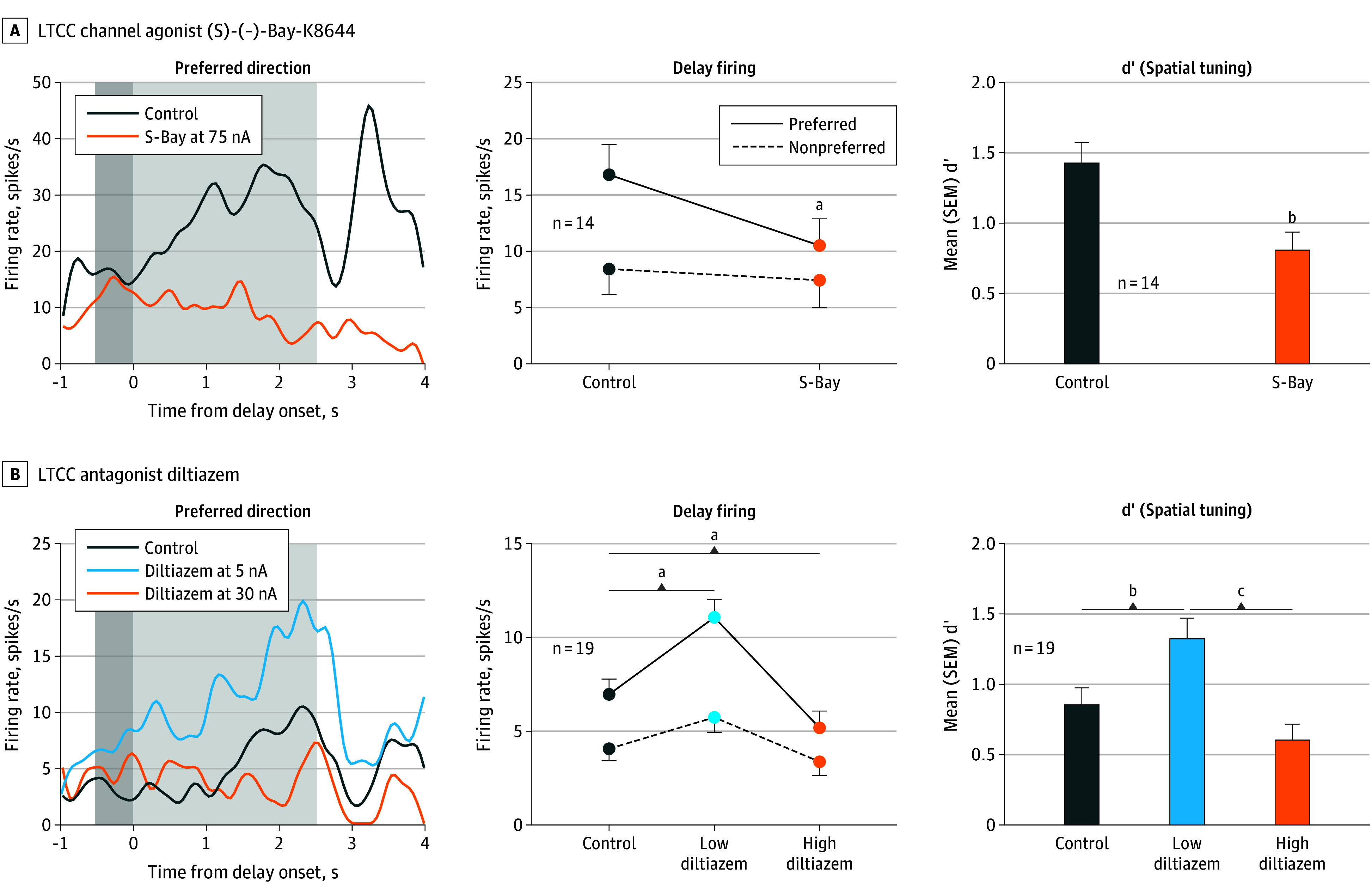
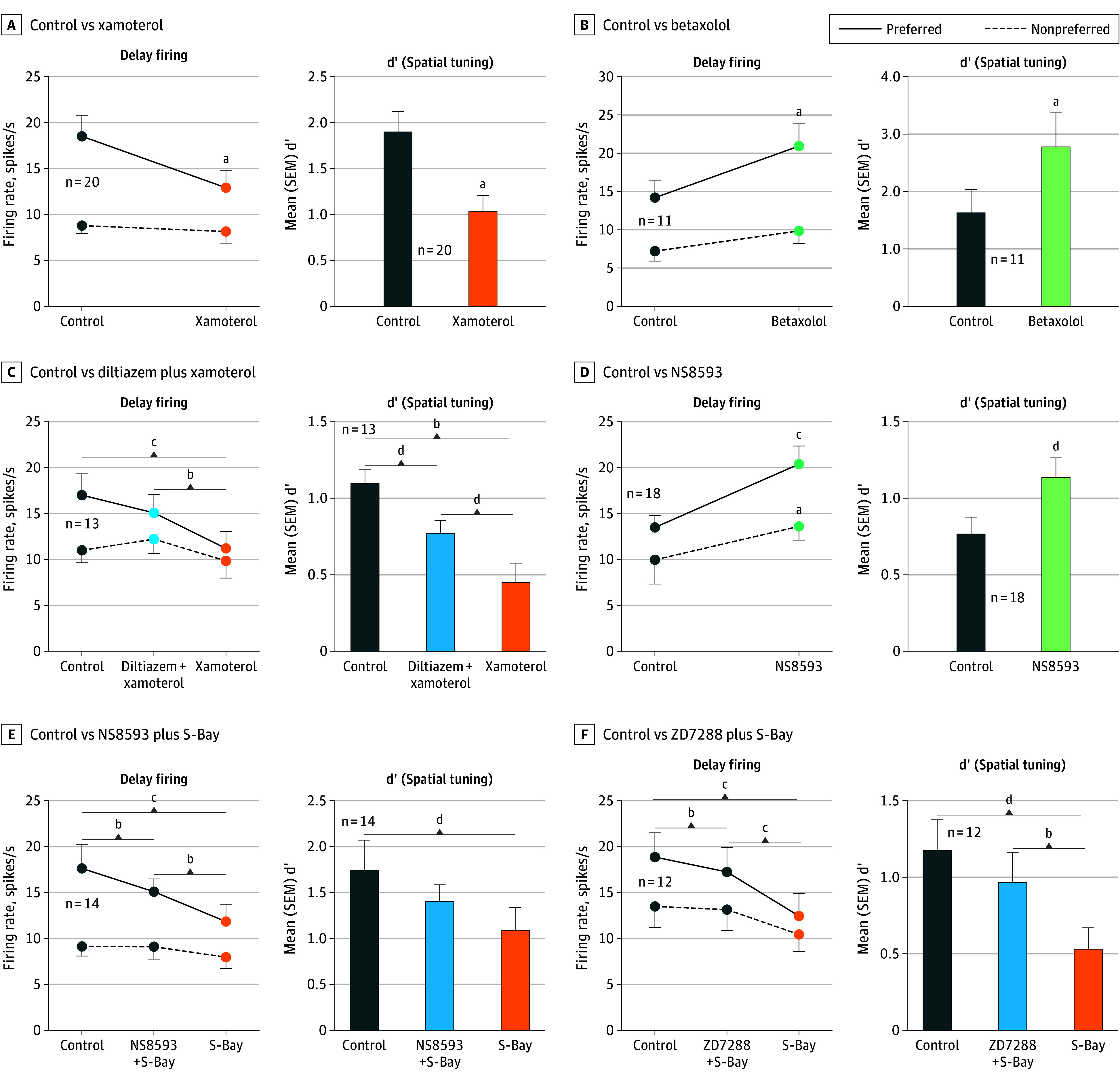
Results: Layer III pyramidal cells in dorsolateral prefrontal cortex coexpress a constellation of calcium-related proteins, delineated by CALB1 (calbindin), and high levels of CACNA1C (Cav1.2), GRIN2B (NMDA receptor GluN2B), and KCNN3 (SK3 potassium channel), concentrated in dendritic spines near the calcium-storing smooth endoplasmic reticulum. L-type calcium channels influenced neuronal firing needed for working memory, where either blockade or increased drive by β1-adrenoceptors, reduced neuronal firing by a mean (SD) 37.3% (5.5%) or 40% (6.3%), respectively, the latter via SK potassium channel opening. An L-type calcium channel blocker or β1-adrenoceptor antagonist protected working memory from stress.

Conclusions and relevance: The layer III pyramidal cells in the dorsolateral prefrontal cortex especially vulnerable in cognitive disorders differentially express calbindin and a constellation of calcium-related proteins including L-type calcium channels Cav1.2 (CACNA1C), GluN2B-NMDA receptors (GRIN2B), and SK3 potassium channels (KCNN3), which influence memory-related neuronal firing. The finding that either inadequate or excessive L-type calcium channel activation reduced neuronal firing explains why either loss- or gain-of-function variants in CACNA1C were associated with increased risk of cognitive disorders. The selective expression of calbindin in these pyramidal cells highlights the importance of regulatory mechanisms in neurons with high calcium signaling, consistent with Alzheimer tau pathology emerging when calbindin is lost with age and/or inflammation.


Conflict of interest statement

Conflict of Interest Disclosures: Drs Joyce, Arnsten, Yang, and Wang reported US provisional patent application No. 63/424,811 pending (“Methods of Treating, Ameliorating, and/or Preventing Stress-Related Disorder”). Dr McCarroll reported grants from the National Science Foundation and the Stanley Center for Psychiatric Research during the conduct of the study. Dr Lewis reported grants from Merck outside the submitted work. Dr Arnsten reported grants from the National Science Foundation, the National Institute of Mental Health, and the National Institute on Drug Abuse during the conduct of the study. No other disclosures were reported.

Figures

Figure 1.
Figure 1.. Transcriptomic Analyses of Excitatory Neurons in the Dorsolateral Prefrontal Cortex (dlPFC) of Humans and Macaques
A, Transcriptomic analyses of human dlPFC pyramidal cells, showing expression levels of CALB1 (encoding the calcium-binding protein calbindin) and KCNN3 (encoding the SK potassium channel opened by calcium, SK3). Population sizes are expressed as number of nuclei out of 20 000. The CUX2-expressing cells have higher CALB1 than other excitatory cells and very high levels of calcium-related genes, including KCNN3. B, Bar graph quantification of CALB1, CACNA1C, KCNN3, GRIN2B (encoding the GluN2B subunit of the NMDA receptor that fluxes high levels of calcium), ADRB1 (encoding the β1-adrenoceptor [β1-AR], which drives Cav1.2 actions in the heart during stress exposure), CACNA1D (encoding the LTCC Cav1.3), HCN1 (encoding the hyperpolarization-activated and cyclic nucleotide–gated channel opened by cyclic adenosine monophosphate), and CHP1 (encoding the calcineurin inhibitor calcineurin homologous protein 1), in the 3 CUX2 (CUX2 A-C) dlPFC populations in the human dlPFC. C, Same as panel A but in the macaque dlPFC. D, Same as panel B but in the macaque dlPFC. Note lower levels of CACNA1D vs CACNA1C in both species. Expression values are normalized counts of the number of transcripts per 100 000 in each cell type.
Figure 2.
Figure 2.. Calbindin-Expressing Pyramidal Cells in Layer III of the Dorsolateral Prefrontal Cortex (dlPFC) of Macaques and NMDA Receptor–GluN2B, Cav1.2 Channels, SK3 Channels, and β1-Adrenoceptor (β1-AR)
A, Expression of the calcium-binding protein calbindin (CB), across the cortical column in the dlPFC of macaques. Pyramidal cells (examples in white rectangles) are concentrated in layer III and have modest calbindin expression (Aa-Ac; white arrowheads), while interneurons are throughout all layers, especially in layer II, and have intense calbindin expression (Ad-Ae; yellow arrowheads). B-E, Calbindin-expressing pyramidal cells coexpress the following proteins: B, NMDA receptor (NMDAR) GluN2B; C, Cav1.2 (amplified with biotin-streptavidin); D, β1-adrenoceptor (β1-AR) (amplified with biotin-streptavidin); and E, SK3. The intensely labeled, bright red neurons (eg, in E) are calbindin-expressing GABAergic interneurons, which have higher levels of calbindin than pyramidal cells. The percentage of calbindin-expressing pyramidal cells coexpressing each protein is shown on the right. Where present, blue labeling is the Hoechst nuclear counterstain.
Figure 3.
Figure 3.. Immuno-Electron Microscopy Ultrastructural Localization of Cav1.2 Channels, SK3 Channels, and β1-Adrenoceptor (β1-AR) in Layer III of the Dorsolateral Prefrontal Cortex (dlPFC) in Macaques
Cav1.2 (A, teal arrowheads), β1-AR (B, red arrowheads), and SK3 channels (C, orange arrowheadss) can be seen in dendritic spines receiving asymmetric (presumed glutamatergic) synapses, often localized on the plasma membrane near the calcium-storing and -releasing smooth endoplasmic reticulum (SER; termed the spine apparatus when it is elaborated in the dendritic spine). The SER spine apparatus is highlighted with pink pseudocoloring. Note that the Cav1.2 labeling can be seen near the postsynaptic density (PSD) and near the SER; additional examples can be seen in eFigures 3 and 4 in Supplement 1. Calbindin was not examined, as it is a cytosolic protein with diffuse labeling. D-F, Examples of measurements from center of membrane-associated diaminobenzidine (DAB) label to the SER spine apparatus. G, Shortest distance measured from center of membrane-associated DAB Cav1.2 (teal), β1-AR (red), and SK3 (orange) channel label to the SER spine apparatus; each dot represents a spine, and the black bar depicts the mean, with error bars indicating SEMs. Note that the DAB label may have obscured the SER spine apparatus in some instances; thus, the proteins on the plasma membrane may have been even closer than measured. H, A working model of calcium actions in layer III dlPFC dendritic spines, showing a functional calcium-related interactome. Under nonstress conditions, moderate Cav1.2 L-type voltage-gated Ca2+ channel actions, including potential calcium-mediated calcium release through ryanodine receptors (RYR) on the SER spine apparatus, are needed for strong working memory delay-related firing, possibly by depolarizing the postsynaptic density (PSD) to permit NMDA receptor (NMDAR) neurotransmission. Previous research has shown that the PSD contains NMDA receptors with GluN2B subunits, and that delay cell firing during working memory depends on NMDA receptor–GluN2B neurotransmission. Under stressful conditions, high levels of norepinephrine stimulate β1-AR to activate a large number of L-type calcium channel (LTCC)/Cav1.2 calcium channels. This may induce high levels of calcium-mediated calcium release from the SER, as occurs with the stress response in the heart. High levels of calcium would open large numbers of SK potassium (K+) channels, rapidly reducing neuronal firing. Feedforward, calcium–cyclic adenosine monophosphate signaling would also open cyclic adenosine monophosphate–sensitive channels on spines (eg, hyperpolarization-activated and cyclic nucleotide–gated slack [K+]) channels. As dlPFC delay cell firing is needed for working memory, these intracellular signaling events lead to cognitive impairment. Sustained reductions in neuronal firing or high levels of cytosolic calcium would also lead to degeneration, especially when the protective effects of calbindin are lost with age and/or inflammation. Black arrows indicate synapses. Ax indicates axon terminal (pseudocolored blue); Mit, mitochondria; Sp, spine (pseudocolored yellow). Scale bars = 200 nm.
Figure 4.
Figure 4.. Recordings From the Dorsolateral Prefrontal Cortex (dlPFC) in Macaques During Working Memory: the Effects of Manipulating LTCC Activity on Delay Cell Firing in the dlPFC of Macaques
The left subpanels show an example neuron’s delay-related firing under control and drug conditions with dark gray indicating the cue period and light gray indicating the delay period; the middle subpanels show the mean (SEM) delay-related firing for all delay cells under control vs drug conditions; the right subpanels show the mean (SEM) d’ measure of spatial tuning for all delay cells under control vs drug conditions. A, The LTCC channel agonist, (S)-(-)-Bay-K8644 (S-Bay; orange), was associated with reduced delay-related firing (R 2-way analysis of variance, F1,13 = 20.06; P < .001) and decreased d’ (paired t test, t13 = 4.16; P = .001). B, The LTCC antagonist diltiazem (dil) produced an inverted-U dose-response, with low doses of diltiazem (5-20nA, light blue) increasing delay-related firing and spatial tuning, while high doses (30-50nA, orange) were associated with reduced delay firing and spatial tuning (firing rate: R 2-way analysis of variance, F2,26 = 26.90, P < .001; spatial tuning: R 1-way analysis of variance, F1.786,32.15 = 13.86, P < .001). aP < .0001. bP < .01. cP < .005.
Figure 5.
Figure 5.. Roles of β1-Adrenoceptor (β1-AR), L-Type Calcium Channel (LTCC), and SK Channels in Dorsolateral Prefrontal Cortex (dlPFC) Delay Cell Firing
The left subpanels show the mean (SEM) delay-related firing for all delay cells under control vs drug conditions; the right subpanels show the mean (SEM) d’ measure of spatial tuning for all delay cells under control vs drug conditions. A, The β1-AR agonist xamoterol (orange) was associated with reduced delay-related firing of dlPFC delay cells for the neurons’ preferred direction but not for nonpreferred directions (R 2-way analysis of variance, F1,19 = 12.99; P = .002), leading to a significant reduction in the d’ measure of spatial tuning (paired t test, t19 = 4.68; P < .001). B, Conversely, the β1-AR antagonist betaxolol (green) was associated with enhanced delay-related firing (R 2-way analysis of variance, F1,10 = 6.42; P = .03), and increased d’ measures of spatial tuning (paired t test, t10 = 3.36; P = .007). C, The reducing effects of the β1-AR agonist xamoterol were blocked by pretreatment with the LTCC antagonist, diltiazem (R 2-way analysis of variance, F2,24 = 5.90; P = .008; Tukey multiple comparisons: preferred direction, control vs diltiazem plus xamoterol; P = .16; control vs xamoterol, P < .001; diltiazem plus xamoterol vs xamoterol, P = .002). D, The SK channel blocker NS8593 was significantly associated with increased delay firing for the neurons’ preferred direction, as well as a smaller increase for nonpreferred directions (R 2-way analysis of variance, F1,17 = 12.13; P = .003), leading to a significant increase in d’ measure of spatial tuning (paired t test, t17 = 2.18; P = .04). E, The mean firing rate of 14 dlPFC delay cells, showing that the reducing effects of the LTCC agonist S-Bay were blocked by pretreatment with the SK channel antagonist NS8593 (R 2-way analysis of variance, F2,26 = 5.89; P = .008). F, The mean firing rate of 12 dlPFC delay cells, showing that the reducing effects of the LTCC agonist S-Bay were blocked by pretreatment with the hyperpolarization-activated and cyclic nucleotide–gated channel antagonist ZD7288 (R 2-way analysis of variance, F2,22 = 13.47; P < .001). aP < .001. bP < .005. cP < .0001. dP < .05.

References

    1. Bigos KL, Mattay VS, Callicott JH, et al. . Genetic variation in CACNA1C affects brain circuitries related to mental illness. Arch Gen Psychiatry. 2010;67(9):939-945. doi:10.1001/archgenpsychiatry.2010.96 - DOI - PMC - PubMed
    1. Cosgrove D, Mothersill O, Kendall K, et al. ; Wellcome Trust Case Control Consortium . Cognitive characterization of schizophrenia risk variants involved in synaptic transmission: evidence of CACNA1C’s role in working memory. Neuropsychopharmacology. 2017;42(13):2612-2622. doi:10.1038/npp.2017.123 - DOI - PMC - PubMed
    1. Koran ME, Hohman TJ, Thornton-Wells TA. Genetic interactions found between calcium channel genes modulate amyloid load measured by positron emission tomography. Hum Genet. 2014;133(1):85-93. doi:10.1007/s00439-013-1354-8 - DOI - PMC - PubMed
    1. Krzyzewska IM, Ensink JBM, Nawijn L, et al. . Genetic variant in CACNA1C is associated with PTSD in traumatized police officers. Eur J Hum Genet. 2018;26(2):247-257. doi:10.1038/s41431-017-0059-1 - DOI - PMC - PubMed
    1. Gordovez FJA, McMahon FJ. The genetics of bipolar disorder. Mol Psychiatry. 2020;25(3):544-559. doi:10.1038/s41380-019-0634-7 - DOI - PubMed

Substances