. 2024 Aug 18;33(17):1524-1539.
doi: 10.1093/hmg/ddae087.

Single nuclei RNA-seq reveals a medium spiny neuron glutamate excitotoxicity signature prior to the onset of neuronal death in an ovine Huntington's disease model

Affiliations

Single nuclei RNA-seq reveals a medium spiny neuron glutamate excitotoxicity signature prior to the onset of neuronal death in an ovine Huntington's disease model

Andrew Jiang et al. Hum Mol Genet. .

Abstract

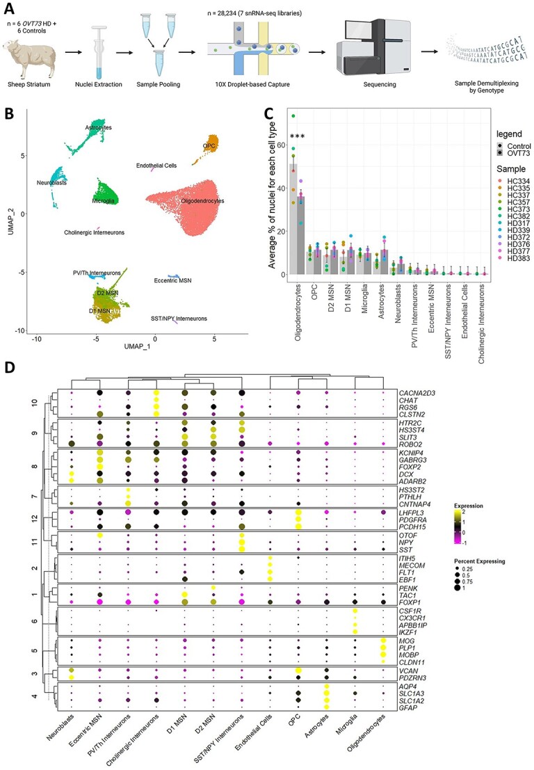
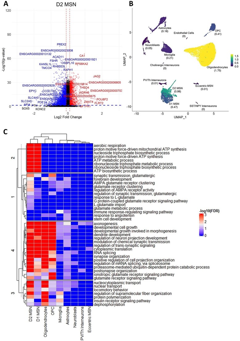
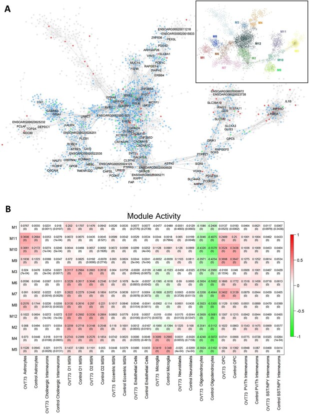
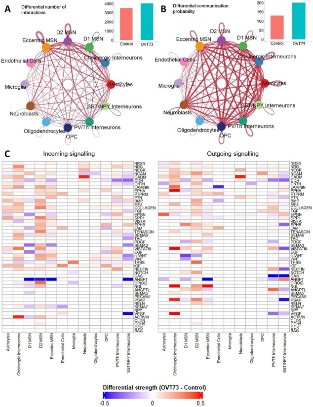
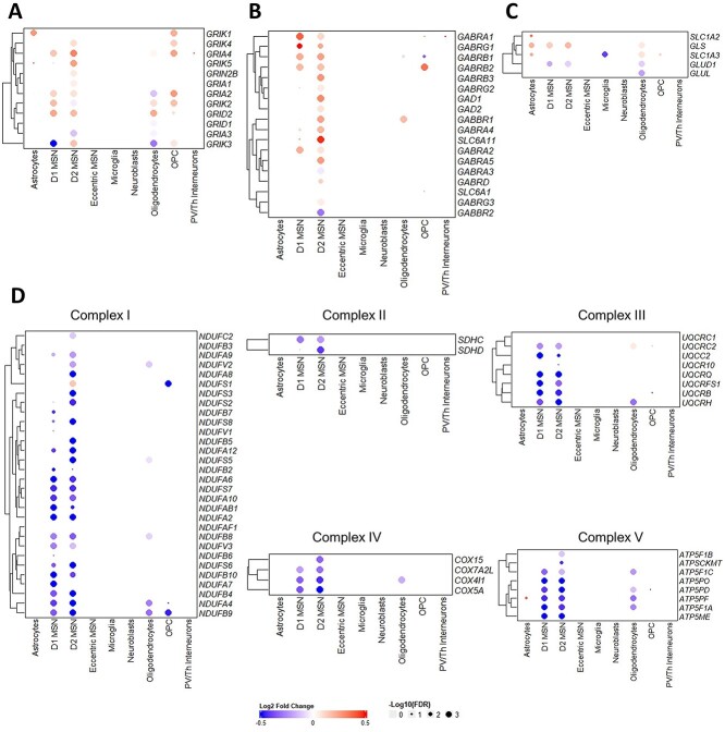
Huntington's disease (HD) is a neurodegenerative genetic disorder caused by an expansion in the CAG repeat tract of the huntingtin (HTT) gene resulting in behavioural, cognitive, and motor defects. Current knowledge of disease pathogenesis remains incomplete, and no disease course-modifying interventions are in clinical use. We have previously reported the development and characterisation of the OVT73 transgenic sheep model of HD. The 73 polyglutamine repeat is somatically stable and therefore likely captures a prodromal phase of the disease with an absence of motor symptomatology even at 5-years of age and no detectable striatal cell loss. To better understand the disease-initiating events we have undertaken a single nuclei transcriptome study of the striatum of an extensively studied cohort of 5-year-old OVT73 HD sheep and age matched wild-type controls. We have identified transcriptional upregulation of genes encoding N-methyl-D-aspartate (NMDA), α-amino-3-hydroxy-5-methyl-4-isoxazolepropionic acid (AMPA) and kainate receptors in medium spiny neurons, the cell type preferentially lost early in HD. Further, we observed an upregulation of astrocytic glutamate uptake transporters and medium spiny neuron GABAA receptors, which may maintain glutamate homeostasis. Taken together, these observations support the glutamate excitotoxicity hypothesis as an early neurodegeneration cascade-initiating process but the threshold of toxicity may be regulated by several protective mechanisms. Addressing this biochemical defect early may prevent neuronal loss and avoid the more complex secondary consequences precipitated by cell death.

Keywords: Huntington's disease; glutamate excitotoxicity; prodromal; single nuclei RNA-seq.


Conflict of interest statement

J.F.G. was a founder and scientific advisory board member with a financial interest in Triplet Therapeutics Inc. His NIH-funded project is using genetic and genomic approaches to uncover other genes that significantly influence when diagnosable symptoms emerge and how rapidly they worsen in Huntington’s disease. The company was developing new therapeutic approaches to address triplet repeat disorders such Huntington’s disease, myotonic dystrophy and spinocerebellar ataxias. His interests were reviewed and are managed by Massachusetts General Hospital and Mass General Brigham in accordance with their conflict of interest policies. J.F.G. has also been a consultant for Wave Life Sciences USA Inc., Biogen Inc. and Pfizer Inc.

All other authors have declared that they have no competing interests.

Figures

Figure 1
Figure 1
Single nuclei RNA-seq of the OVT73 sheep striatum. (A) Experimental design. Nuclei were extracted from the striatum of 12 sheep (6 OVT73 and 6 controls) and pooled to form 7 nuclei suspensions. Single nuclei RNA libraries were generated from the sample multiplexed suspensions and sequenced. Reads were demultiplexed based on the natural genetic variation between pooled samples. Panel image created with BioRender.com. (B) UMAP visualisation of clusters identified and annotated by cell marker gene expression. (C) Proportions of identified cell types across the 12 animals. A significant decrease in oligodendrocytes between OVT73 and control cases was observed (***ANOVA, P < 0.001) (D) dot plot of selected cell-type enriched markers for annotated cell types in the sheep striatum.
Figure 2
Figure 2
Differentially expressed genes in OVT73 versus control sheep striatum. (A) Volcano plot of differentially expressed genes (DEGs) identified in D2 medium spiny neurons (OVT73 D2 MSN vs control D2 MSN). Horizontal blue line shown at P = 0.05, vertical red lines shown at log2 fold change of −0.1 and 0.1. (B) Proportion of DEGs over the median number of expressed genes in each cell type. (C) Heatmap of most significant gene ontology terms (ordered by lowest FDR adjusted p-values) in cell types.
Figure 3
Figure 3
Co-expression gene modules. (A) Co-expression modules generated using the multiscale embedded gene Co-expression network analysis (MEGENA). A total of 12 modules were identified with the structure outlined in the top right. module hub genes are labelled with connected genes represented as dots. Genes differentially expressed between OVT73 and controls are coloured in blue. Genes with evidence to support an interaction with HTT as curated by the HDinHD database [38] were coloured in red. genes that were both differentially expressed and a known HTT interactor were coloured in green. (B) Co-expression module activity in OVT73 and control cell types. Module activity in cell types were determined by computing the module eigengene (first principal component) using normalised expression values of module genes. Eigengene values are shown with p-values of the correlation shown in parentheses in each square. Higher eigengene values indicate higher gene expression of module genes within the cell type.
Figure 4
Figure 4
CellChat cell–cell communication networks inferred from expression of ligand-receptor pairs. Circle plot showcasing the (A) differential number of ligand-receptor interactions and (B) differential communication probability between OVT73 and controls for any two cell types. Red arrows indicate increased number/communication probability in OVT73, blue arrows indicate decreased number/communication probability in OVT73. Thickness of line indicates greater number/communication probability. The total number of ligand-receptor interactions and total interaction strength for OVT73 and controls is also shown as bar graphs on the top right. (C) Differential information flow between OVT73 and control cell types for outgoing (ligand to receptor) and incoming (receptor to ligand) signalling pathways. The information flow for a given signalling pathway is defined as the sum of communication probabilities of all ligand receptor pairs in the pathway. A positive value indicates more communication in OVT73.
Figure 5
Figure 5
Heatmap of log2 fold change of selected genes between OVT73 and control cell types implicated in glutamate signalling. Differential gene transcriptional regulation of (A) glutamate receptors (NMDAR, AMPAR, Kainate, delta receptors), (B) GABAA receptors and glutamate to GABA conversion genes, (C) glutamate uptake transporters and glutamine-glutamate cycle genes and (D) genes encoding oxidative phosphorylation complexes.
Figure 6
Figure 6
Gene regulatory networks show reduced CREB regulon activity in OVT73. (A) CREB-related transcription factor regulated gene modules (regulons) network. The associated transcription factor regulon is shown as triangles with gene members of the regulon connected as dots. The two panels on the right show the log2 fold change of differentially expressed gene members of the regulon between OVT73 and controls in D1 MSNs and D2 MSNs. (B) Differential regulon activity was computed by subtraction of regulon activity between OVT73 and controls. A randomised permutation test with 2000 permutations was performed to determine significant differential regulon activity between OVT73 and control cell types. P-values of the randomised permutation test are shown in the parentheses.

Similar articles

Cited by

References

    1. The Huntington's Disease Collaborative Research Group . A novel gene containing a trinucleotide repeat that is expanded and unstable on Huntington's disease chromosomes. The Huntington's disease collaborative research group. Cell 1993;72:971–983. - PubMed
    1. Snell RG, MacMillan JC, Cheadle JP. et al. Relationship between trinucleotide repeat expansion and phenotypic variation in Huntington's disease. Nat Genet 1993;4:393–397. - PubMed
    1. Jiang A, Handley RR, Lehnert K. et al. From pathogenesis to therapeutics: a review of 150 years of Huntington's disease research. Int J Mol Sci 2023;24:13021. - PMC - PubMed
    1. Doble A. The role of excitotoxicity in neurodegenerative disease: implications for therapy. Pharmacol Ther 1999;81:163–221. - PubMed
    1. Salinska E, Danysz W, Lazarewicz JW. The role of excitotoxicity in neurodegeneration. Folia Neuropathol 2005;43:322–339. - PubMed

Publication types

MeSH terms