. 2024 Jun;300(6):107393.
doi: 10.1016/j.jbc.2024.107393. Epub 2024 May 21.

A novel gain-of-function phosphorylation site modulates PTPN22 inhibition of TCR signaling

Affiliations

A novel gain-of-function phosphorylation site modulates PTPN22 inhibition of TCR signaling

Chuling Zhuang et al. J Biol Chem. 2024 Jun.

Abstract

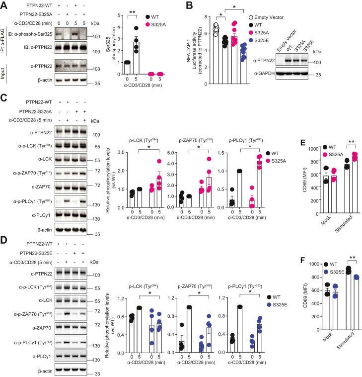
Protein tyrosine phosphatase nonreceptor type 22 (PTPN22) is encoded by a major autoimmunity gene and is a known inhibitor of T cell receptor (TCR) signaling and drug target for cancer immunotherapy. However, little is known about PTPN22 posttranslational regulation. Here, we characterize a phosphorylation site at Ser325 situated C terminal to the catalytic domain of PTPN22 and its roles in altering protein function. In human T cells, Ser325 is phosphorylated by glycogen synthase kinase-3 (GSK3) following TCR stimulation, which promotes its TCR-inhibitory activity. Signaling through the major TCR-dependent pathway under PTPN22 control was enhanced by CRISPR/Cas9-mediated suppression of Ser325 phosphorylation and inhibited by mimicking it via glutamic acid substitution. Global phospho-mass spectrometry showed Ser325 phosphorylation state alters downstream transcriptional activity through enrichment of Swi3p, Rsc8p, and Moira domain binding proteins, and next-generation sequencing revealed it differentially regulates the expression of chemokines and T cell activation pathways. Moreover, in vitro kinetic data suggest the modulation of activity depends on a cellular context. Finally, we begin to address the structural and mechanistic basis for the influence of Ser325 phosphorylation on the protein's properties by deuterium exchange mass spectrometry and NMR spectroscopy. In conclusion, this study explores the function of a novel phosphorylation site of PTPN22 that is involved in complex regulation of TCR signaling and provides details that might inform the future development of allosteric modulators of PTPN22.

Keywords: PTPN22; T cell receptor; allosteric regulation; autoimmunity; catalysis; intrinsically disordered region; phosphoproteomics; phosphorylation; transcriptomics; tyrosine phosphatase.


Conflict of interest statement

Conflict of interest The authors declare that they have no conflicts of interest with the contents of this article.

Figures

Figure 1
Figure 1
PTPN22 Ser325is an inducible GSK3 phosphorylation site in human T cells.A, schematic illustration of 3× FLAG PTPN22 protein purification (left panel) and mass spectra (right panel) indicating Ser325 phosphorylation in immunoprecipitated PTPN22 from 3× FLAG PTPN22 Jurkat WT cells cross-linked with antibodies against human CD3/CD28. Data is representative of three independent biological replicates. Peptide MS2 fragmentation pattern shown displaying m/z and peptide spectral match intensity. B, Western blot analysis obtained using a phospho-Ser325–specific antibody in immunoprecipitated PTPN22 from the lysates of PTPN22 KO Jurkat cells overexpressing 3× FLAG WT or S325A PTPN22 and treated with antibodies against human CD3/CD28 for the indicated time (left panel). Quantification of the phospho-Ser325/total PTPN22 ratio normalized to nonstimulated conditions in four independent experiments (right panel). Statistical significance was assessed using the two-way ANOVA test followed by Bonferroni’s post hoc test, ∗ p < 0.05. C, endogenous Ser325 phosphorylation was analyzed by Western blotting in PTPN22 immunoprecipitated from lysates of 3× FLAG WT PTPN22 Jurkat cells stimulated with antibodies against human CD3/CD28 for the indicated times (left panel). Quantifications of the phospho-Ser325/total PTPN22 ratio normalized to nonstimulated conditions in four independent experiments (right panel). Statistical significance was assessed using the Kruskal–Wallis test, ∗p < 0.05. D, immunoprecipitation analysis of endogenous PTPN22 Ser325 phosphorylation in lysates of human primary CD4+ effector T cells stimulated with antibodies against human CD3/CD28 for the indicated time (left panel). Quantification of the phospho-Ser325/total PTPN22 ratio normalized to nonstimulated conditions in five independent experiments (right panel). Statistical significance was assessed by using the Kolmogorov–Smirnov test, ∗∗ p < 0.01. E, prediction of potential kinases responsible for PTPN22 Ser325 phosphorylation in descending order from left to right. F, immunoprecipitation analysis of phospho-PTPN22 Ser325 in lysates of 3× FLAG PTPN22 WT Jurkat cells with or without incubation with 5 μM GSK3 inhibitor IX and stimulation with antibodies against human CD3/CD28 (left panel). Quantifications of phospho-Ser325/total PTPN22 ratio from Western blot analysis of four independent experiments (right panel). Statistical significance was assessed using the two-way ANOVA test followed by Bonferroni’s post hoc test, ∗∗p < 0.01. G, immunoprecipitation analysis of PTPN22 and GSK3 interaction in lysates of BioID2 Jurkat cells (left panel). Histograms shows quantifications of immunoprecipitated GSK3 α/β based on the Western blot analysis and are representative of four independent experiments (right panel). Statistical significance was assessed using the Kolmogorov–Smirnov test, ∗p < 0.05. CD, cluster of differentiation; GSK3, glycogen synthase kinase 3; PTPN22, protein tyrosine phosphatase nonreceptor type 22.
Figure 2
Figure 2
Phosphorylation of PTPN22 Ser325enhances the inhibitory effect of PTPN22 on T cell receptor signaling.A, immunoprecipitation analysis of PTPN22 Ser325 phosphorylation in lysates of 3× FLAG PTPN22 WT and CRISPR/Cas9-mediated S325A KI Jurkat cells stimulated with antibodies against human CD3/CD28 for the indicated time by Western blotting (left panel). Histogram shows quantifications of the phospho-Ser325/total PTPN22 ratio normalized to nonstimulated condition and is representative of four independent experiments (right panel). Statistical significance was assessed using the two-way ANOVA followed by Bonferroni’s post hoc test, ∗∗p < 0.01. B, dual-luciferase reporter assay analysis of full-length PTPN22 inhibition of TCR signaling in PTPN22 KO Jurkat cells overexpressing full-length 3× FLAG WT, S325E, or S325A PTPN22 together with NFAT/AP-1 firefly and Renilla luciferase reporters and stimulated with antibodies against human CD3/CD28. Luciferase activity was measured (left panel), and the numbers on the y-axis indicate NFAT/AP-1 firefly luciferase activity normalized first to Renilla luciferase activity in each group (KO, WT, or S325 mutant), then to the amount of PTPN22 relative to that of GAPDH as assessed by Western blotting (right panel). Mean ± SEM are shown from three independent experiments each with three replicates per condition. Statistical significance was assessed by using the Kruskal–Wallis test, ∗p < 0.05. C and D, Western blot analysis of TCR signaling in 3× FLAG PTPN22 WT, and CRISPR/Cas9 mediated S325A (C) or S325E (D) KI Jurkat cells treated with antibodies against human CD3/CD28 for indicated time, followed by detection of phosphorylated LCK (Tyr394), ZAP70 (Tyr319), and PLC-γ (Tyr783) in lysates (left panels). Histograms show quantification of phosphorylated LCK, ZAP70, and PLC-γ normalized to relative total protein by four independent experiments (right panels). Statistical significance was assessed using the Kolmogorov–Smirnov test, ∗p < 0.05. E and F, flow cytometry analysis of TCR-induced CD69 expression in 3× FLAG PTPN22 WT, S325A (E), or S325E (F) KI Jurkat cells treated with (stimulated) or without (mock) antibodies against human CD3/CD28 for 4 h. Histograms show median fluorescent intensity (MFI) from seven independent experiments. Statistical significance was assessed using two-way ANOVA, followed by Bonferroni’s post hoc test, ∗∗p < 0.01. AP-1, activator protein-1; CD, cluster of differentiation; GSK3, glycogen synthase kinase 3; LCK, lymphocyte-specific protein tyrosine kinase; NFAT, nuclear factor of activated T cells; PLC, phospholipase C; PTPN22, protein tyrosine phosphatase nonreceptor type 22; TCR, T cell receptor; ZAP70, zeta-chain–associated protein kinase 70.
Figure 3
Figure 3
Signaling pathway changes in global phospho-MS and bulk RNA-seq.A, STRING-DB–generated protein–protein interaction networks of proteins with enriched phosphopeptides from stimulated S325E/A conditions further controlled by removing features with similar increases under mock stimulation. Only terms >2 in strength and an adjusted p value <0.05 were considered. B, STRING-DB–generated protein–protein interaction networks of proteins increased in stimulated S325E. Of note, S325A exhibited no enrichments. C, overrepresentation analysis for reactome pathways. Five hundred six DEGs (≥2-fold and adjusted p value < 0.05: stimulated versus mock) in both S325A and S325E. D, GO functional enrichment for biological process of top genes from 506 DEGs in C ranked by ΔΔlog2FoldChange (99 genes: ΔΔlog2FoldChange>1). Selected GO terms containing “immune” and “T cell activation” terms displayed. E, Δlog2(FoldChange) values for genes from GO terms “immune response” (gray), “T cell activation” (green), or both (black). F, volcano plots (stimulated versus mock) for S325A, S325E, and their WT counterparts. Exemplar genes CD74 and CXCL10 are shown. DEG, differentially expressed gene; GO, gene ontology; MS, mass spectrometry.
Figure 4
Figure 4
In vitro catalytic activity of PTPN22 and its variants.A and B, phosphatase activity assays were performed using immunoprecipitated PTPN22 from PTPN22 WT, or S325A, S325E KI Jurkat cells, and DiFMUP as a substrate. Histograms (left panel) show quantification of initial rates of reaction normalized to the amount of PTPN22 WT as assessed by Western blotting (right panel). Mean ± SEM are shown from five (A) and ten (B) independent experiments. Statistical significance was assessed by using the Kolmogorov–Smirnov test, ∗∗ p < 0.01, ∗∗∗ p < 0.001. C and D, dual-luciferase reporter assay analysis of truncated WT and S325E inhibition of TCR signaling in PTPN22 KO Jurkat cells overexpressing 3 × FLAG PTPN221-340 (C), or PTPN221-330 (D) WT, and S325E together with NFAT/AP-1 firefly and Renilla luciferase reporters and stimulated with antibodies against human CD3/CD28. Luciferase activity was measured and quantified as described in Figure 2B. Means ± SEM are shown from three or four independent experiments each with three replicates per condition. Statistical significance was assessed by using the Kruskal–Wallis test, ∗p < 0.05, ∗∗ p < 0.01. E, representative SDS-PAGE of final purified samples of PTPN221-330 WT and S325E recombinant proteins. F and G, phosphatase activity assays were performed by using recombinant PTPN221-330 WT or S325E and DiFMUP as a substrate. F, representative Michaelis–Menten curve of eight independent experiments each with three technical replicates and representative SDS PAGE of 1 μM PTPN221-330 WT and S325E recombinant proteins. G, dot plot showing kcatand KM. Each data point represents one of eight independent experiments performed as in (F). Statistical significance was assessed using two-tailed Mann–Whitney test, ∗ p < 0.05, ∗∗ p < 0.01. AP-1, activator protein-1; CD, cluster of differentiation; DiFMUP, 6,8-difluoro-4-methylumbelliferyl phosphate; NFAT, nuclear factor of activated T cells; PTPN22, protein tyrosine phosphatase nonreceptor type 22; TCR, T cell receptor.
Figure 5
Figure 5
Effect of Ser325phosphorylation mimic on the deuterium exchange rates of PTPN22 1 to 330.A, rainbow plot showing percent differences in deuterium exchange between PTPN221-330 WT and S325E. Deuterium exchange was assessed for five different time points from 10 to 100000 s. The numbering of the polypeptide chain follows the sequence of the protein as used in the experiment, which has a 17-amino acid N-terminal leader preceding the native methionine 1. Data are representative of two independent experiments. B, the averages of number of deuterium exchanged (#D) shown over time for two of the peptides with the greatest changes. C, ribbon representation of PTPN22 (PDB code 2P6X) annotated with the location of the active site and of polypeptide regions mentioned in the text. For reference, the ribbon color matches the bottom band in the sequence view in A. D, solvent accessible surface representation of the PTP domain in two orthogonal views colored according to the percent difference in the exchange rate at 10,000 s as shown in A. Key residues are indicated. The molecular graphics objects in (C) and (D) were generated with UCSF Chimera (57). Ni-NTA, nickel-nitrilotriacetic acid; PTP, protein tyrosine phosphatase; PTPN22, PTP nonreceptor type 22.
Figure 6
Figure 6
Effect of mimicking Ser325phosphorylation on the interaction between the PTPN22 interdomain and catalytic domain.A, 2D [1H, 15N] HSQC spectrum of 15N-labeled PTPN22 interdomain (residues 299–360, black) and S325E (blue). S325 and E325 residues are annotated. B, 2D [1H, 15N] HSQC spectrum of 15N-labeled PTPN22 interdomain (residues 299–360) alone (black) and in complex with the PTPN22 catalytic domain (residues 1–299; red). Peaks with changing intensities are annotated. C, 2D [1H, 15N] HSQC spectrum of 15N-labeled PTPN22 S325E interdomain (residues 299–360) alone (blue) and in complex with the PTPN22 catalytic domain (residues 1–299; red). Peaks with changing intensities are annotated. HSQC, heteronuclear single quantum coherence; PTPN22, protein tyrosine phosphatase nonreceptor type 22.

References

    1. Tonks N.K. Protein tyrosine phosphatases: from genes, to function, to disease. Nat. Rev. Mol. Cel. Biol. 2006;7:833–846. - PubMed
    1. Bottini N., Peterson E.J. Tyrosine phosphatase PTPN22: multifunctional regulator of immune signaling, development, and disease. Annu. Rev. Immunol. 2014;32:83–119. - PMC - PubMed
    1. Maine C.J., Teijaro J.R., Marquardt K., Sherman L.A. PTPN22 contributes to exhaustion of T lymphocytes during chronic viral infection. Proc. Natl. Acad. Sci. U. S. A. 2016;113:E7231–E7239. - PMC - PubMed
    1. Crabtree J.N., He W., Guan W., Flage M., Miller M.S., Peterson E.J. Autoimmune variant PTPN22 C1858T is associated with impaired responses to Influenza vaccination. J. Infect. Dis. 2016;214:248–257. - PMC - PubMed
    1. Hasegawa K., Martin F., Huang G., Tumas D., Diehl L., Chan A.C. PEST domain-enriched tyrosine phosphatase (PEP) regulation of effector/memory T cells. Science. 2004;303:685–689. - PubMed

Publication types

MeSH terms

Substances