. 2024 Nov-Dec;50(6):1192-1207.
doi: 10.1002/biof.2077. Epub 2024 May 22.

Nuclear receptor 4A1 (NR4A1) upregulated by n-butylidenephthalide via the mitogen-activated protein kinase (MAPK) pathway ameliorates drug-induced gingival enlargement

Affiliations

Nuclear receptor 4A1 (NR4A1) upregulated by n-butylidenephthalide via the mitogen-activated protein kinase (MAPK) pathway ameliorates drug-induced gingival enlargement

Tomoya Ueda et al. Biofactors. 2024 Nov-Dec.

Abstract

Drug-induced gingival enlargement (DIGE) is a side effect of ciclosporin, calcium channel blockers, and phenytoin. DIGE is a serious disease that leads to masticatory and esthetic disorders, severe caries, and periodontitis but currently has no standard treatment. We recently reported that nuclear receptor 4A1 (NR4A1) is a potential therapeutic target for DIGE. This study aimed to evaluate the therapeutic effects of n-butylidenephthalide (BP), which increases the expression of NR4A1, on DIGE. In this study, NR4A1 mRNA expression was analyzed in the patients with periodontal disease (PD) and DIGE. We evaluated the effect of BP on NR4A1 expression in gingival fibroblasts and in a DIGE mouse model. RNA sequencing (RNA-seq) was conducted to identify the mechanisms by which BP increases NR4A1 expression. The results showed that NR4A1 mRNA expression in the patients with DIGE was significantly lower than the patients with PD. BP suppressed the upregulation of COL1A1 expression, which was upregulated by TGF-β. BP also ameliorated gingival overgrowth in DIGE mice and reduced Col1a1 and Pai1 expression. BP also decreased Il1β mRNA expression in gingival tissue in DIGE. RNA-seq results showed an increase in the expression of several genes related to mitogen-activated protein kinase including DUSP genes in gingival fibroblasts stimulated by BP. Treatment with ERK and JNK inhibitors suppressed the BP-induced increase in NR4A1 expression. In addition, BP promoted the phosphorylation of ERK in gingival fibroblasts. In conclusion, BP increases NR4A1 expression in gingival fibroblasts through ERK and JNK signaling, demonstrating its potential as a preventive and therapeutic agent against DIGE.

Keywords: COL1A1; ERK; MAPK; NR4A1; drug‐induced gingival enlargement; n‐butylidenephthalide.


Conflict of interest statement

There are no conflicts of interest to disclose.

Figures
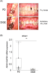
FIGURE 1
FIGURE 1
(A). Intraoral photographs of periodontitis (PD) and drug‐induced gingival hyperplasia (DIGE). On the left is a view of the maxillary right molar taken from the occlusal plane. On the right is a frontal view in occlusion; the PD patient is patient no. 6 and the DIGE patient is patient no. 10. (B). PD and gene expression of NR4A1 in gingival tissue from DIGE patients (n = 10), Box plot, median with interquartile range; Dot, the expression in each patient, **p < 0.01.
FIGURE 2
FIGURE 2
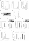
The effect of n‐butylidenephthalide (BP; dose‐dependent (left graph) or time‐dependent (right graph) manner course) on the expression of NR4A1 in GT1 (A) and primary human gingival fibroblasts (B). BP was applied for 1 h at the indicated dose in dose‐dependent manner course and 25 μg/mL BP was applied for indicated time in time‐dependent manner course. (C) The protein levels of NR4A1 in GT1. (D) Relative protein levels of NR4A1 standardized by β actin (n = 3). (E) The effect of BP on transforming growth factor‐β (TGF‐β) induced COL1A1 mRNA levels and protein COL1A1 levels (F; western blotting bands and relative protein levels standardized by β Actin) in GT1. BP (25 μg/mL) was applied to GT1 simultaneously with TGF‐β (10 ng/mL) application (n = 3). (G) mRNA expression of NR4A1. BP was applied for 12 h to GT1 12 h after TGF‐β stimulation (n = 3). (H) Protein COL1A1 levels in BP post treatment (western blotting bands and relative protein levels standardized by β Actin, n = 3). (I) The expression of Nr4a1 induced by BP in mouse gingival fibroblast (mGF) derived from wild type (WT) and NR4A1 knockout mouse (n = 3). J. Evaluation of the role of NR4A1 in the suppression of Col1a1 mRNA expression by BP in gingival fibroblasts derived from WT and NR4A1 knockout mice. BP was applied for 1 h at the indicated dose (n = 3). Bar graph, the mean of mRNA expression and standard deviation (SD), *p < 0.05, **p < 0.01; N.S., not significant. Cohen's d: 0.68; †††1.66.
FIGURE 3
FIGURE 3
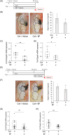
The effect of n‐butylidenephthalide (BP) in the drug‐induced gingival enlargement (DIGE) mouse model. (A). Prevention model schedule. Ciclosporin A (CsA; 50 mg/kg/day) was applied intraperitoneally. BP was topically applied to gingival tissue. (B) The degree of gingival overgrowth was assessed (n = 33–34/group). (C) Col1a1 mRNA expression levels in gingival tissue with or without BP in the prevention model (n = 10–11/group). (D). Pai1 mRNA expression levels in gingival tissue with or without BP in the prevention model (n = 10‐11/group). (E). Treatment model schedule. BP was applied after developing DIGE. (F). Photo of gingival tissue. The degree of gingival overgrowth was assessed (n = 24‐27/group). (G). Col1a1 mRNA expression levels in gingival tissue with or without BP in the treatment model (n = 13‐15/group). (H). Pai1 mRNA expression levels in gingival tissue with or without BP in the treatment model (n = 13‐15/group). Bar graph; mean (standard deviation, SD), Box plot; median (interquartile range), *p < 0.05; **p < 0.01.
FIGURE 4
FIGURE 4
(A) Hematoxylin and eosin (HE) staining of gingival tissue in drug‐induced gingival enlargement (DIGE) mice in the prevention group with or without stimulation with n‐butylidenephthalide (BP; schedule; Figure 3A, n = 9). (B) Picro Sirius red staining of gingival tissue in DIGE mice in the prevention group with or without stimulation with BP (n = 9). (C) HE staining of gingival tissue in DIGE mice in the treatment group with or without stimulation with BP (schedule indicated in Figure 3E, n = 10). (D) Picro Sirius red staining of gingival tissue in DIGE mice in the treatment group with or without stimulation with BP. Connective tissue area was evaluated (n = 10). Bar graph; mean (standard deviation, SD), *p < 0.05; **p < 0.01. CsA, ciclosporin A.
FIGURE 5
FIGURE 5
(A) Evaluation of the effect of n‐butylidenephthalide (BP) in inflammatory cells infiltrating the gingival tissue of drug‐induced gingival enlargement (DIGE) mice. The squared area was 100 μm × 100 μm. Red arrowheads indicate inflammatory cells. (B) Comparison of inflammatory cell number in gingival tissue area in the DIGE prevention model (schedule indicated in Figure 3A, n = 9). (C) The mRNA expression of Il1β, Il6, and Tnfα in gingival tissue in the DIGE prevention model (schedule indicated in Figure 3A, n = 11). Bar graph; mean (standard deviation, SD), Box plot; median (interquartile range), *p < 0.05, N.S., not significant. CsA, ciclosporin A.
FIGURE 6
FIGURE 6
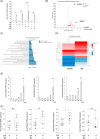
(A) The effect of n‐butylidenephthalide (BP) on NR4A1 expression with ciclosporin A (CsA), NIF, or PHT in GT1 (n = 3). (B) Volcano map of differentially expressed genes in GT1 applied with BP or vehicle. Arrows indicate DUSP1 and NR4A1. (C) Gene ontology enrichment analysis of the differentially expressed genes; molecular function. (D) Heatmap of genes associated with mitogen‐activated protein kinase signaling. (E) The mRNA expression of DUSP1, 5, and 10 stimulated by BP (25 μg/mL) in GT1 for indicated times. (F) The mRNA expression of Nr4a1, Dusp1, 5, and 10 in gingival tissue in the drug‐induced gingival enlargement prevention model (schedule indicated in Figure 3A, n = 11). Bar graph; mean (standard deviation, SD), Box plot; median (interquartile range), *p < 0.05; **p < 0.01 vs. control. N.S.; not significant.
FIGURE 7
FIGURE 7
(A) Inhibitor analysis for JNK (SP600125), p38 (SB203580), ERK1/2 (PD98059), and PKC (Go6983). Inhibitors (5 μM) were applied for 1 h before BP stimulation (n = 3). (B). Western blotting for phosphorylation JNK, total JNK, phosphorylation ERK and total ERK. n‐Butylidenephthalide (BP; 25 μg/mL) was applied for the indicated time. (C). Relative protein levels of p‐JNK standardized by total JNK (n = 5). (D). Relative protein levels of p‐ERK (n = 5). Bar graph; mean (standard deviation, SD), **p < 0.01 versus control. †† p < 0.01 versus BP only application.

References

    1. Hatano S, Matsuda S, Okanobu A, Furutama D, Memida T, Kajiya M, et al. The role of nuclear receptor 4A1 (NR4A1) in drug‐induced gingival overgrowth. FASEB J. 2021;35:e21693. - PubMed
    1. Chen J, Jia J, Ma L, Li B, Qin Q, Qian J, et al. Nur77 deficiency exacerbates cardiac fibrosis after myocardial infarction by promoting endothelial‐to‐mesenchymal transition. J Cell Physiol. 2021;236:495–506. - PubMed
    1. Fuchs CD, Claudel T, Mlitz V, Riva A, Menz M, Brusilovskaya K, et al. GLP‐2 improves hepatic inflammation and fibrosis in Mdr2−/− mice via activation of NR4a1/Nur77 in hepatic stellate cells and intestinal FXR signaling. Cell Mol Gastroenterol Hepatol. 2023;16:847–856. - PMC - PubMed
    1. Ma G, Chen F, Liu Y, Zheng L, Jiang X, Tian H, et al. Nur77 ameliorates age‐related renal tubulointerstitial fibrosis by suppressing the TGF‐β/Smads signaling pathway. FASEB J. 2022;36:e22124. - PubMed
    1. Palumbo‐Zerr K, Zerr P, Distler A, Fliehr J, Mancuso R, Huang J, et al. Orphan nuclear receptor NR4A1 regulates transforming growth factor‐β signaling and fibrosis. Nat Med. 2015;21:150–158. - PubMed

MeSH terms