. 2024 Nov 25;16(6):mjae024.
doi: 10.1093/jmcb/mjae024.

Phase separation of SPIN1 through its IDR facilitates histone methylation readout and tumorigenesis

Affiliations

Phase separation of SPIN1 through its IDR facilitates histone methylation readout and tumorigenesis

Yukun Wang et al. J Mol Cell Biol. .

Abstract

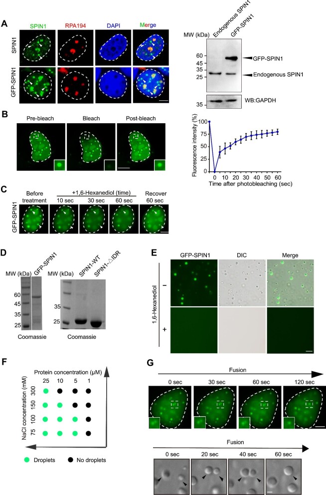
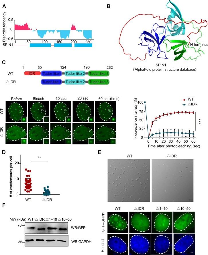
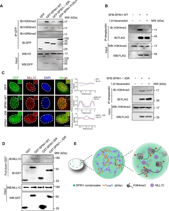
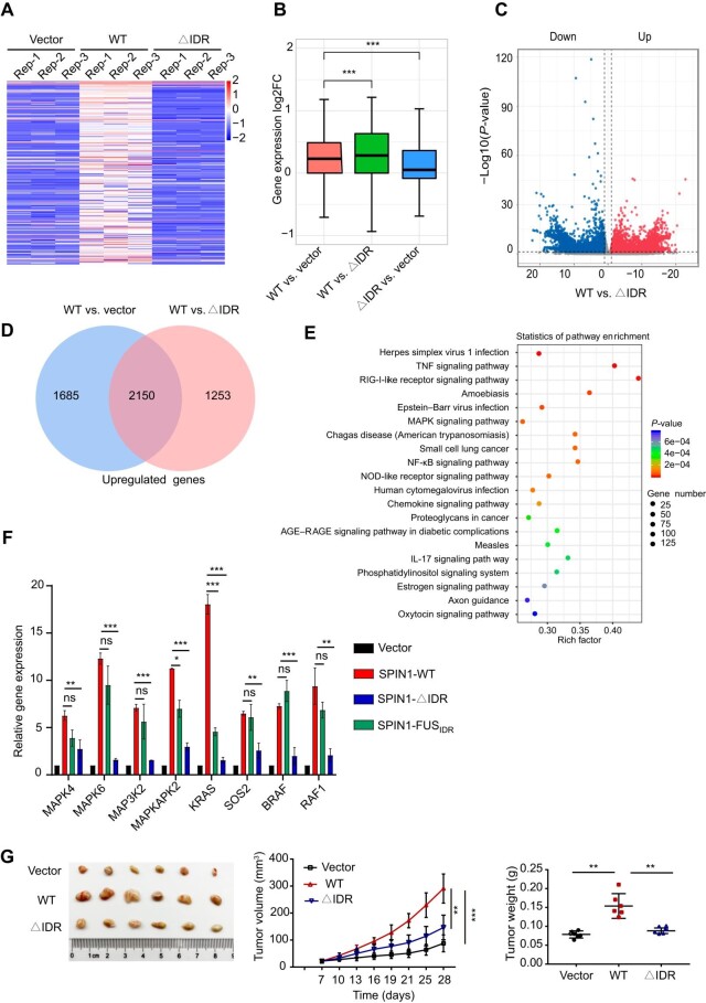
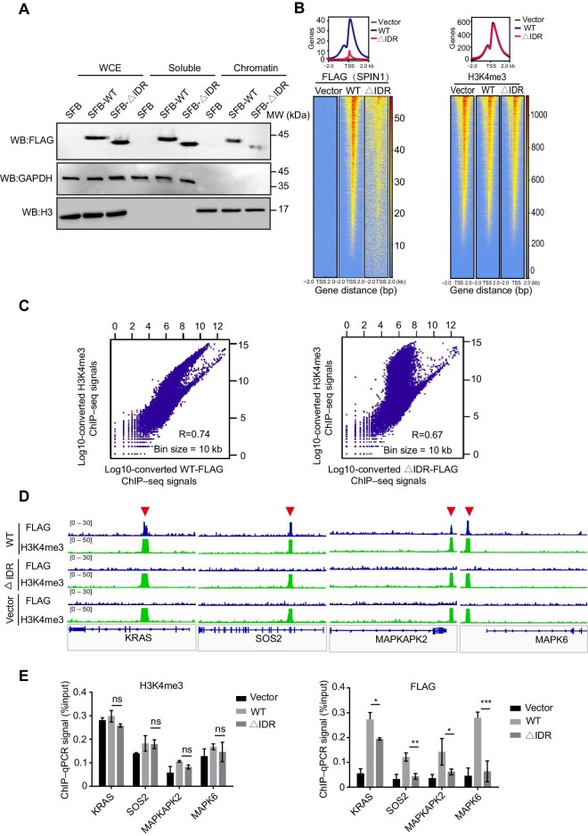
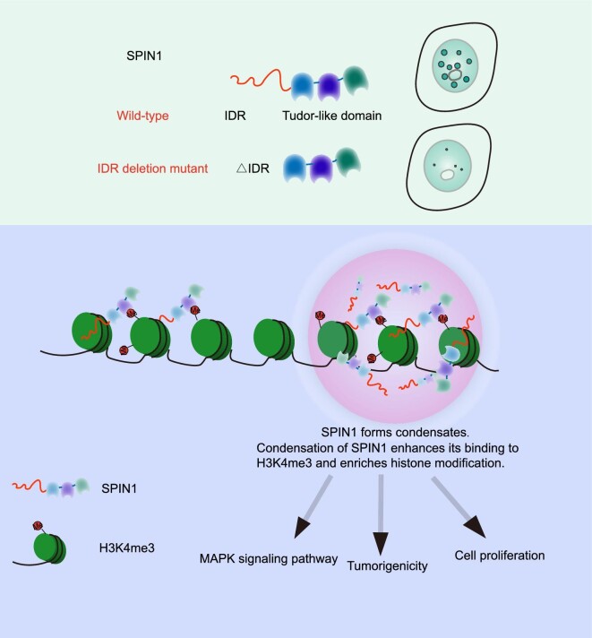
Spindlin1 (SPIN1) is a unique multivalent histone modification reader that plays a role in ribosomal RNA transcription, chromosome segregation, and tumorigenesis. However, the function of the extended N-terminal region of SPIN1 remains unclear. Here, we demonstrated that SPIN1 can form phase-separated and liquid-like condensates both in vitro and in vivo through its N-terminal intrinsically disordered region (IDR). The phase separation of SPIN1 recruits the histone methyltransferase MLL1 to the same condensates and enriches the H3K4 methylation marks. This process also facilitates the binding of SPIN1 to H3K4me3 and activates tumorigenesis-related genes. Moreover, SPIN1-IDR enhances the genome-wide chromatin binding of SPIN1 and facilitates its localization to genes associated with the MAPK signaling pathway. These findings provide new insights into the biological function of the IDR in regulating SPIN1 activity and reveal a previously unrecognized role of SPIN1-IDR in histone methylation readout. Our study uncovers the crucial role of appropriate biophysical properties of SPIN1 in facilitating gene expression and links phase separation to tumorigenesis, which provides a new perspective for understanding the function of SPIN1.

Keywords: IDR; SPIN1; histone methylation reader; phase separation; tumorigenesis.


Figures

Figure 1
Figure 1
SPIN1 forms reversible, dynamic phase-separated condensates in the nucleus. (A) SPIN1 forms condensates in the nucleus. Left: representative co-immunostaining images of endogenous SPIN1 (top) or exogenous GFP-tagged SPIN1 (bottom) with the nucleolus marker RPA194 in U2OS cells. Scale bar, 5 μm. Right: the expression levels of endogenous or exogenous SPIN1. (B) Fluorescence recovery kinetics of GFP-SPIN1 analyzed by the FRAP assay. Left: representative images of the condensates formed by SPIN1 before and after photobleaching. The photobleached region is boxed and amplified. Scale bar, 5 μm. Right: statistical analysis of the FRAP assay from three independent experiments. Data are shown as mean ± SEM. (C) 1,6-Hexanediol treatment disrupts GFP-SPIN1 droplets. (D) GFP-SPIN1, SPIN1-WT, and SPIN1-∆IDR proteins purified from E. coli and examined by Coomassie blue staining. (E) Images of the droplets formed by 10 μM GFP-SPIN1 in the presence or absence of 2.5% 1,6-hexanediol. Scale bar, 20 μm. (F) Phase diagrams of the SPIN1 protein at the concentration ranging from 1 μM to 25 μM in ‘condensation buffer’. (G) Representative droplet fusion events at the indicated time points. Rapid fusion of GFP-SPIN1 in U2OS cell nucleus is boxed and amplified (top). The arrowhead indicates the two-droplet fusion in vitro (bottom). Scale bar, 5 μm.
Figure 2
Figure 2
The IDR of SPIN1 is essential for its phase separation. (A) The disorder tendency of the SPIN1 protein sequence analyzed by IUPred3. (B) The SPIN1 structure analyzed by AlphaFold. SPIN1-IDR is highlighted in red. (C) The FRAP assay of condensates formed by GFP-SPIN1-WT or GFP-SPIN1-∆IDR. The schematic diagram on the top shows the domains of SPIN1-WT and the SPIN1-∆IDR mutant. Left: representative images of condensates at the indicated time points. Scale bar, 5 μm. Right: statistical analysis of the FRAP assay from three independent observations. (D) Quantification of the condensates formed by GFP-SPIN1-WT or GFP-SPIN1-∆IDR (50 individual cells, mean  ±  SD). (E) DIC images of the phase-separated condensates formed by 10 μM SPIN1-WT or SPIN1-∆IDR in the presence of 10% (w/v) PEG5000. Scale bar, 20 μm. (F) Left: the expression levels of SPIN1-WT and SPIN1 mutants. Right: the localization of SPIN1-WT and SPIN1 mutants. Scale bar, 5 μm.
Figure 3
Figure 3
SPIN1-IDR promotes the recognition of H3K4me3 by SPIN1. (A) Loss of SPIN1-IDR compromises the binding of SPIN1 to H3K4me3 but does not affect the binding to H3K9me3 or H3R8me2. SGC7901 cells were transfected with GFP or the SPIN1 mutants. Then, the whole-cell lysates were immunoprecipitated with GFP antibody. (B) 1,6-Hexanediol impairs the binding of SPIN1 to H3K4me3. SGC7901 cells stably expressing SFB-SPIN1-WT (top) or SFB-SPIN1-∆IDR (bottom) were treated with or without 1% 1,6-hexanediol before being harvested for IP and western blotting. (C) MLL1C is partially recruited to the condensates formed by SPIN1. Left: co-staining images of SPIN1-WT or SPIN1-∆IDR with MLL1C. Scale bar, 5 μm. Right: statistical analysis of the fluorescence signals. (D) The interaction between GST-SPIN1 or the mutants and endogenous MLL1C. The recombinant proteins were pretreated with 10% (w/v) PEG5000 to facilitate the formation of condensates and then incubated with cell lysates. (E) A proposed model illustrating the regulatory role of SPIN1 condensation in facilitating the local enrichment of MLL1C and promoting the binding ability of SPIN1 to histone methylation.
Figure 4
Figure 4
SPIN1-IDR regulates gene expression and tumor formation. (A) Heatmap of the top 300 upregulated genes in cells expressing SPIN1-WT compared with cells expressing the empty vector or SPIN1-∆IDR. (B) Box plots showing the relative expression of the top 300 genes in A. (C) A volcano plot showing the statistically upregulated (right) and downregulated (left) genes. (D) Venn diagrams showing the numbers of uniquely or commonly upregulated genes in SPIN1-WT vs. vector and SPIN1-WT vs. SPIN1-∆IDR. (E) KEGG enrichment analysis of the differentially expressed genes in cells expressing SPIN1-WT vs. SPIN1-∆IDR. (F) Relative mRNA levels of the indicated genes associated with the MAPK signaling pathway. Data from triplicate experiments are presented as mean ± SD. (G) SPIN1-WT but not SPIN1-∆IDR significantly facilitates xenograft tumor growth. Left: images of the xenograft tumors. Middle: curves of tumor volume. Right: tumor weight. n = 6/group.
Figure 5
Figure 5
SPIN1-IDR occupies the promoter region of genes involved in the MAPK signaling pathway. (A) Loss of IDR significantly reduces the chromatin-binding ability of SPIN1. Whole-cell extracts (WCE), along with soluble and chromatin-bound SPIN1, were isolated from SGC7901 cells. (B) Heatmap (lower) and composite plot (upper) for SPIN1 or H3K4me3 peaks in the indicated cells. Each row represents a peak called for FLAG or H3K4me3 occupancy at gene loci ±2 kb around the TSS. (C) Scatterplots showing the correlation between the ChIP–seq signals of SPIN1-WT (left) or SPIN1-∆IDR (right) and H3K4me3. (D) Representative integrative genomics viewer browser tracks for the indicated genes associated with the MAPK signaling pathway. (E) ChIP–qPCR validations of the binding sites in D with anti-FLAG or anti-H3K4me3 antibodies.
Figure 6
Figure 6
A proposed model illustrating that SPIN1 forms condensates via LLPS to facilitate the recognition of H3K4me3. SPIN1 forms phase-separated and liquid-like condensates through its N-terminal IDR. This process facilitates the binding of SPIN1 to H3K4me3 and activates tumorigenesis-related genes.

Similar articles

Cited by

References

    1. Ahn J.H., Davis E.S., Daugird T.A. et al. (2021). Phase separation drives aberrant chromatin looping and cancer development. Nature 595, 591–595. - PMC - PubMed
    1. Alberti S., Dormann D. (2019). Liquid–liquid phase separation in disease. Annu. Rev. Genet. 53, 171–194. - PubMed
    1. Alberti S., Gladfelter A., Mittag T. (2019). Considerations and challenges in studying liquid–liquid phase separation and biomolecular condensates. Cell 176, 419–434. - PMC - PubMed
    1. Bae N., Gao M., Li X. et al. (2017a). A transcriptional coregulator, SPIN·DOC, attenuates the coactivator activity of Spindlin1. J. Biol. Chem. 292, 20808–20817. - PMC - PubMed
    1. Bae N., Viviano M., Su X. et al. (2017b). Developing Spindlin1 small-molecule inhibitors by using protein microarrays. Nat. Chem. Biol. 13, 750–756. - PMC - PubMed

Publication types

MeSH terms