. 2024 Jul 1;132(1):96-107.
doi: 10.1152/jn.00001.2024. Epub 2024 May 22.

Surprising multifunctionality of a Tritonia swim CPG neuron: C2 drives the early phase of postswim crawling despite being silent during the behavior

Affiliations

Surprising multifunctionality of a Tritonia swim CPG neuron: C2 drives the early phase of postswim crawling despite being silent during the behavior

Evan S Hill et al. J Neurophysiol. .

Abstract

In response to a suitably aversive skin stimulus, the marine mollusk Tritonia diomedea launches an escape swim followed by several minutes of high-speed crawling. The two escape behaviors are highly dissimilar: whereas the swim is a muscular behavior involving alternating ventral and dorsal whole body flexions, the crawl is a nonrhythmic gliding behavior mediated by the beating of foot cilia. The serotonergic dorsal swim interneurons (DSIs) are members of the swim central pattern generator (CPG) and also strongly drive crawling. Although the swim network is very well understood, the Tritonia crawling network to date comprises only three neurons: the DSIs and pedal neurons 5 and 21 (Pd5 and Pd21). Since Tritonia's swim network has been suggested to have arisen from a preexisting crawling network, we examined the possible role that another swim CPG neuron, C2, may play in crawling. Because of its complete silence in the postswim crawling period, C2 had not previously been considered to play a role in driving crawling. However, semi-intact preparation experiments demonstrated that a brief C2 spike train surprisingly and strongly drives the foot cilia for ∼30 s, something that cannot be explained by its synaptic connections to Pd5 and Pd21. Voltage-sensitive dye (VSD) imaging in the pedal ganglion identified many candidate crawling motor neurons that fire at an elevated rate after the swim and also revealed several pedal neurons that are strongly excited by C2. It is intriguing that unlike the DSIs, which fire tonically after the swim to drive crawling, C2 does so despite its postswim silence.NEW & NOTEWORTHY Tritonia swim central pattern generator (CPG) neuron C2 surprisingly and strongly drives the early phase of postswim crawling despite being silent during this period. In decades of research, C2 had not been suspected of driving crawling because of its complete silence after the swim. Voltage-sensitive dye imaging revealed that the Tritonia crawling motor network may be much larger than previously known and also revealed that many candidate crawling neurons are excited by C2.

Keywords: invertebrate; multifunctional; neural network.


Conflict of interest statement

No conflicts of interest, financial or otherwise, are declared by the authors.

Figures

None
Graphical abstract
Figure 1.
Figure 1.
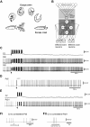
A: illustration of the Tritonia escape swim and crawl behaviors. An escape swim is always followed by an escape crawl. B: diagram of the Tritonia swim and crawl circuitry. The swim circuit is well understood from the levels of the sensory neurons to swim command and central pattern generator (CPG) neurons. In addition to being members of the swim CPG, the dorsal swim interneurons (DSIs) are known to also drive crawling by exciting the only known crawling motor neurons, Pd5 and Pd21. Solid lines represent monosynaptic connections, and dashed lines represent polysynaptic connections. Bars represent excitatory synapses, and circles denote inhibitory synapses. DFN, dorsal flexion neuron; DRI, dorsal ramp interneuron; VFN, ventral flexion neuron; VSI, ventral swim interneuron. C: intracellular recording in the isolated Tritonia central nervous system (CNS) from C2 and 2 DSIs during the escape swim motor program and during the postswim period, showing that whereas the DSIs increase their firing rate after the swim, C2 is completely silent. D: intracellular recording from Pd5 during the swim motor program (indicated by the dashed gray line) and postswim period, showing that Pd5 is inhibited during the swim and then fires at an elevated rate in the postswim period. E: intracellular recording from C2 and Pd21 during the swim and postswim period, showing that Pd21 fires at an elevated rate postswim while C2 is completely silent. Fi: a C2 spike train (10 Hz, 2 s, gray bar) inhibits Pd5 for many seconds. Fii: a C2 spike train (10 Hz, 5 s, gray bar) weakly excites Pd21. The arrows in C–E denote when a stimulus (10 V, 10 Hz, 2 s, 5-ms pulses) was delivered to pedal nerve 3 (which contains sensory neuron axons) to elicit a swim motor program.
Figure 2.
Figure 2.
Experiments in semi-intact preparations demonstrated that C2 strongly drives carbon particle movement on the foot, i.e., ciliary beating. A: schematic of the experimental setup. B: movement of carbon particles on the foot was significantly increased after the termination of a 10-Hz, 5-s C2 spike train (gray bar), with the effect lasting ∼30 s.
Figure 3.
Figure 3.
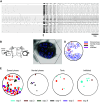
Voltage-sensitive dye (VSD) imaging in the dorsal pedal ganglion revealed that many neurons increased their spiking rate postswim. A: neurons that fired >150% of the spikes postswim that they fired preswim (33 neurons out of a total of 41 neurons recorded) are shown. A 10-V, 10-Hz, 2-s stimulus was given to the contralateral pedal nerve 3 (PdN3) at the arrow to elicit a swim motor program. B: schematic of the Tritonia central nervous system (CNS) showing the location of the photodiode array (PDA) (hexagon) over the right dorsal pedal ganglion and placement of the stimulating extracellular electrode. Ce, cerebral ganglion; Pd, pedal ganglion; Pl, pleural ganglion. C: map of the ganglion locations of the neurons shown in A, color-coded by their activity during the swim (key on right of D). The hexagon corresponds to the PDA’s field of view. D: composite map showing neurons from 7 preparations that fired >150% the number of spikes postswim compared to preswim. The black oval represents the outline of the right pedal ganglion. E: neurons from the 7 preparations that fired >150% the number of spikes after swim vs. before swim are shown by type (dorsal phase, ventral phase, double burster, tonic, inhibited), color-coded by experiment (exp). All of the types of neurons appear to be distributed throughout the ganglion without any clear spatial segregation.
Figure 4.
Figure 4.
Voltage-sensitive dye (VSD) imaging revealed that many neurons in the ventral pedal ganglion also increased their spiking rate after the swim. A: neurons that fired >150% of the spikes after the swim that they fired before swim (28 neurons out of a total of 37 neurons recorded) are shown. A 10-V, 10-Hz, 2-s stimulus to the contralateral pedal nerve 3 (PdN3) was given at the arrow. B: schematic of the experimental setup showing the location of the photodiode array (PDA) (hexagon) over the right ventral pedal ganglion. Ce, cerebral ganglion; Pd, pedal ganglion; Pl, pleural ganglion. C: map of the ganglion locations of the neurons shown in A, color-coded by their activity during the swim (key on right of D). D: composite map showing neurons from 6 preparations that fired >150% the number of spikes postswim compared to preswim. The black oval represents the outline of the right pedal ganglion. E: neurons from the 6 preparations that fired >150% the number of spikes postswim vs. preswim shown by type (dorsal phase, ventral phase, tonic, inhibited), color-coded by experiment (exp). Whereas the dorsal-phase neurons are arranged in a “C” shape along the periphery of the ganglion, there is no clear spatial segregation of the other types of neurons. The large dorsal-phase neurons in the top right quadrant (outlined by a dashed rectangle) are likely to be Pd21 (based on their location, size, and activity during and after the swim) recorded in 5 separate experiments.
Figure 5.
Figure 5.
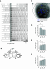
Voltage-sensitive dye (VSD) imaging combined with intracellular stimulation shows that C2 excites many neurons in the contralateral pedal ganglion. A: many neurons in the contralateral dorsal pedal ganglion were excited by a 10-Hz, 2-s C2 spike train (gray bar) to fire at >150% the spike rate they fired at before the C2 spike train. Other neurons in the contralateral pedal ganglion were either inhibited or not affected by the C2 spike train (data not shown). B: schematic of the experimental setup. The hexagon shows the location of the photodiode array. Ce, cerebral ganglion; Pd, pedal ganglion; Pl, pleural ganglion. C: map of the ganglion locations of the neurons excited by the C2 spike train color-coded according to their activity during the swim: blue, neurons that fired during the dorsal phase; black, neurons that were tonically active during the swim. D: bar graph showing the significant effect of the C2 spike train on the spike rate of the neurons shown in A for three 5-s bins (i.e., for the duration of the recording) following the C2 spike train. E and F: bar graphs showing similar significant effects of the C2 spike train on neurons in the contralateral dorsal pedal ganglion in 2 other experiments. In DF * represent significant differences (P < 0.05).
Figure 6.
Figure 6.
The data suggest that C2 and the dorsal swim interneurons (DSIs) work together to drive the initial phase of the escape crawl, after which extended crawling, which can last for many tens of minutes, is driven solely by the DSIs. A: C2 and the DSIs first generate the swim behavior as central pattern generator (CPG) members and then both drive the early phase of the escape crawl by different mechanisms: C2 by slow, long-lasting excitation and the DSIs by elevated tonic firing. Prolonged crawling is then driven solely by the DSIs. B: the Tritonia crawling network may be much bigger than was previously thought, with many hypothesized unidentified crawling motor neurons receiving fast inhibitory followed by slow excitatory input from C2. Solid lines represent monosynaptic connections, and dashed lines represent polysynaptic connections. Bars represent excitatory synapses, and circles denote inhibitory synapses. The red line between C2 and the hypothesized unidentified efferent crawl neurons signifies that although we speculate that the connection is monosynaptic, it has not yet been shown to be. DFN, dorsal flexion neuron; DRI, dorsal ramp interneuron; VFN, ventral flexion neuron; VSI, ventral swim interneuron.

Comment in

Similar articles

Cited by

References

    1. Audesirk G. Central neuronal control of cilia in Tritonia diomedea. Nature 272: 541–543, 1978. doi: 10.1038/272541a0. - DOI - PubMed
    1. Willows AO. Giant brain cells in mollusks. Sci Am 224: 68–75, 1971. doi: 10.1038/scientificamerican0271-68. - DOI - PubMed
    1. Popescu IR, Frost WN. Highly dissimilar behaviors mediated by a multifunctional network in the marine mollusk Tritonia diomedea. J Neurosci 22: 1985–1993, 2002. doi: 10.1523/JNEUROSCI.22-05-01985.2002. - DOI - PMC - PubMed
    1. Getting PA. Emerging principles governing the operation of neural networks. Annu Rev Neurosci 12: 185–204, 1989. doi: 10.1146/annurev.ne.12.030189.001153. - DOI - PubMed
    1. Frost WN, Hoppe TA, Wang J, Tian LM. Swim initiation neurons in Tritonia diomedea. Am Zool 41: 952–961, 2001. doi: 10.1093/icb/41.4.952. - DOI

Publication types

MeSH terms

LinkOut - more resources